


Abstract
18F-2β-Carbomethoxy-3β-(4-chlorophenyl)-8-(2-fluoroethyl)nortropane (18F-FECNT), a PET radioligand for the dopamine transporter (DAT), generates a radiometabolite that enters the rat brain. The aims of this study were to characterize this radiometabolite and to determine whether a similar phenomenon occurs in human and nonhuman primate brains by examining the stability of the apparent distribution volume in DAT-rich (striatum) and DAT-poor (cerebellum) regions of the brain. Methods: Two rats were infused with 18F-FECNT and sacrificed at 60 min. Extracts of brain and plasma were analyzed by high-performance liquid chromatography (HPLC) and liquid chromatography–mass spectrometric (LC-MS) techniques. Two human participants and 3 rhesus monkeys were injected with 18F-FECNT and scanned kinetically, with serial arterial blood analysis. Results: At 60 min after the injection of rats, 18F-FECNT accumulated to levels about 7 times higher in the striatum than in the cortex and cerebellum. The radiometabolite was distributed at equal concentrations in all brain regions. The LC-MS techniques identified N-dealkylated FECNT as a major metabolite in the rat brain, and reverse-phase HPLC detected an equivalent amount of radiometabolite eluting with the void volume. The radiometabolite likely was 18F-fluoroacetaldehyde, the product expected from the N-dealkylation of 18F-FECNT, or its oxidation product, 18F-fluoroacetic acid. The distribution volume in the cerebellum increased up to 1.7-fold in humans between 60 and 300 min after injection and 2.0 ± 0.1-fold (mean ± SD; n = 3) in nonhuman primates between 60 and 240 min after injection. Conclusion: An 18F-fluoroalkyl metabolite of 18F-FECNT originating in the periphery confounded the measurements of DAT in the rat brain with a reference tissue model. Its uniform distribution across brain regions suggests that it has negligible affinity for DAT (i.e., it is an inactive radiometabolite). Consistent with the rodent data, the apparent distribution volume in the cerebellum of both humans and nonhuman primates showed a continual increase at late times after injection, a result that may be attributed to entry of the radiometabolite into the brain. Thus, reference tissue modeling of 18F-FECNT will be prone to more errors than analysis with a measured arterial input function.
- in vivo metabolism
- dopamine transporter
- receptor
- 18F-2β-carbomethoxy-3β-(4-chlorophenyl)-8-(2-fluoroethyl)nortropane
Noninvasive imaging of the brain dopamine transporter (DAT) has been successfully used to assess pathophysiology, disease progression, and effects of therapy in Parkinson's disease (1). 18F-2β-Carbomethoxy-3β-(4-chlorophenyl)-8-(2-fluoroethyl)nortropane (18F-FECNT) appears to be a good PET probe for DAT, because of its high binding affinity and selectivity and its favorable time course in attaining a binding equilibrium (2,3). However, using a rat model of Parkinson's disease (4), Lu et al. observed the effect of an accumulating radiometabolite of 18F-FECNT in the brain as a time-dependent increase in the distribution volume. This phenomenon was confirmed in preliminary work (5) in which a dual kinetic model was shown to be suitable for dealing with this situation (6). In the present study, we present a rat brain ex vivo analysis confirming the presence of a polar radiometabolite and its nonspecific distribution in various brain regions, and we offer a hypothesis on its origin and method of entry into the brain. We used regional brain time–activity curves with distribution volume analysis to demonstrate the presence of a polar radiometabolite in the human and nonhuman primate brains similar to that in the rat brain, and we suggest how to deal with this situation.
MATERIALS AND METHODS
Materials
FECNT and (2β-carbomethoxy-3β-(4-chlorophenyl)-8-nortropane) (nor-CCT) (7) were purchased from Dr. Mark Goodman (Emory University) and were fully characterized by 1H nuclear magnetic resonance, melting point, mass spectrometry (MS), and elemental analyses; they were of greater than 99% chemical purity, as assessed by high-performance liquid chromatography (HPLC). All other reagents and solvents were obtained in analytical grade and were used as received unless otherwise stated. Water was obtained from a reverse osmosis–electrodeionization purifier (model Milli-Q Synthesis A 10, RiOs; Millipore).
General Methods
The labeling agent, 18F-1-fluoro-2-tosyloxyethane, was purified by HPLC with a stainless steel Xterra C18 column (7.8 × 300 mm; Waters Corp.) eluted with an acetonitrile:water gradient (system A) at 6 mL/min. A second gradient system, system B, consisting of CH3CN:ammonium hydroxide (0.1 mol/L) (pH 10), was used to purify 18F-FECNT with the same Xterra C18 column eluted at 5 mL/min. A third system, system C, consisting of a Novapak C18 column (100 × 8 mm; Waters Corp.) with radial compression module RCM-100 and a mobile phase of methanol:H2O:triethylamine (80:20:0.1, v/v) at 1.5 mL/min, was used for radiometabolite analysis. All biologic samples were prefiltered through nylon filters (13 mm × 0.45 μm; Iso-Disk; Supelco). The HPLC system consisted of Beckman Gold (Beckman Coulter, Inc.) analytic pumps equipped with an in-line photodiode-array detector and a flowthrough NaI(Tl) scintillation detector–rate meter (Bioscan). Data from the analysis of radiometabolites were collected and stored with Bio-Chrome Lite software (Bioscan) and analyzed after decay correction of radiochromatograms. Ex vivo measurements of radioactivity were performed by use of a calibrated automatic well-type γ-counter (model 1480 Wizard; Perkin-Elmer) with an electronic window set at between 360 and 1,800 keV (counting efficiency, 51.9%). Decay corrections were performed at a half-life of 109.77 min.
Animal experiments were performed in accordance with the Guide for the Care and Use of Laboratory Animals (8) and were approved by the National Institute of Mental Health Animal Care and Use Committee.
Radiopharmaceutical Preparation
18F-FECNT for injection was prepared as previously described (2) but with modifications in the HPLC purification (system A) of the intermediate, 18F-1-fluoro-2-tosyloxyethane (9). The 18F-labeled alkylating agent was reacted with nor-CCT (0.75 mg) by evaporating the CH3CN under helium sweep gas (10 mL/min) in an open vessel heated at 110°C for 10 min. 18F-FECNT was purified by reverse-phase HPLC (system B).The chemical purity, radiochemical purity, and identity of 18F-FECNT were assessed by HPLC (system A) at the end of each preparation. The absence of precursor from a preparation also was monitored. Radiochemical purity and identity tests were repeated at the time of radiometabolite analysis (system C). Quantification of the carrier present in each preparation, so as not to exceed 3.5 μg per dose, was performed by LC-MS techniques (10).
Rat Brain Biodistribution Study
For ex vivo brain analyses, female Sprague–Dawley rats (weight, 250–350 g) were anesthetized by inhalation of 1.5% isoflurane in O2. The rats were infused (through the tail vein) with no-carrier-added 18F-FECNT (31.5 MBq/h, at a rate of 1.5 mL/h for 1.0 h). At the end of the infusion, a blood sample (2 mL) was removed by cardiac puncture, and the rat was sacrificed with an intravenous administration of potassium chloride (0.5 mL, 20 mEq/10 mL; Abbott Laboratories). Plasma samples (200 μL) were isolated from whole blood by centrifugation at 1,800g for 3 min and placed in CH3CN (700 μL) spiked with standard FECNT (20 μg). The whole cerebellum, striatum, and cortex each were resected, weighed, and placed in CH3CN (1–2 mL) that had been spiked with FECNT (50 μg), and activity was determined with the γ-counter. The brain tissues were homogenized by use of a Tissue Tearor (model 985-370; BioSpec Products Inc.) with sham washings between homogenizations in 3 consecutive vessels, each containing pure CH3CN; the homogenizer then was wiped dry to prevent cross-contamination. Samples were centrifuged at 9,400g for 2 min (Allegra 21R; Beckman Coulter). The supernatants were subjected to HPLC analysis, and the activities in the resulting precipitates were used to calculate the percent recovery of activity in the CH3CN supernatants. The composition of the activity in each tissue sample was determined by HPLC.
In vitro rat brain and blood stability studies were performed with 2 rats. The first rat was anesthetized by inhalation of 1.5% isoflurane in O2; the second was anesthetized with a mixture of ketamine (75 mg per kilogram of body weight; Ketaset III; Wyeth) and xylazine hydrochloride (13 mg per kilogram of body weight; Xyla-Ject; Phoenix Scientific Inc.) (0.2 mL intraperitoneally). Blood (2 mL) was removed by cardiac puncture from each rat, which then was sacrificed. The first rat was perfused with cold (4°C) 0.9% NaCl (10 mL) containing heparin at 100 U/mL (heparin sodium, 1,000 U/mL; Elkins Sinn Inc.). The brain was placed in cold heparinized 0.9% NaCl (5 mL). The second rat was decapitated. The brains were homogenized in an ice bath at the highest speed with three 30-s pulses and 5-min pauses and then cooled over ice. 18F-FECNT (∼740 kBq) was added to each brain homogenate, and 74 kBq was added to each 1 mL of whole blood; these samples then were incubated at 37°C in a reciprocating shaker water bath (model 25; Precision Scientific) at 60 oscillations per minute. Brain homogenate samples (50 μL; n = 11) were periodically removed and placed in CH3CN (300 μL) for up to 90 min for HPLC analysis. At the end of the 90-min period, 18F-FECNT (10 kBq) was added to each brain homogenate, and the mixture was incubated for an additional 2–4 h. Whole blood was incubated with 18F-FECNT for 10 min, and then plasma was isolated. Each sample of brain homogenate (50 μL) or plasma (200 μL) was placed in CH3CN (300 or 700 μL, respectively) containing FECNT (20 μg) and mixed well. Next, H2O (100 or 200 μL) was added to the brain-CH3CN or plasma-CH3CN mixture, respectively, and mixed well. The activity in the samples was measured with the γ-counter, the samples were centrifuged at 9,400g for 2 min, and the supernatants were injected into the HPLC column. The recovery of radioactivity from the HPLC column was monitored either by γ-counter measurements of the radioactivity in collected eluates for comparison with the radioactivity injected or by injection of 2 mL of absolute methanol at the end of radiochromatography with continued monitoring for radioactive peak elution.
The distribution of 18F-FECNT between the plasma and the cellular component of the blood was calculated with the following formula: (radioactivity in plasma or cellular blood fraction/total radioactivity) × 100.
Characterization of Rat Brain Radiometabolite
Two male Sprague–Dawley rats (280 and 443 g) that had been given ketamine:xylazine anesthesia each were infused through the penile vein with 18F-FECNT for 60 min (53 and 34 MBq/h plus 120 and 150 μg of carrier FECNT at rates of 1.6 and 1.8 mL/h for 1.0 h, respectively). Body temperature was maintained at between 36.5 and 37.0°C. At the end of the infusion time, urine was collected. The brain was excised, homogenized in 1.5 times its weight of CH3CN, diluted with H2O (500 μL), and rehomogenized. These homogenates were divided into 4 samples, activity was counted with the γ-counter, and the samples were centrifuged at 9,400g for 2 min. The supernatants then were injected into the HPLC column. The eluates corresponding to the polar radiometabolite, the parent radioligand, and the intermediate region (between these peaks) were collected, and ∼2-mL samples of each fraction were evaporated with a SpeedVac concentrator (Thermo Electron Corp.). The residue was reconstituted in CH3CN:H2O (50:50, v/v; 100 μL), and the resulting solution (5 μL) was injected into the LC-MS instrument, which was equipped with an electrospray ionization interface (model LCQ Deca; Thermo Electron Corp.). Urine was centrifuged at 9,400g for 5 min, and aliquots (10 μL) were injected directly into the LC instrument. LC separation was performed with a reverse-phase C18 column (2 × 150 mm, 5 μm; Luna; Phenomenex). The LC mobile phase consisted of a mixture of A (aqueous ammonium formate at 10 mmol/L) and B (CH3CN) run as a linear gradient at 150 μL/min from 80% of A to 20% of A over 8 min and then isocratically at 20% of A for 4 min (system D). The source voltage for the electrospray was 4.5 kV, and the capillary voltage was 16 V. The capillary temperature was 260°C. In the MS mode, the LC-MS instrument was set up to acquire ions between m/z 150 and m/z 500. For MS-MS acquisition, the molecular ions (including the 37Cl isotope) of FECNT and the metabolite were isolated (isolation width of 5 amu) and energized at 38% collision energy.
A similar analysis was performed with synthetic standards of FECNT and the metabolite (nor-CCT). The excised brain from a control rat (466 g) was processed in a manner similar to the brains treated with the carrier and was used to provide a baseline pattern. Urine analysis was performed after samples were centrifuged (5,000g, 5 min), and the supernatants (2 μL) were injected into the LC-MS instrument. After LC separation, full-scan mass spectral analysis revealed m/z 280 and m/z 282 ions for the metabolite nor-CCT and m/z 312 and m/z 314 ions for the metabolite FECNT carboxylic acid; these mass spectra were identical to the corresponding mass spectra of the synthetic standards.
Nonhuman Primate Studies
Six rhesus monkeys (Macaca mulatta) that had fasted and that weighed 13.3 ± 1.7 kg (mean ± SD) were used for plasma analysis studies of 18F-FECNT. Three of them (13.8 ± 0.8 kg) were imaged for 240 min. Animals were immobilized with ketamine and maintained under anesthesia with 1.6% isoflurane in O2. A blood sample (2 mL) was withdrawn before radiopharmaceutical administration for determination of plasma protein binding and for construction of standards A and B for plasma analysis. Standard A was whole blood incubated at room temperature with 18F-FECNT for the duration of the study to confirm its stability. Standard B was plasma mixed with a sample of the radiopharmaceutical and processed in a manner similar to that used for the rest of the sample to determine the efficiency of the analysis method. One sample was used immediately for the protein binding assay. The radiopharmaceutical was injected intravenously at 115 ± 22 MBq (4.5 mL), and PET serial dynamic image data were acquired with an Advance PET scanner (GE Healthcare) for 300 min. Acquisition was done for 57 frames with a scan duration ranging from 30 s to 5 min. A second, intra-arterial line in the contralateral limb was infused with 0.9% NaCl solution (11 mL/kg/h) to obtain 21 blood samples. For analysis of plasma 18F-FECNT, 8 samples (0.5 mL each) were drawn at 15-s intervals until 2 min, and then samples (1 mL) were collected at 2.5, 3, 6, 10, 20, 40, 60, 90, 120, 150, 180, 210, 240, and 300 min in heparin-treated syringes. PET images were coregistered to magnetic resonance images, and time–activity curves were obtained for irregular regions of interest for the striatum and the cerebellum.
Human Studies
Two healthy humans (men; 24 and 33 y; 93 and 99 kg) participated in this study. One venous antecubital catheter was placed for radioligand injection, and an arterial catheter was placed in the contralateral radial artery for blood sampling. Just before radioligand injection, blood samples (4 mL) were drawn for protein binding determinations and for construction of standards A and B for plasma radiochromatographic analysis. This step was followed by an 8-min transmission scan, performed with a 68Ge rotating pin source, to provide a measured correction of photon attenuation of the head. 18F-FECNT doses of 360 and 396 MBq were administered over 60 s. Simultaneously with the radioligand administration, a series of 2-h emission scans were started: 6 for 30 s, 3 for 1 min, 2 for 2 min, and 22 for 5 min. After a 1-h interval, another set of emission scans (24 for 5 min) were obtained from 3 to 5 h. To measure the plasma radiochromatographic analysis. of 18F-FECNT, 10 samples were drawn at 15-s intervals until 2.5 min, and then samples (1.5 mL) were collected at 3, 4, 6, 8, 10, 15, 20, 30, 40, 50, 60, 75, 90, 115, and 150 min in heparin-treated syringes.
Plasma 18F-FECNT was used as the input function for compartmental nonlinear least squares analysis. For time points before the plasma 18F-FECNT peak, the measured values were used. After the peak, the measured values were fitted biexponentially, and their estimates were used in subsequent calculations. Brain data were weighted on the basis of noise-equivalent counts. An unconstrained 2-tissue compartment model was applied to calculate the total distribution; the rate constants k1, k2, k3, and k4 were used as previously described (11) using PMOD2.55. Throughout this article, brain and blood volumes are reported in cubic centimeters and milliliters, respectively, to distinguish between data sources. Tomographic images were reconstructed by 3-dimensional filtered backprojection with a Hanning filter. Volumes of interest were drawn on coregistered magnetic resonance images.
Plasma Analysis
Arterial blood samples collected from the 2 humans and the 6 nonhuman primates were centrifuged for 2 min at 1,800g. Each plasma sample was added to 1.5 times its volume of CH3CN. The mixture was agitated, H2O was added at 9% the final volume, and mixing was done again. Activity measurements for these samples were decay corrected to time of dose administration and expressed as disintegrations per minute per milliliter. Plasma samples were centrifuged at 9,400g for 2 min, and the supernatants were analyzed by HPLC (system B). Recounting was done for the precipitates to monitor the extraction efficiency. The parent activity concentration in the plasma was calculated as the product of the HPLC fractional parent activity and the total plasma disintegrations per minute per milliliter. Activities in the brain and plasma also were expressed as percent standardized uptake values, which were calculated with the following formula: [(activity per gram of tissue)/injected activity] × grams of body weight × 100.
The plasma free fraction of 18F-FECNT (the non–protein-bound fraction) was measured in duplicate for each study by ultrafiltration with Amicon Centrifree units (Millipore; molecular weight cutoff, 30,000) at 3,000g for 20 min as described previously (12).
RESULTS
Radiopharmaceutical
18F-FECNT was prepared from the 18F-fluoride ion with an overall decay-corrected radiochemical yield of 18.8% ± 6.7% (mean ± SD; n = 11). The radiochemical purity was 98.7% ± 1.4% (n = 11). The specific activity, decay corrected to the time of injection, was 98.5 ± 46.5 GBq/μmol (n = 10).
Rat Brain Biodistribution
The composition of the activity in the rat brain and plasma was measured after 60 and 120 min of constant infusion (CI) of 18F-FECNT. As expected, all tissues showed a higher level of accumulation of the radiometabolite at 120 min than at 60 min. However, the percentage of activity that represented the parent radioligand decreased in that time interval. 18F-FECNT decreased from 90.2% at 60 min after injection to 71.0% at 120 min in the rat striatum and from 59.3% to 12.6% in the cerebellum (Table 1). The recovery of radioactivity from the various tissues and fluids into the CH3CN that was injected into the HPLC column was more than 90%. The recovery of radioactivity from the HPLC column was complete. Thus, the efficiency of the radiometabolite analysis was not less than 90%.
18F-FECNT in Various Rat Brain Tissues After CI of 18F-FECNT for 60 or 120 Minutes, as Analyzed by HPLC
The absolute concentrations of 18F-FECNT and the radiometabolite across brain regions and in plasma after 60 min of CI were compared (Fig. 1). The concentrations of 18F-FECNT varied across brain regions and were highest in the striatum, being about 7-fold higher than those in the cerebellum. In contrast, the concentrations of the radiometabolite were uniform across brain regions, consistent with it being an inactive metabolite, that is, one that does not accumulate in DAT-rich brain regions. The concentrations of 18F-FECNT were higher in all brain regions than in plasma. Ratios of 18F-FECNT concentrations in brain tissues to those in plasma ranged from 5 (cerebellum to plasma) to as high as 34 (striatum to plasma). Conversely, the concentrations of the radiometabolite were higher in plasma than in all brain regions. The ratio of radiometabolite concentrations in all brain regions to those in plasma was 0.5. These data are consistent with the generation of the metabolite in the periphery, with crossing of the blood–brain barrier.
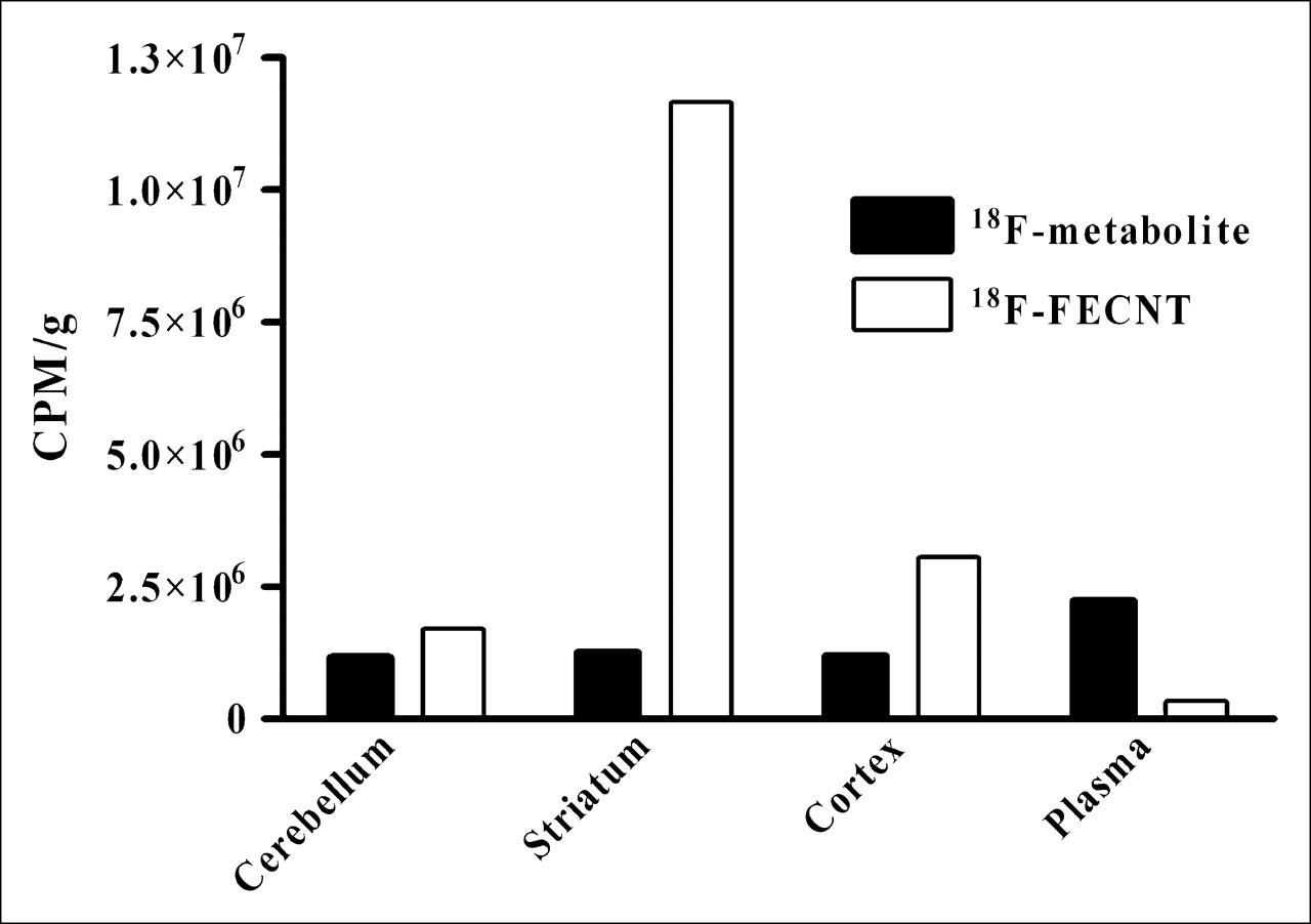
Characteristic distribution of 18F-FECNT and its radiometabolite, presented as their specific concentrations in rat brain regions and plasma after 1 h of intravenous CI of 18F-FECNT.
18F-FECNT was shown to be metabolically stable in both rat whole blood and rat brain homogenates in vitro. In rat whole blood, 18F-FECNT was distributed 59% to cellular blood elements and 41% to plasma. The radioligand was stable for up to 4 h (>99%) in rat brain homogenates maintained at 37°C. The in vitro homogenate incubations were performed with brains obtained from 2 rats given 2 different forms of anesthesia (i.e., isoflurane and ketamine:xylazine) to determine whether anesthesia had any effect on the in vitro metabolism of 18F-FECNT. Both brains showed no degradation of the parent radioligand, as analyzed with multiple and periodic monitoring of aliquots by reverse-phase HPLC. Similarly, perfusion of the rat brain to remove residual blood did not affect the in vitro metabolism of the radioligand.
Characterization of Rat Brain Radiometabolite
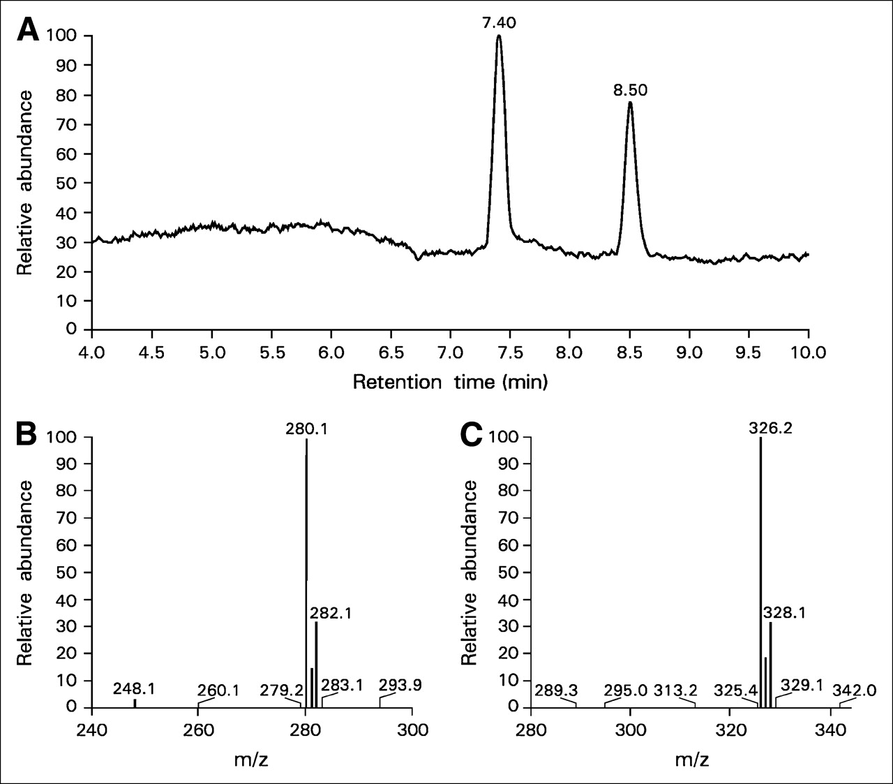
The brains from 2 rats that were infused with carrier-added 18F-FECNT as well as the brain from a control rat were used to identify the metabolite(s) in the brain. Brain extracts from each carrier-treated rat were divided into 3 samples and analyzed sequentially. HPLC (system C) showed that 67.3% ± 0.7% (n = 3) of the activity was associated with the polar radiometabolite and that the remaining 32% ± 0.1% of the activity was associated with the parent. These data constituted a ratio of 2.1 ± 0.01 (n = 3) for polar radiometabolite activity to parent activity in the carrier-treated rat brains. Samples from the carrier-treated and control rat brains were subjected to LC-MS and MS-MS analyses; this step was followed by comparison of ion chromatograms. Figure 2A is an ion chromatogram (encompassing m/z 275 through m/z 330 ions; system D) for a carrier-treated sample (obtained with HPLC system C; the fraction containing the radioactive parent radioligand) showing parent FECNT eluting at a retention time of 8.5 min and the major metabolite eluting at a retention time of 7.4 min. The mass spectrum for the metabolite peak is shown in Figure 2B, and that for FECNT is shown in Figure 2C. The electrospray ionization of the metabolite generated m/z 280 and m/z 282 ions (37Cl isotope) whereas, as expected, FECNT generated the m/z 326 molecular ion (and the m/z 328 ion for the 37Cl isotope). The relative abundances of the 37Cl isotope in FECNT and nor-CCT in brain extracts were 31.7% and 32.3%, respectively. These measured values are consistent with a 32.5% natural abundance of 37Cl. Authentic FECNT and nor-CCT had similar elution times of 6.2 and 6.6 min, respectively, with system C.

(A) Partial-ion chromatogram (between m/z 275 and m/z 330) from LC-MS analysis of brain extracts showing peaks for metabolite nor-CCT (retention time, 7.4 min) and for FECNT (retention time, 8.5 min). (B and C) Mass spectra for nor-CCT peak (B) and for FECNT peak (C).
The identities of both the metabolite nor-CCT (N-dealkylated) and FECNT in rat brains were confirmed by the finding that their LC retention times (Fig. 2A), mass spectra (Figs. 2B and 2C), and MS-MS product ion spectra were identical to those of synthetic standards (data not shown). However, the fluoroalkyl moiety itself was not detectable by existing LC-MS techniques. A different method of chromatographic separation and chemical modification (not available at the time) would have been needed to detect and identify this molecule. The 2.1 molar ratio of nor-CCT to FECNT, as measured by LC-MS (system D), was similar to that of the radiometabolite to 18F-FECNT (2.1 ± 0.01; n = 3), as measured by the HPLC (system C). Two major molecules were detected in rat urine, parent FECNT and nor-CCT, and traces of the β-carboxylic acid of FECNT also were present.
Primate Studies
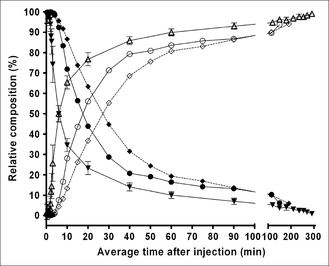
Analysis of arterial plasma samples from both humans and nonhuman primates showed composition profiles similar to those of the rats. Two main radioactivity peaks were detected in each plasma sample. One peak eluted in the void volume, and the other eluted at a retention time corresponding to that of parent 18F-FECNT. The plasma free fractions of 18F-FECNT were 9.4% ± 2.7% (n = 6) in monkeys and 4.7% ± 1.3% (n = 4) in humans. The relative compositions of the activity in plasma over time were similar in the 6 monkeys and the 2 humans (Fig. 3), although the appearance of radiometabolites was somewhat delayed in the humans. At about 6 min after the administration of 18F-FECNT, monkey plasma was composed of 50% radiometabolite and 50% parent radioligand, with little variability, as indicated by the small SD error bars (Fig. 3). The 50% radiometabolite:50% parent composition occurred at 18 and 27 min after injection in humans, as shown in Figure 3.

Arterial plasma compositions of 18F-FECNT and its polar radiometabolite after intravenous administration. Data are shown individually for 2 human participants, human 1 (♦) and human 2 (•) for 18F-FECNT and human 1 (⋄) and human 2 (○) for radiometabolite, and collectively (mean ± SD) for 6 rhesus monkeys for 18F-FECNT (▾) and radiometabolite (▵).
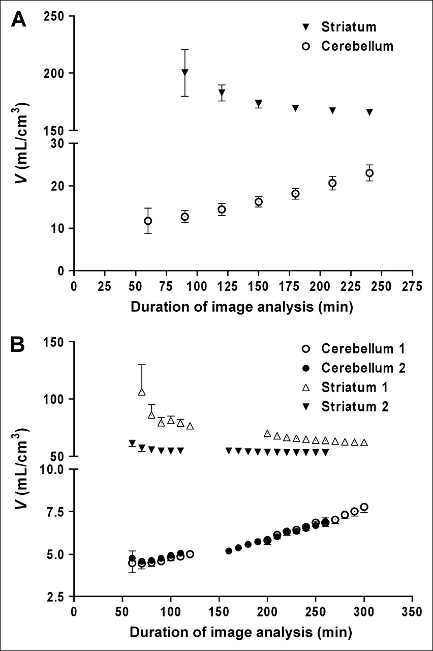
The uptake and washout of brain activity were similar in monkeys and humans, with peak uptake in the striatum at about 100 min and then relatively slow washout (Fig. 4). Although activity in the striatum showed a gradual decline after about 100 min, the activity in the cerebellum was virtually stable. Such differential washout rates in target and background regions can be attributable to the effect of the accumulation of a radiolabeled metabolite(s) in the brain. Although this differential washout can be demonstrated by a time-dependent increase in the ratio of the activity in the striatum to that in the cerebellum, a better parameter for evaluating this effect is distribution volume assessed relative to that of the parent compound in plasma. Compartmental analysis with an arterial input function provides the ratio for each region to plasma (rather than the ratio for region to region) and uses an integrated area under the curve (rather than a single time point measurement). The cerebellar distribution volume for both monkey and human studies increased as a function of the duration of image analysis. In monkeys, the distribution volume increased 2.0-fold ± 0.2 (n = 3) between 60 and 240 min (Fig. 5A), whereas in humans, the increases were 1.7-fold between 60 and 300 min and 1.5-fold between 60 and 260 min (Fig. 5B). However, the distribution volume in the striatum did not increase during the same periods, indicating a smaller contribution of the radiometabolite to the total activity (specific binding plus nondisplaceable activity). The distribution volume in the striatum became stable after 180 min, with negligible changes after 160 and 200 min in the 2 participants.
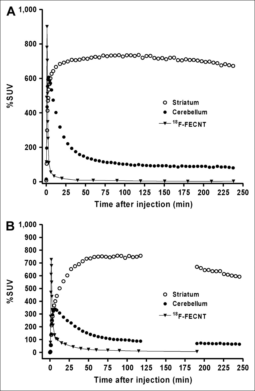
Time–activity curves for striatum and cerebellum for rhesus monkeys (A) and humans (B). Data are reported as percent standardized uptake values (%SUV). Arterial plasma 18F-FECNT input function was corrected for radiometabolites.

(A) Average cerebellar and striatal distribution volumes (V) for 3 rhesus monkeys and corresponding percent coefficients of variation, determined by use of increasing amounts of available data from each study, after intravenous administration of 18F-FECNT at 114.7 ± 22.3 MBq. (B) Cerebellar and striatal distribution volumes (V) for 2 humans and percent coefficients of variation, determined by use of variable portions of data from time 0 to time shown on x-axis, after administration of 359.3 and 396.1 MBq of 18F-FECNT. Error bars indicate percent coefficient of variation.
DISCUSSION
The major findings of the present study are as follows. First, 18F-FECNT is metabolized in rats, monkeys, and humans in vivo to yield a polar radiometabolite in plasma. Second, strong evidence indicates that an 18F-labeled 2-carbon metabolite enters the rat brain to a significant degree. As shown in Table 1, after 120 min of CI of the radioligand, 87.4% of the activity in the cerebellum represented the radiometabolite, with the remainder 12.6% being 18F-FECNT. Third, this radiometabolite is distributed nonspecifically in the brain and constitutes a higher percentage of total activity in the background (e.g., the cerebellum) than in the target (e.g., the striatum). Fourth, although we did not obtain primate tissue samples, the time–activity curves for brain uptake in both monkeys and humans were consistent with the accumulation of a radiometabolite, because the distribution volume in the cerebellum increased during the course of the imaging study. Six monkey imaging studies were performed. The initial 3 were acquired with short acquisition times, and the data did not converge. Hence, the last 3 studies were carried out with a more appropriate design (Materials and Methods) that resulted in useful information. However, plasma data from all 6 animal studies were compatible because they were acquired under identical conditions. Thus, we included all plasma data to better support our statements regarding the metabolism of 18F-FECNT.
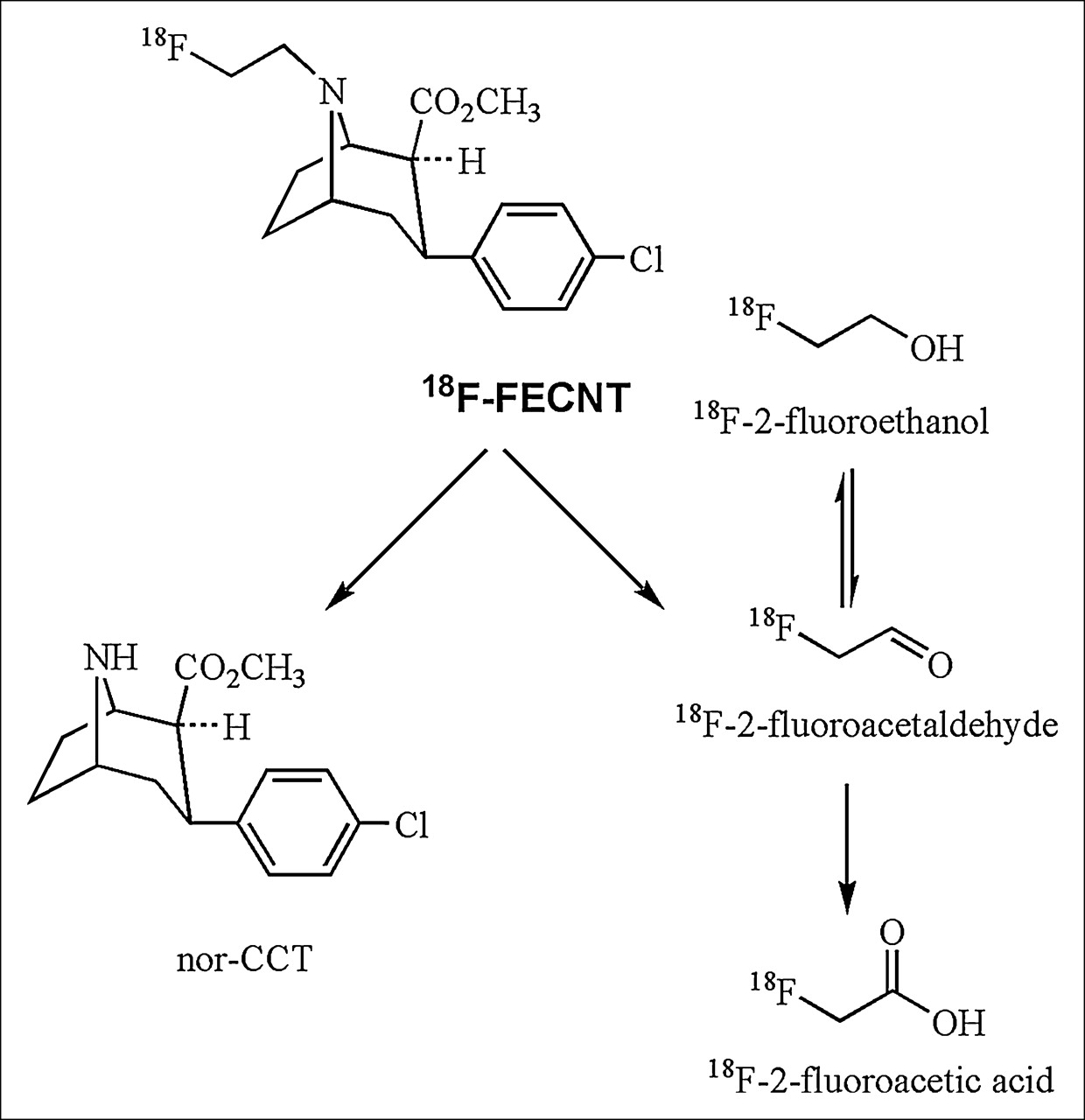
Subsequent to the administration of carrier-added 18F-FECNT, the concentration of radioactivity of the polar radiometabolite was twice that of the parent 18F-FECNT, whereas in the same rat brain samples, the concentration of nor-CCT was twice that of FECNT, as detected by LC-MS technique. FECNT is analogous to the cocaine molecule. The initial and major step of the oxidative metabolism of cocaine is its cytochrome P-450–catalyzed N-dealkylation to yield norcocaine and formaldehyde (13,14). This phase I biotransformation is characteristic of the membrane-bound cytochrome P-450 liver enzymes, which include those for monooxygenase–, oxygen reductase–, or substrate reductase–mediated reactions (15). Therefore, FECNT may undergo similar oxidative metabolism and generate nor-CCT and an equivalent amount of fluoroacetaldehyde, as shown in the metabolic scheme in Figure 6. Because 18F-FECNT is stable in vitro in rat brain homogenates, its metabolism presumably occurs in the periphery. Because nor-CCT has significant lipophilicity (clogP = 3.1) that is similar to that of FECNT, as judged by its chromatographic behavior, it may cross the blood–brain barrier but will not interfere with PET imaging measurements at no-carrier-added levels because it is nonradioactive. All 3 of the possible 2-carbon residues containing 18F (alcohol, aldehyde, and carboxylate) are expected to be metabolically interchangeable in vivo and to be able to gain access through the blood–brain barrier (Fig. 6). For 18F-fluoroacetate, the rate of entry is similar to that of 18F-FDG (16,17). Acetates are used in the glutamate pool in the brain glial compartment as energy sources under different pathologic conditions (18). The polar radiometabolite showed time-dependent accumulation in the brain (Table 1) with a uniform distribution and therefore no apparent affinity for DAT (Fig. 1). In the radiometabolite analysis, the recovery of radioactivity from the HPLC column was invariably quantitative. Hence, the limiting factor was the efficiency of extraction of radioactivity from the various tissues and fluids. More than 90% of the radioactivity present in the rat brain homogenates was extracted into acetonitrile, which has a dielectric constant of 37.5. HPLC analysis of these extracts revealed the presence of this radiometabolite of 18F-FECNT in the void volume of the column, thus characterizing it as polar. Diethyl ether, with a dielectric constant of 4.3, is not expected to extract this radiometabolite; consequently, HPLC analysis of such extracts would not detect its presence. This fact may help in clarifying the reason for the difference between the findings reported in the present study, in which CH3CN was used, and those reported by Goodman et al. (2), who used diethyl ether.

Possible route for in vivo oxidative metabolism of 18F-FECNT mediated by liver cytochrome P-450 enzymes.
Arterial plasma showed similar radiochromatographic profiles in rats, monkeys, and humans, suggesting similar metabolic fates for 18F-FECNT in these 3 species. Time–activity curves for the striatum and the cerebellum were similar for humans and monkeys and consistent with the prior demonstration of the accumulation of a radiometabolite in rat brains. The washout of activity from the striatum but the stable levels in the cerebellum could have been caused by the accumulation of a radiometabolite(s) in the brain. An inactive metabolite (i.e., with no affinity for the target DAT) would be distributed uniformly in all brain regions. Because target regions have much higher levels of the parent than do background areas, the inactive metabolite represents a larger percentage of activity in the cerebellum than in the striatum. The decreasing activity in the striatum presumably represents the sum of 2 opposite trends: the washout of the parent radioligand and the accumulation of the radiometabolite. The net effect in the striatum is negative, as indicated by the decreasing activity. However, the net effect in the cerebellum is almost zero, in part because the radiometabolite represents a much larger component of the total activity in the cerebellum than in the striatum because of the continuous replenishment of the radiometabolite from the periphery. Furthermore, the rate of metabolism of 18F-FECNT was higher in monkeys than in humans (Fig. 3). The human cerebellar distribution volume increased 1.5- and 1.7-fold at 260 and 300 min after administration, respectively. This increase was consistent with a lower metabolic rate in humans than in monkeys, which showed about a 2-fold increase at 240 min after administration (Fig. 5). The continual change in the cerebellar distribution volume in humans is not expected to be reproducible, because the production of the radiometabolite and its consequent brain accumulation are dependent on an individual's basal metabolic rate which, as shown in Figure 4, is highly variable.
CONCLUSION
Assuming that an inactive radiometabolite of 18F-FECNT enters the human brain, what will its impact on quantification be? As an inactive radiometabolite, it will have a much greater effect in the cerebellum than in the striatum. Because this radiometabolite represents a relatively small percentage of activity in the primate striatum, the errors in measuring the distribution volume relative to an input function will be relatively small. However, if the distribution volume is measured essentially as a ratio of activity in the striatum to that in the cerebellum, then the high level of contamination of cerebellar activity will have a direct impact on such an outcome measure. For this reason, and in light of the large variability in the rate of metabolism of 18F-FECNT in humans, analysis of 18F-FECNT images probably should not use a reference tissue model.
Acknowledgments
We gratefully acknowledge Peter Herscovitch, MD, and the staff of the Clinical Center Department, NIH, for assistance in the successful completion of this study and Douglas Vines, BSc, for his excellent technical support. We also extend our sincere appreciation to Jian-Qiang Lu, MD, for his support in the rat studies. We also thank Cyrill Burger, PhD, Piotr Rudnicki, PhD, Krzysztof Mikolajczyk PhD, and Michal Grodzki, PhD, for providing PMOD2.55. The Intramural Research Program of NIMH, NIH, supported this research.
References
- Received for publication August 30, 2005.
- Accepted for publication November 18, 2005.


Jump to section
Related Articles
Cited By...
- [11C]ZTP-1: An Effective Short-Lived Radioligand for PET of Rat and Monkey Brain Phosphodiesterase Type 4 Subtype B
- PET Quantification in Healthy Humans of Cyclooxygenase-2, a Potential Biomarker of Neuroinflammation
- [11C]PS13 Demonstrates Pharmacologically Selective and Substantial Binding to Cyclooxygenase-1 in the Human Brain
- Robust Quantification of Phosphodiesterase-4D in Monkey Brain with PET and 11C-Labeled Radioligands That Avoid Radiometabolite Contamination
- Whole-Body PET Imaging in Humans Shows That 11C-PS13 Is Selective for Cyclooxygenase-1 and Can Measure the In Vivo Potency of Nonsteroidal Antiinflammatory Drugs
- First-in-Human Evaluation of 18F-PF-06445974, a PET Radioligand That Preferentially Labels Phosphodiesterase-4B
- In Vivo Evaluation of 6 Analogs of 11C-ER176 as Candidate 18F-Labeled Radioligands for 18-kDa Translocator Protein
- PET ligands [18F]LSN3316612 and [11C]LSN3316612 quantify O-linked-{beta}-N-acetyl-glucosamine hydrolase in the brain
- Evaluation of a PET Radioligand to Image O-GlcNAcase in Brain and Periphery of Rhesus Monkey and Knock-Out Mouse
- Evaluation of Two Potent and Selective PET Radioligands to Image COX-1 and COX-2 in Rhesus Monkeys
- Whole-Body Biodistribution and Dosimetry of the Dopamine Transporter Radioligand 18F-FE-PE2I in Human Subjects
- 11C-ER176, a Radioligand for 18-kDa Translocator Protein, Has Adequate Sensitivity to Robustly Image All Three Affinity Genotypes in Human Brain
- The PET Radioligand 18F-FIMX Images and Quantifies Metabotropic Glutamate Receptor 1 in Proportion to the Regional Density of Its Gene Transcript in Human Brain
- 11C-CUMI-101, a PET Radioligand, Behaves as a Serotonin 1A Receptor Antagonist and Also Binds to {alpha}1 Adrenoceptors in Brain
- Brain and Whole-Body Imaging in Rhesus Monkeys of 11C-NOP-1A, a Promising PET Radioligand for Nociceptin/Orphanin FQ Peptide Receptors
- Imaging of the Striatal and Extrastriatal Dopamine Transporter with 18F-LBT-999: Quantification, Biodistribution, and Radiation Dosimetry in Nonhuman Primates
- Preclinical Evaluation of 18F-JNJ41510417 as a Radioligand for PET Imaging of Phosphodiesterase-10A in the Brain
- Imaging and Quantitation of Cannabinoid CB1 Receptors in Human and Monkey Brains Using 18F-Labeled Inverse Agonist Radioligands
- Quantification of Translocator Protein (18 kDa) in the Human Brain with PET and a Novel Radioligand, 18F-PBR06
- Human Brain Imaging and Radiation Dosimetry of 11C-N-Desmethyl-Loperamide, a PET Radiotracer to Measure the Function of P-Glycoprotein
- PET Measurement of the In Vivo Affinity of 11C-(R)-Rolipram and the Density of Its Target, Phosphodiesterase-4, in the Brains of Conscious and Anesthetized Rats
- 11C-Loperamide and Its N-Desmethyl Radiometabolite Are Avid Substrates for Brain Permeability-Glycoprotein Efflux