


Abstract
Pretargeting involves administration of a tumor-targeting monoclonal antibody (mAb) covalently linked to a molecule having a high-affinity binding site for a rapidly distributed radiolabeled effector molecule. The aim of this study was to compare pretargeting to a conventionally labeled antibody for tumor targeting of the intermediate-lived radionuclide 64Cu, which has shown promise for PET imaging and radioimmunotherapy of cancer. Methods: DOTA-biotin (where DOTA is 1,4,7,10-tetraazacyclododecane-N,N′,N″,N‴-tetraacetic acid) and the intact immunoconjugate DOTA-NR-LU-10 were labeled to high specific activities with 64Cu, and the serum stabilities and target binding capabilities of each agent were assayed in vitro. Nude mice bearing SW1222 human colorectal carcinoma xenografts were administered 64Cu-DOTA-biotin, with and without pretreatment with the mAb-streptavidin conjugate NR-LU-10/SA and the synthetic clearing agent Biotin-GalNAc16, or injected with 64Cu-DOTA-NR-LU-10. Biodistributions of both agents were obtained from 5 min to 48 h after injection. Results: Both 64Cu-DOTA-biotin and 64Cu-DOTA-NR-LU-10 were 100% stable in serum in vitro. 64Cu-DOTA-biotin exhibited >98% specific binding to immobilized streptavidin, whereas the immunoreactivity of 64Cu-DOTA-NR-LU-10 averaged nearly 80%. Biodistributions in SW1222-bearing mice showed that NR-LU-10/SA–pretargeted 64Cu-DOTA-biotin attained a peak tumor uptake of 18.9 percentage injected dose per gram (%ID/g) at 1 h, with concomitant rapid disappearance from blood and renal excretion. In the absence of pretargeting, 64Cu-DOTA-biotin had very similar biodistribution and clearance properties, except with extremely low nonspecific tumor uptake. In contrast, 64Cu-DOTA-NR-LU-10 reached 80.3 %ID/g in tumor tissue, after 48 h, whereas blood clearance was considerably slower than pretargeted 64Cu-DOTA-biotin. Comparison of the time–activity curves for tumor uptake and blood clearance of pretargeted 64Cu and the 64Cu-labeled antibody revealed that the maximum tumor accumulations of radioactivity were similar for each agent, 17.9 percentage injected activity per gram (%IA/g) and 20.7 %IA/g, respectively. However, the tumor-to-blood ratio of areas under the curves was 14 times higher for pretargeted 64Cu-DOTA-biotin because of the substantial increase in blood clearance of the small effector molecule. Conclusion: The extremely rapid tumor uptake and blood clearance of pretargeted 64Cu-DOTA-biotin should afford markedly superior PET imaging contrast and therapeutic efficacy, compared with conventionally labeled 64Cu-DOTA-NR-LU-10. Further comparison of the therapeutic efficacy, toxicity, and dosimetry of these 2 agents is warranted.
Radiolabeled monoclonal antibodies (mAbs) have shown considerable promise for tumor imaging and radioimmunotherapy (RIT) of cancer. However, because of their slow blood clearance, radiolabeled mAbs generally do not localize to solid tumors in sufficient quantities to provide high-contrast imaging or consistent therapeutic efficacy without significant bone marrow toxicity. Further improvements in targeting solid tumors will likely require implementation of several innovative approaches, including the use of new targeting molecules and novel delivery systems.
Antibody pretargeting is an approach in which an unlabeled mAb-receptor construct is first administered and allowed to accumulate in tumors, and then radionuclide imaging or therapy is given in the form of a small effector molecule that binds rapidly with high affinity to the mAb-receptor construct at the tumor site. In some cases, an intermediate clearing step is performed to reduce levels of the antibody-receptor construct in circulation. When successful, this process results in immediate accumulation of radioactivity in the tumor, causing substantial increases in tumor-to-blood ratio and tumor absorbed dose. Thus, pretargeting combines the desirable properties of high tumor uptake of antibodies with rapid pharmacokinetics and fast whole-body clearance of radioactivity of radiolabeled small molecules.
Several types of receptor/effector approaches have been developed for pretargeted RIT, including mAb/hapten (1–7), biotin/avidin (8–13), and oligonucleotide/antisense oligonucleotide (14,15) systems. The high-affinity noncovalent binding of biotin to streptavidin (∼1013 mol/L−1) makes this system attractive for mAb pretargeting methods. A streptavidin conjugate of the anti-Ep-CAM mAb NR-LU-10 (NR-LU-10/SA), which binds 4 molecules of radiolabeled biotin, has been prepared and evaluated in nude mice bearing breast and small cell lung carcinoma xenografts (11,13) and in patients (16,17) for pretargeted RIT of metastatic colon cancer. In mouse models, NR-LU-10/SA exhibited tumor uptake and blood clearance equivalent to unmodified intact mAb. Treatment with the synthetic clearing agent, Biotin-GalNAc16, removed 90%–95% of circulating NR-LU-10/SA. The effector molecule, 90Y-DOTA-biotin (where DOTA is 1,4,7,10-tetraazacyclododecane-N,N′,N″,N‴-tetraacetic acid), showed rapid disappearance from blood and low normal organ uptake, with urinary excretion of 90 percentage injected dose (%ID) in 2 h. In tumor-bearing mice, sequential administration of these agents resulted in stable, high-efficiency delivery of >20 %ID per gram (%ID/g) of 90Y to tumor, with whole-body excretion and nontarget organ uptake similar to that of 90Y-DOTA-biotin alone.
64Cu (half-life = 12.7 h; β+ 655 keV [17.4%]; β− 573 keV [39%]) is an attractive radionuclide for PET imaging and targeted radiotherapy of cancer. 64Cu-Labeled antibodies (18–20) and peptides (21–23) have shown promise for tumor targeting in animal models and patients. Compared with covalently labeled mAbs, pretargeting is an appealing strategy for delivery of intermediate-lived radionuclides to tumors. Even antibody fragments take hours to localize to tumors, during which a significant amount of 64Cu will have decayed. Pretargeting of 64Cu should allow immediate accumulation of radioactivity in the tumor, resulting in a substantial increase in tumor-to-blood ratios and tumor absorbed dose. The objective of these studies was to compare the tumor targeting capability of 64Cu-DOTA-biotin, after pretargeting with NR-LU-10/SA, to that of the intact mAb NR-LU-10, conjugated to DOTA and labeled with 64Cu.
MATERIALS AND METHODS
Materials
64Cu was produced by previously published methods (24) on a Cyclotron Corp. CS-15 cyclotron at Washington University School of Medicine. DOTA-biotin (25), Biotin-GalNAc16 (26,27), and NR-LU-10/SA conjugate (13) were prepared as described. All solutions were prepared using ultrapure water (18 MΩ-cm resistivity). Unless otherwise noted, all chemicals were purchased from Aldrich Chemical Co. Fast protein liquid chromatography (FPLC) was performed on a Pharmacia FPLC System, using a Superose 12 HR 10/30 column, 20 mmol/L N-(2-hydroxyethyl)piperazine-N′-(2-ethanesulfonic acid)/150 mmol/L NaCl, pH 7.3, as the mobile phase, and a flow rate of 0.4 mL/min. Reversed-phase thin-layer chromatography (TLC) was performed on Whatman MKC18F plates, using 10% ammonium acetate:methanol (60:40) as the mobile phase. Radio-TLC detection was accomplished using a Bioscan System 200 imaging scanner. Radioactive samples were counted on a Beckman 8000 γ-counter. Outbred female nu/nu mice (4–6 wk of age) were obtained from Harlan Bioproducts. Animals were maintained on a biotin-deficient diet (Purina Test Diet) for 5 d before radiopharmaceutical administration and throughout the course of biodistribution and excretion studies.
Preparation of 64Cu-DOTA-Biotin
Representative conditions for labeling DOTA-biotin with 64Cu at low specific activities are given here. To 102 MBq (2.76 mCi) of 64Cu in 164 μL of 0.2 mol/L ammonium acetate, pH 5.0, containing 1 mg/mL gentisic acid, was added 92.0 μg (0.114 μmol) of DOTA-biotin in 46.0 μL of 0.2 mol/L ammonium acetate, pH 5.0. The reaction mixture was incubated at 80°C for 1 h, after which TLC showed that 64Cu incorporation was 100%.
In the case of high specific activity labeling, where 64Cu incorporation was <95%, the radiolabeled conjugate was purified as follows. After incubation at 80°C for 1 h, an aliquot of one ninth of a reaction volume of 10 mmol/L ethylenediaminetetraacetic acid, pH 5.5, was added, and the reaction mixture was incubated at room temperature for 5 min. The reaction mixture was then applied to a C18 SepPak cartridge, which was washed with 6 mL of 0.2 mol/L ammonium acetate, pH 5.0, and eluted with 1 mL of ethanol. Successive fractions of 100, 300, and 600 μL of ethanol were collected, and purified 64Cu-DOTA-biotin was obtained in the second fraction.
Preparation of 64Cu-DOTA-NR-LU-10
NR-LU-10 was conjugated with 20 theoretic equivalents of N-hydroxysulfosuccinimidyl DOTA, according to a method described previously (28). Representative conditions for labeling DOTA-NR-LU-10 with 64Cu are given here. To 169 MBq (4.58 mCi) of 64Cu in 310 μL of 0.1 mol/L ammonium citrate, pH 5.5, was added 1.0 mg of DOTA-NR-LU-10 in 125 μL of 0.1 mol/L ammonium citrate, pH 5.5. The reaction mixture was incubated at 43°C for 1 h, after which 48.4 μL of 10 mmol/L DTPA, pH 6.0, was added. The reaction mixture was incubated at room temperature for 15 min, and then the radiolabeled conjugate was purified using a 3-mL Sephadex G-25-50 spin column, equilibrated with 0.1 mol/L ammonium citrate, pH 6.6 (18,29). The column was eluted by centrifugation at 2,500 rpm for 4 min in a tabletop centrifuge. Immunoreactivity of 64Cu-DOTA-NR-LU-10 was assessed by the method of Lindmo et al. (30).
Serum Stability Studies
To 36.1 MBq (976 μCi) of 64Cu-DOTA-biotin was added 500 μL of rat serum (Sigma Chemical Co.). The resulting mixture was incubated at 37°C for 48 h, and 1.5-μL aliquots were analyzed by radio-TLC after 0, 0.25, 0.5, 1, 2, 4, 24, and 48 h of incubation to determine conjugate stability. An aliquot of 6.48 MBq (175 μCi) of 64Cu-DOTA-NR-LU-10 was added to 500 μL of rat serum. This mixture was incubated at 37°C for 48 h. At 0, 2, 4, 24, and 48 h, 25-μL aliquots of the serum were analyzed by FPLC.
Streptavidin Binding Assay
DOTA-biotin was labeled with 64Cu at specific activities of 962 MBq/μmol (26.0 mCi/μmol) and 24.3 GBq/μmol (657 mCi/μmol). Aliquots of 200 μL of 4% streptavidin-agarose beads (Sigma Chemical Co.) were washed twice with 200 μL of phosphate-buffered saline (PBS), pH 7.5, and the beads were drained by centrifugation at 3,800 rpm for 2 min in a tabletop centrifuge. Then 1.27 MBq (34.3 μCi) of 64Cu-DOTA-biotin in 500 μL of PBS, pH 7.5, was added, and the beads were incubated at room temperature with continuous end-over-end mixing for 10 min. The beads and supernatant were separated by centrifugation at 3,800 rpm for 2 min, after which the beads were washed twice with 200 μL of PBS, pH 7.5. The beads were drained by centrifugation at 3,800 rpm for 2 min, and then the beads and combined supernatant were counted in the γ-counter. Streptavidin binding was calculated as the percentage of the total radioactivity bound to the SA-agarose beads after elution.
Biodistribution Studies
All animal experiments were conducted in compliance with guidelines established by the Washington University Animal Studies Committee. Athymic nude mice were implanted subcutaneously in the hind flank with 5 × 10⁶ SW1222 human colorectal carcinoma cell suspensions (0.15 mL) with >90% viability. Radiopharmaceuticals were injected intravenously via the tail vein 2 wk after tumor implantation, when tumors had grown to 100–400 mg, with an average size of ∼250 mg. For all biodistribution studies, groups of 5 mice at each time point were randomized, such that mice in each group carried uniform tumors with the weight range and approximate mean weight given above.
For pretargeting studies, mice were injected intravenously with 400 μg of NR-LU-10/SA in 100 μL of normal saline. After 22 h, the mice were administered an intravenous dose of 100 μg of the synthetic clearing agent, Biotin-GalNAc16, in 100 μL of normal saline. Six hours after injection of the clearing agent, the mice were injected with 2.2 MBq (60 μCi)/2.0 μg of 64Cu-DOTA-biotin in 100 μL of saline. Biodistributions were obtained at 5 min, 30 min, and 1, 2, 4, 24, and 48 h after injection. Tissues harvested included blood, lung, liver, spleen, kidney, muscle, fat, heart, bone, uterus, ovaries, bladder, gallbladder, stomach, small intestine, upper large intestine, lower large intestine, and tumor. Tissues were drained of blood, weighed, and counted in the γ-counter with a standard of the injected dose, such that decay-corrected uptakes were calculated as the %ID/g of tissue and the %ID per organ (%ID/organ). Biodistributions of 64Cu-DOTA-biotin without pretreatment with NR-LU-10/SA and Biotin-GalNAc16 were obtained in an identical manner.
Biodistributions of 64Cu-DOTA-NR-LU-10 were obtained at 5 min and 3, 6, 18, 24, and 48 h after intravenous injection of 2.6 MBq (70 μCi)/45.5 μg of the radiolabeled mAb in 100 μL of saline. The tissues listed above were drained of blood, weighed, and counted in the γ-counter with a standard of the injected dose to determine the %ID/g and the %ID/organ.
Excretion Studies
Groups of 6 nude mice bearing uniform SW1222 tumors (100–400 mg; mean weight, ∼250 mg) were injected intravenously with 64Cu-DOTA-biotin as described above, with or without pretreatment with NR-LU-10/SA and Biotin-GalNAc16. Immediately after radiopharmaceutical injection, mice were placed in metabolism cages. Urine and feces were collected together at 5 min, 30 min, and 1, 2, 4, 24, and 48 h after injection. Radioactivity was counted in the γ-counter with a standard of the injected dose, and the %ID for each sample was calculated.
Statistical Analysis
To compare biodistribution differences between NR-LU-10/SA–pretargeted 64Cu-DOTA-biotin and 64Cu-DOTA-NR-LU-10, as well as differences in organ uptakes of each agent at various time points, a Student t test was performed. Differences at the 95% confidence level (P < 0.05) were considered significant.
RESULTS
64Cu Labeling Studies
64Cu-Labeled DOTA-biotin and DOTA-NR-LU-10 were both radiolabeled and purified at specific activities suitable for both preclinical and clinical studies. DOTA-biotin was labeled with 64Cu using modifications of a previously reported method (31) for radiometal labeling of DOTA-octreotide conjugates. After incubation with 64Cu acetate at 80°C and pH 5.0, DOTA-biotin could be labeled with 64Cu at specific activities as high as 74 MBq/μg ([2 mCi/μg] 1,742 Ci/mmol) and purified using a SepPak cartridge, as described (21). Radiochemical purity of 64Cu-DOTA-biotin, determined by TLC, was 95%–100%. DOTA-NR-LU-10 was labeled with 64Cu using modifications of published procedures (18,28,29). The DOTA-conjugated mAb was incubated with 64Cu citrate at 43°C and pH 5.5 and purified by gel-filtration spin column chromatography. 64Cu labeling of DOTA-NR-LU-10 averaged 85.2%, and the average radiochemical purity of the 64Cu-labeled mAb, determined by FPLC, was 99.5%. Typically, DOTA-NR-LU-10 could be labeled to a maximum specific activity of 403 MBq/mg ([10.9 mCi/mg] 1,635 Ci/mmol). The immunoreactivity of 64Cu-DOTA-NR-LU-10 averaged 78.5%.
Serum Stability Studies
64Cu-Labeled DOTA-biotin and DOTA-NR-LU-10 were incubated in rat serum for 48 h at 37°C, and aliquots of the resulting mixtures were analyzed to determine the kinetic stability of the radiolabeled conjugates. No loss of 64Cu from either bioconjugate was observed during the course of the studies, and the radiochemical purity of each agent remained at 100% for 48 h under physiologic conditions.
Streptavidin Binding Assay
64Cu-DOTA-biotin was assayed for streptavidin binding by mixing the radiolabeled compound with 4% streptavidin-agarose beads for 10 min at room temperature. After separating the supernatant and washing the beads, it was found that 98.4%–98.8% of 64Cu-DOTA-biotin bound specifically to the immobilized streptavidin.
Effect of Specific Activity on Biodistribution of 64Cu-DOTA-Biotin
The biodistributions of 1.1 MBq (30 μCi) of NR-LU-10/SA–pretargeted 64Cu-DOTA-biotin, diluted respectively to total masses of 0.5, 1.0, 2.0, and 5.0 μg with unlabeled DOTA-biotin, are shown in Figure 1. At 2 h after injection, no significant differences (P > 0.05) in organ uptakes were observed at doses of <1.0 μg. However, as the mass of DOTA-biotin was increased to 2.0–5.0 μg, significant differences (P < 0.05) in target and nontarget organ uptakes were observed. Tumor uptake of 64Cu-DOTA-biotin decreased when the mass of compound was increased from 1.0 to 5.0 μg. Similar decreases in bone, fat, and muscle were observed when 5.0 μg of 64Cu-DOTA-biotin was administered, compared with 0.5–1.0 μg. As expected, the amount of 64Cu-DOTA-biotin remaining in the blood decreased significantly (P < 0.05) when the mass was increased from 1.0 to 5.0 μg. At 2 h, the only significant difference in clearance organ (liver and kidney) uptake was observed in the kidney at doses between 1.0 and 2.0 μg, where renal clearance increased with increasing mass (P = 0.007). At 24 h after injection, significant decreases in tumor (24.6 %ID/g to 13.3 %ID/g; P = 0.002) and bone (0.380 %ID/g to 0.303 %ID/g; P = 0.006) uptakes were observed as the mass of DOTA-biotin was increased from 0.5 to 5.0 μg. The amount of 64Cu activity remaining in the blood at 24 h also decreased significantly from 0.5–1.0 μg to 2.0–5.0 μg of DOTA-biotin. The only significant difference in clearance organ uptake at 24 h was a slight decrease in kidney uptake between the 1.0- and 5.0-μg doses (P = 0.03).
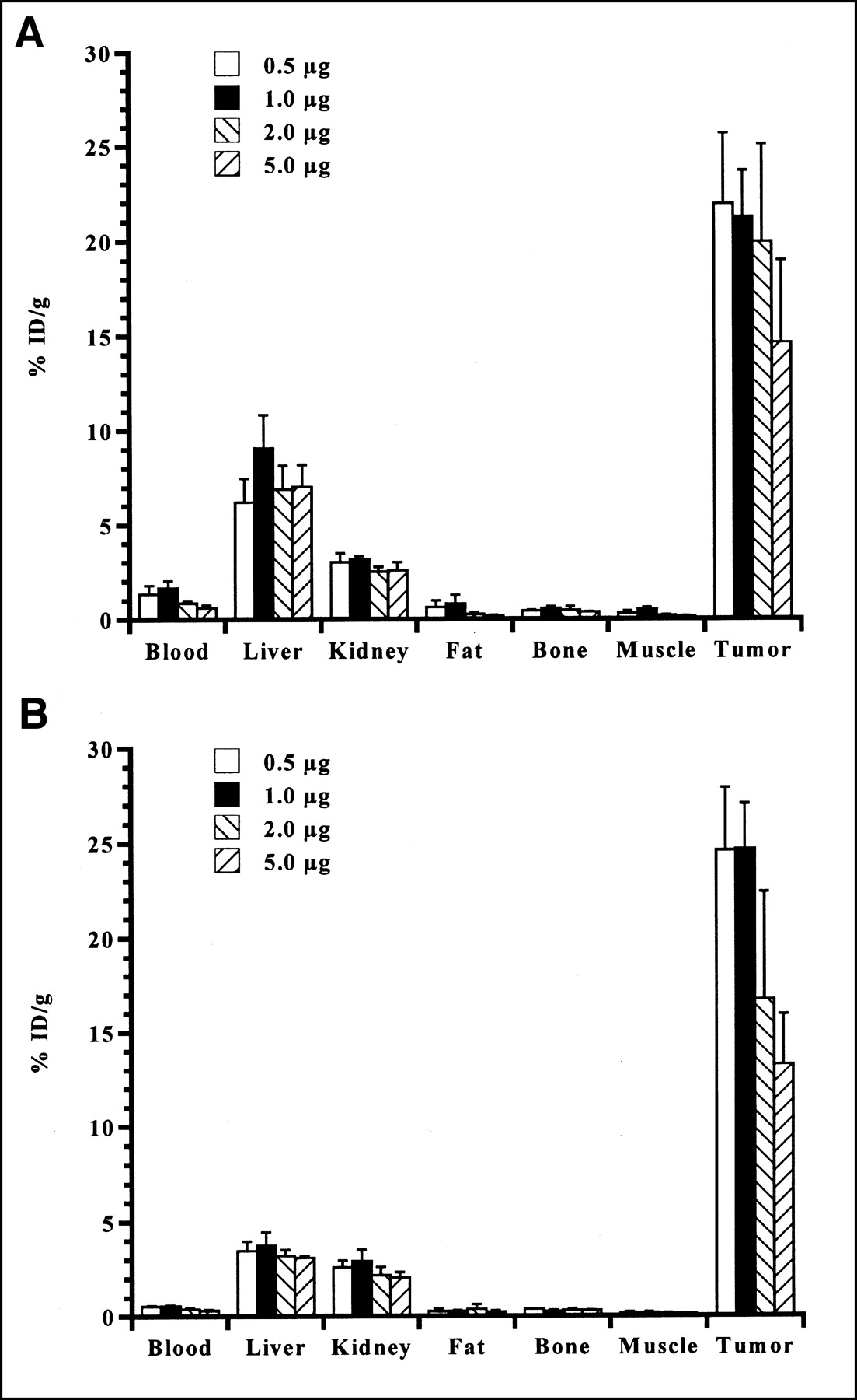
Biodistributions of 1.1 MBq (30 μCi) of NR-LU-10/SA–pretargeted 64Cu-DOTA-biotin in SW1222-bearing nude mice at 2 h (A) and 24 h (B) after injection, administered at doses of 0.5, 1.0, 2.0, and 5.0 μg of compound.
Biodistributions of 64Cu-DOTA-Biotin and 64Cu-DOTA-NR-LU-10
The biodistributions of 64Cu-DOTA-biotin, with and without prior administration of NR-LU-10/SA and Biotin-GalNAc16, are given in Tables 1 and 2, respectively. In these experiments, injected doses of 2.0 μg of 64Cu-labeled DOTA-biotin were used to approximate the mass anticipated to be used in a pretargeted RIT study in the same animal model. When pretargeted by NR-LU-10/SA, accumulation of 64Cu-DOTA-biotin in tumor was extremely rapid and, by 5 min after injection, the tumor-to-blood ratio had already reached 1.37. Afterward, tumor uptake increased significantly (P < 0.0055) at time points of 30 min and later. Peak tumor uptake of pretargeted 64Cu occurred at 1 h after injection, but this value of 18.9 %ID/g was only statistically different from the uptakes at 5 min and 48 h. Therefore, tumor retention of pretargeted 64Cu-DOTA-biotin was consistently high throughout the course of the study. The activity in the blood decreased extremely rapidly and following 5 min after injection (P < 0.0001). At 5 min, blood uptake of pretargeted 64Cu was 6.53 %ID/g, but this value decreased to 1.00 %ID/g by 1 h and to 0.28 %ID/g by 48 h. This blood clearance was accompanied by extremely rapid renal clearance and excretion of the residual radioactivity. Although kidney accumulation was initially 20.8 %ID/g at 5 min, this uptake decreased significantly to 3.40 %ID/g (P = 0.002) by 1 h and to 2.13 %ID/g (P = 0.0007) by 48 h. Cumulative total urinary and fecal excretion of 64Cu-DOTA-biotin, with and without NR-LU-10/SA pretargeting, is presented in Table 3. Throughout the first 4 h of the study, excretion was nearly identical between the pretargeted and nonpretargeted animals. At 24 and 48 h, excretion in the pretargeted animals was slightly lower than that in the nonpretargeted group, presumably due to retention of radioactivity in the tumor. Other tissues that exhibited significant clearance of pretargeted 64Cu-DOTA-biotin (Table 1) from 5 min to 48 h were bone (P = 0.003), muscle (P < 0.0001), and small intestine (P = 0.001). Liver clearance of pretargeted 64Cu was slower and less dramatic. It took 24 h (P = 0.005) to 48 h (P = 0.001) for liver uptakes of NR-LU-10/SA–pretargeted 64Cu-DOTA-biotin to decrease significantly from the value at 5 min.
Biodistribution (%ID/g ± SD) of 2.0 μg of NR-LU-10/SA–Pretargeted 64Cu-DOTA-Biotin in Athymic Nude Mice Bearing SW1222 Human Colorectal Carcinoma Xenografts
Biodistribution (%ID/g ± SD) of 2.0 μg of Nonpretargeted 64Cu-DOTA-Biotin in Athymic Nude Mice Bearing SW1222 Human Colorectal Carcinoma Xenografts
Cumulative Excretion (%ID ± SD) of 2.0 μg of NR-LU-10/SA–Pretargeted and Nonpretargeted 64Cu-DOTA-Biotin in Athymic Nude Mice Bearing SW1222 Human Colorectal Carcinoma Xenografts
When 64Cu-DOTA-biotin was not pretargeted by NR-LU-10/SA (Table 2), the biodistribution was very similar to that obtained after pretargeting, with the exception of extremely low nonspecific accumulation of radioactivity in the tumor. Tumor uptake of nonpretargeted 64Cu-DOTA-biotin decreased from 2.34 %ID/g at 5 min to 0.53 %ID/g at 48 h. Not only was tumor uptake of nonpretargeted 64Cu below uniform distribution at 5 min after injection but it also was never >1 %ID/g at any other time point. Throughout the course of the study, tumor uptake of 64Cu-DOTA-biotin was significantly lower (P < 0.0001) in the absence of pretargeting, compared with NR-LU-10/SA for prelocalization. From 1 to 24 h after injection, blood clearance of nonpretargeted 64Cu was faster (P < 0.02) than that when pretargeting was used. Between 4 and 48 h, other tissues that showed faster clearance of nonpretargeted 64Cu-DOTA-biotin were kidney (P < 0.04) and small intestine (P < 0.03).
The biodistribution of 64Cu-DOTA-NR-LU-10 is shown in Table 4. In contrast to the pretargeting system, tumor uptake of the 64Cu-labeled mAb was considerably slower, taking 24–48 h to achieve maximum uptake. However, accumulation of 64Cu-DOTA-NR-LU-10 in the SW1222 xenograft was ultimately much higher, reaching 62.2 %ID/g at 24 h and 80.3 %ID/g at 48 h after injection. Tumor uptake of the radiolabeled antibody increased significantly (P < 0.001) from 5 min to 48 h, but the values at 24 and 48 h were not statistically different. This high tumor uptake was accompanied by relatively slow disappearance from blood. Although blood concentrations of 64Cu-DOTA-NR-LU-10 decreased significantly (P < 0.0001) at time points later than 5 min (60.8 %ID/g), 20.4 %ID/g still remained in circulation at 48 h. After tumor and blood, the next major organ of uptake for the 64Cu-labeled mAb was the liver. 64Cu activity in the liver diminished significantly at time points later than 5 min, but this clearance was slow and modest, decreasing from 10.8 %ID/g at 3 h to 8.99 %ID/g at 6 h (P = 0.0239). Liver activity did not decrease significantly again until it reached 8.39 %ID/g at 48 h after injection (P = 0.0006 compared with that at 3 h). Similarly, renal accumulation of 64Cu-DOTA-NR-LU-10 decreased significantly (P ≤ 0.0112) from 10.7 %ID/g at 5 min. Like hepatobiliary clearance, kidney clearance was slow and modest. In fact, renal uptake of the 64Cu-labeled antibody actually increased significantly (P = 0.0083) from 6.78 %ID/g at 6 h to 8.23 %ID/g at 18 h, after which it decreased significantly (P = 0.0005) to 6.61 %ID/g at 48 h. Bone uptake of 64Cu-DOTA-NR-LU-10 decreased significantly (P = 0.0003) from 5 min to 48 h after injection, but small (P = 0.048), upper large (P = 0.0251), and lower large (P = 0.001) intestines showed significant increases in uptake of 64Cu, consistent with hepatobiliary excretion of the radiolabeled antibody.
Biodistribution (%ID/g ± SD) of 17.1 μg of 64Cu-DOTA-NR-LU-10 in Athymic Nude Mice Bearing SW1222 Human Colorectal Carcinoma Xenografts
Tumor-to-Blood Ratios of Pretargeted 64Cu-DOTA-Biotin and 64Cu-DOTA-NR-LU-10
The biodistribution data obtained using NR-LU-10/SA–pretargeted 64Cu-DOTA-biotin and 64Cu-DOTA-NR-LU-10 were used to generate time–activity curves for tumor uptake and blood clearance of the 2 radiopharmaceuticals. Physical decay was included, such that uptakes were calculated in percentage injected activity per gram (%IA/g). The ratio of the areas under the curves (AUCs) for tumor versus blood, shown in Figure 2, was used to estimate the anticipated therapeutic index of each agent. The tumor AUC for 64Cu-DOTA-NR-LU-10 was 783 %IA/g-h, and the blood AUC was 621 %IA/g-h, giving a tumor-to-blood ratio of only 1.26, due to the slow blood clearance of the radiolabeled mAb. In contrast, pretargeted 64Cu-DOTA-biotin gave a tumor AUC of 307 %IA/g-h and a blood AUC of only 17.4 %IA/g-h. The extremely rapid tumor uptake and blood clearance of pretargeted 64Cu-DOTA-biotin afforded a tumor-to-blood ratio of 17.6, a value 14 times higher than that of the 64Cu-labeled antibody.

Tumor and blood time–activity curves of 64Cu-DOTA-NR-LU-10 (A) and NR-LU-10/SA–pretargeted 64Cu-DOTA-biotin (B) in SW1222-bearing nude mice. Note difference in y-axis scales.
DISCUSSION
One of the goals of these studies was to test the hypothesis that a rapid targeting system, such as antibody pretargeting, will allow efficient delivery of intermediate half-life radionuclides such as 64Cu to tumors in vivo. 64Cu-Labeled antibodies have demonstrated utility for PET in animal models and in humans (18,19) as well as for RIT in tumor-bearing rodents (20). However, conventionally labeled mAbs typically take 1–2 d to reach maximum tumor uptake, even in mouse models. Although they display more rapid pharmacokinetic properties, enzymatically generated antibody fragments (32) or bioengineered single-chain antibodies (33) require ∼24 h to reach maximum tumor-to-blood ratios in rodents. During these protracted uptake times, much 64Cu will have decayed in circulation. 64Cu-Labeled peptides have also shown promise for PET and targeted radiotherapy in tumor-bearing animals (21,22) and for diagnostic imaging of patients (23). Although radiolabeled peptides accumulate in tumors within a few hours, absolute tumor uptake of radioactivity is generally considerably less than that with labeled antibodies. Using an unlabeled mAb-receptor construct and a radiolabeled effector molecule, pretargeting combines the desired properties of the high tumor uptake of antibodies with the rapid tumor targeting and clearance properties of small molecules, creating the potential to deliver substantial quantities of 64Cu to mouse tumor xenografts within minutes.
We prepared the radiopharmaceuticals 64Cu-DOTA-biotin and 64Cu-DOTA-NR-LU-10 and compared their biodistribution properties in nude mice bearing SW1222 human colorectal carcinoma xenografts. Both agents could be labeled to comparable high specific activities with 64Cu (59.2–64.8 TBq/mmol [1,600–1,750 Ci/mmol]), sufficient for both PET and RIT applications. Using the macrocyclic chelator DOTA, 64Cu could be complexed to both biotin and NR-LU-10, and both radiolabeled conjugates were completely inert to radiometal loss in serum over a 48-h period at 37°C. Both 64Cu-labeled agents retained antigen or receptor binding activity in vitro. More than 98% of 64Cu-DOTA-biotin bound specifically to immobilized streptavidin, and the average immunoreactivity of 64Cu-DOTA-NR-LU-10 was nearly 80% with Ep-CAM–positive tumor cells, a value approaching the upper limit for the cell binding assay used.
To assess the potential of 64Cu-DOTA-biotin for pretargeted RIT, the biodistribution of this agent was determined as a function of the mass of compound injected. As the mass of 64Cu-DOTA-biotin was increased from 2.0 to 5.0 μg, uptakes in tumor, bone, fat, and muscle decreased, while disappearance from the blood and renal excretion of the compound increased. These observations were consistent with the notion that as the mass increased, the protein-bound fraction of the radiopharmaceutical in plasma decreased and the unbound fraction increased, leading to higher urinary clearance. Furthermore, the decreased tumor uptake at 2.0–5.0 μg of 64Cu-DOTA-biotin was consistent with the fact that, under the dose conditions used, ∼15 μg of the agent would completely saturate the NR-LU-10/SA conjugate taken up by a 0.1- to 0.4-g SW1222 xenograft. Therefore, using 64Cu-DOTA-biotin at the highest possible specific activity may improve its therapeutic efficacy. Subsequent biodistribution studies were performed with 2.0 μg of 64Cu-DOTA-biotin, a mass anticipated to be needed to deliver a therapeutic quantity of 64Cu (≥74 MBq [≥2 mCi]) to SW1222 tumors in the mouse model.
The rapid blood clearance of NR-LU-10/SA–pretargeted 64Cu-DOTA-biotin was accompanied by concomitant rapid renal filtration. Nearly half of the injected dose was excreted within 1 h. Although urine and feces were collected together in these studies, it is likely that the vast majority of the dose was excreted in the urine, on the basis of previous work by Axworthy et al. (13) using 90Y-DOTA-biotin. Although renal filtration of pretargeted 64Cu-DOTA-biotin was extremely rapid, approximately 2–2.75 %ID/g was retained in the kidneys at time points later than 1 h. Similar, albeit significantly lower, values were obtained when 64Cu-DOTA-biotin alone was injected, with kidney uptakes ranging from 2.10 %ID/g at 2 h to 0.81 %ID/g at 48 h. Kidney uptake from 64Cu-DOTA-NR-LU-10 was consistently higher than that of the pretargeting system at time points later than 3 h. Previously, Rogers et al. (34) showed that in the kidney >85% of radioactivity retained from copper radiopharmaceuticals was converted to low-molecular-weight metabolites, suggesting that radiocopper-labeled bioconjugates undergo efficient deposition, degradation, and retention in renal cell lysosomes.
However, in this study a more surprising result was that liver uptake of 64Cu from the pretargeting system was considerably higher than had been observed previously with NR-LU-10/SA–pretargeted 90Y-DOTA-biotin (13). In the absence of pretargeting, liver accumulation of 64Cu from the DOTA-biotin conjugate was comparable with that observed when NR-LU-10/SA prelocalization was used. The only significant difference in hepatic accumulation between pretargeted and nonpretargeted 64Cu occurred at 24 h (P = 0.0008). These findings suggested that liver uptake of 64Cu from DOTA-biotin may be attributable largely to the radiopharmaceutical itself and not the pretargeting system per se. Indeed, Bass et al. (35) demonstrated that 64Cu dissociated from macrocyclic chelators in the liver and was transchelated by proteins in high concentrations, particularly by superoxide dismutase, an enzyme with an essential copper cofactor. This mechanism may account for hepatic retention of 64Cu from DOTA-biotin, which cleared only modestly at 24–48 h after injection. Liver uptake of 64Cu from the radiolabeled mAb was consistently higher than that from DOTA-biotin and did not clear significantly until 48 h. It is likely that the same protein transchelation mechanism was responsible for liver retention of 64Cu from DOTA-NR-LU-10, with the higher uptakes resulting from the fact that more of the mAb dose was deposited in the liver.
The biodistribution of NR-LU-10/SA–pretargeted 64Cu-DOTA-biotin revealed that maximum tumor uptake was achieved by 1 h after injection, compared with 48 h for 64Cu-DOTA-NR-LU-10. These results demonstrated that the pretargeting system was capable of nearly instantaneous delivery of 64Cu to SW1222 xenografts. In contrast, 2–4 physical half-lives were required for the 64Cu-labeled antibody to reach its maximum uptake in tumor tissue. However, maximum tumor uptake of 64Cu-DOTA-NR-LU-10 was ∼4 times higher than that of pretargeted 64Cu-DOTA-biotin. When adjusted for physical decay, the maximum concentration of radioactivity in SW1222 tumors was in fact very similar between the 2 targeting systems. As shown in Figure 2, tumor uptake of pretargeted 64Cu peaked at 17.9 %IA/g at 1 h after injection, whereas at 18 h the 64Cu-labeled mAb delivered 20.7 %IA/g to the tumor. Throughout the course of the studies, the tumor AUC for 64Cu-DOTA-NR-LU-10 was ∼2.5-fold higher than that of pretargeted 64Cu-DOTA-biotin. For RIT applications, a larger tumor dose, in terms of mGy/MBq (rad/mCi) injected, could be delivered using 64Cu-DOTA-NR-LU-10, but the potential benefit of this higher tumor dose would have to be weighed against the potential toxicities resulting from the relatively slow clearance of the radiolabeled mAb.
While the absolute tumor uptake of 64Cu-DOTA-NR-LU-10 was considerably higher than that of pretargeted 64Cu-DOTA-biotin, the pretargeting system exhibited much more rapid disappearance of radioactivity from circulation. It took between 6 and 18 h for levels of circulating 64Cu-DOTA-NR-LU-10 (in %ID/g) to drop below the level of tumor uptake, whereas the tumor-to-blood ratio of pretargeted 64Cu-DOTA-biotin had exceeded 1:1 by 5 min after injection. In terms of the %ID/g, the maximum tumor-to-blood ratio of each agent was attained at 48 h, but the value for pretargeted 64Cu (47.5:1) was 12 times higher than that of the 64Cu-labeled mAb (3.94:1). Taking physical decay into account, the blood AUC of pretargeted 64Cu-DOTA-biotin was nearly 36 times lower than that of 64Cu-DOTA-NR-LU-10. The rapid disappearance from the blood of the pretargeting system has profound implications for improvements in PET imaging contrast as well as efficacy and toxicity of RIT, compared with the covalently labeled mAb.
Moreover, the rapid biodistribution and clearance properties of the NR-LU-10/SA pretargeting system resulted in a considerable improvement in the efficiency of tumor targeting, compared with the conventional RIT agent. At 1 h, 16.7% of the pretargeted 64Cu-DOTA-biotin dose remaining in the animal had already accumulated in the tumor. By 48 h, nearly 80% of the radioactivity from pretargeted 64Cu had been eliminated, and 32.8% of the remaining dose had been taken up by the tumor. Assuming minimal excretion of the 64Cu-labeled antibody over the 48-h period, only 15.4% of the injected dose ultimately accumulated in the tumor, with 30.2% of the dose remaining in circulation. Thus, the pretargeting system, by virtue of its fast clearance and high tumor uptake, resulted in a >2-fold more efficient delivery of 64Cu to SW1222 tumor xenografts. This increase in targeting efficiency, coupled with the 14-fold improvement in tumor-to-blood AUC ratio, may allow substantially higher therapeutic doses of pretargeted 64Cu to be administered to tumor-bearing mice, with decreased toxicity compared with that of the covalently labeled mAb.
CONCLUSION
In the work presented here, the biodistribution, clearance, and tumor targeting properties of 2 antibody-based 64Cu radiopharmaceuticals for pretargeted and conventional PET and RIT applications were compared directly in a xenograft-bearing mouse model of human colorectal cancer. The antibody pretargeting strategy, using NR-LU-10/SA and 64Cu-DOTA-biotin, displayed more rapid tumor uptake, substantially faster clearance, and superior tumor-to-normal tissue ratios. Comparison of the areas under the time–activity curves for tumor uptake and blood clearance for the 2 targeting systems revealed that accumulation of radioactivity in the tumor was 2.5 times higher for the intact radioimmunoconjugate 64Cu-DOTA-NR-LU-10, but whole-body radioactivity exposure from blood was nearly 36 times lower for the pretargeting system. Thus, pretargeted 64Cu-DOTA-biotin should afford markedly superior PET imaging contrast and considerably greater efficacy for RIT. However, the maximum tumor uptake of radioactivity from the 2 targeting systems was similar; therefore, further investigation of the therapeutic efficacy of these 2 agents in SW1222-bearing mice is warranted.
Acknowledgments
The authors thank Lynne Jones, Margaret Morris, and Jian Wang for technical support as well as Todd Perkins and Drs. Deborah McCarthy and Michael Welch for providing 64Cu under National Institutes of Health (NIH) Research Resource grant CA86307. This work was supported by an NIH Individual National Research Service Award, grant CA79188, Department of Energy grant DE-FG02-87ER60512, and a Siteman Cancer Center Research Development Award from Washington University School of Medicine.
Footnotes
Received Oct. 11, 2002; revision accepted Mar. 21, 2003.
For correspondence or reprints contact: Carolyn J. Anderson, PhD, Mallinckrodt Institute of Radiology, Washington University School of Medicine, 510 S. Kingshighway Blvd., Campus Box 8225, St. Louis, MO 63110.
E-mail: andersoncj{at}mir.wustl.edu
REFERENCES
In this issue


Jump to section
Related Articles
Cited By...
- 131I-GD2-ch14.18 Scintigraphy to Evaluate Option for Radioimmunotherapy in Patients with Advanced Tumors
- Development of Novel PSMA Ligands for Imaging and Therapy with Copper Isotopes
- Monte Carlo N-Particle (MCNP) Modeling of the Cellular Dosimetry of 64Cu: Comparison with MIRDcell S Values and Implications for Studies of Its Cytotoxic Effects
- Reduced 64Cu Uptake and Tumor Growth Inhibition by Knockdown of Human Copper Transporter 1 in Xenograft Mouse Model of Prostate Cancer
- Diels-Alder Reaction for Tumor Pretargeting: In Vivo Chemistry Can Boost Tumor Radiation Dose Compared with Directly Labeled Antibody
- A Pretargeted PET Imaging Strategy Based on Bioorthogonal Diels-Alder Click Chemistry
- 64Cu-p-NH2-Bn-DOTA-hu14.18K322A, a PET Radiotracer Targeting Neuroblastoma and Melanoma
- PET Imaging of Prostate Cancer Xenografts with a Highly Specific Antibody against the Prostate-Specific Membrane Antigen
- PET Imaging of CCND1 mRNA in Human MCF7 Estrogen Receptor Positive Breast Cancer Xenografts with Oncogene-Specific [64Cu]Chelator-Peptide Nucleic Acid-IGF1 Analog Radiohybridization Probes
- Bispecific Antibody Pretargeting of Radionuclides for Immuno-Single-Photon Emission Computed Tomography and Immuno-Positron Emission Tomography Molecular Imaging: An Update
- Comparative biodistributions of pretargeted radioimmunoconjugates targeting CD20, CD22, and DR molecules on human B-cell lymphomas
- Bispecific Antibody Pretargeting PET (ImmunoPET) with an 124I-Labeled Hapten-Peptide
- Comparison of a tetravalent single-chain antibody-streptavidin fusion protein and an antibody-streptavidin chemical conjugate for pretargeted anti-CD20 radioimmunotherapy of B-cell lymphomas
- Antibody Pretargeting Advances Cancer Radioimmunodetection and Radioimmunotherapy
- Pretargeted Radioimmunotherapy of Mesothelin-Expressing Cancer Using a Tetravalent Single-Chain Fv-Streptavidin Fusion Protein
- Perspectives on Cancer Therapy with Radiolabeled Monoclonal Antibodies
- The Promise of Immuno-PET in Radioimmunotherapy
- Preparation and Biological Evaluation of Copper-64-Labeled Tyr3-Octreotate Using a Cross-Bridged Macrocyclic Chelator
- PET Imaging of Oncogene Overexpression Using 64Cu-Vasoactive Intestinal Peptide (VIP) Analog: Comparison with 99mTc-VIP Analog