


Abstract
The aim of this project was to study a mechanism that might explain the increased uptake of 18F-labeled FDG seen in inflammation. The approach chosen was to examine the effect on 18F-FDG uptake of acute activation of murine lymphocytes by concanavalin A (Con A). Methods: Immunocompetent BALB/c mice and nude mice received an intravenous injection of 10 mg/kg Con A. Twenty-four hours later, the mice received an intravenous injection of 0.74 MBq (20 μCi) 18F-FDG. One hour later, biodistribution was determined. The distribution of the radiolabel in the liver was also evaluated by autoradiography. In vitro 18F-FDG uptake to splenocytes from BALB/c mice with and without Con A pretreatment were determined 30, 60, and 120 min after the splenocytes were mixed with 18F-FDG (0.74 MBq [20 μCi]/200 μL). Results: In immunocompetent BALB/c mice, pretreatment with Con A significantly increased 18F-FDG uptake in the spleen and liver. Autoradiographs of the liver showed that pretreatment with Con A produced a specific localization of 18F-FDG at periportal areas, where numerous sites of cellular infiltration were observed. In vitro binding of 18F-FDG to the splenocytes was significantly higher for Con A-pretreated BALB/c mice than for control mice. Conclusion: This study showed that Con A increased 18F-FDG uptake. Con A-activated lymphocytes actively took up 18F-FDG both in vitro and in vivo, and 18F-FDG specifically accumulated in Con A-mediated acute inflammatory tissues.
Application of PET with 18F-labeled FDG has been widespread in clinical oncology (1–3). However, 18F-FDG is not a cancer-specific agent and is known to accumulate in cases of acute inflammation, in granulomatous diseases, and in autoimmune diseases (4–6). Under such conditions, the suggestion is that 18F-FDG is taken up by infiltrating cells such as macrophages, lymphocytes, and granulocytes. However, only a few studies have described 18F-FDG uptake under these conditions using animal models (7).
Tiegs et al. (8) described a model of experimental liver injury induced by concanavalin A (Con A) in mice. This plant lectin derived from the jack bean is known to mitogenically activate T lymphocytes in vitro to produce lymphokines and monokines such as tumor necrosis factor (9). Two other studies have revealed that Con A induced T cell-dependent acute liver injury in mice (10,11). The purpose of this study was to investigate the effect of acute activation of murine lymphocytes by Con A on 18F-FDG uptake.
MATERIALS AND METHODS
18F-FDG Preparation
18F was produced by a 20Ne (d, α) 18F nuclear reaction, and 18F-FDG was synthesized by the acetyl hypofluorite method (12).
Animal Model and Biodistribution Study
Five-week-old female BALB/c mice and BALB/c nu/nu mice were obtained from Japan SLC Co. Ltd. (Hamamatsu, Japan). Group of mice (n = 5 for each group) received an injection of 10 mg/kg Con A (Sigma, St. Louis, MO) through their tail veins. Twenty-four hours after injection of the Con A, the mice received an intravenous injection of 0.74 MBq (20 μCi) 18F-FDG. The mice were killed 60 min later by ether inhalation, and blood and various organs were removed, weighed, and measured for radioactivity using a γ-counter. The percentage injected dose per gram of tissue was determined and was normalized to a 20-g mouse. All animal experiments were performed in accordance with the regulations of Kyoto University Animal Facility regarding animal care and handling.
Autoradiography and Hematoxylin-Eosin Staining
Distribution of the radiolabel in the liver was evaluated using autoradiography. One hour after 37 MBq (1 mCi) 18F-FDG had been injected into Con A-treated and untreated BALB/c mice, the livers were removed and quickly frozen in an optimal-cutting-temperature compound (Sakura Finetrek U.S.A. Inc., Torrance, CA), from which 15-μm frozen sections were made. Dried sections of the liver were placed in a lighttight cassette directly contacting X-OMAT XAR film (Eastman Kodak, Rochester, NY). After 4 h of exposure at room temperature, the films were processed through an automatic developer. The same sections were then stained with hematoxylin and eosin (H & E) for histologic comparison.
Splenocyte Preparation and In Vitro Binding of 18F-FDG
Groups of BALB/c mice (n = 4 for each group) with and without Con A pretreatment were killed by cervical dislocation, the spleens were excised, and single-cell suspensions of splenocytes were prepared. Splenocytes (2 × 10⁶ cells per 200 μL) were then mixed with 18F-FDG (0.74 MBq [20 μCi]/200 μL). After 30, 60, and 120 min of incubation at room temperature, the cells were spun and were washed twice with phosphate-buffered saline. The radioactivity of the cell pellets was then measured using a γ-counter.
Statistical Analysis
Data were analyzed using the Mann-Whitney U test, and a probability value of <0.05 was considered significant.
RESULTS
Biodistribution of 18F-FDG in Mice That Received Con A
The biodistributions of 18F-FDG in immunocompetent and immunodeficient mice with and without Con A pretreatment are summarized in Figure 1. In immunocompetent BALB/c mice, pretreatment with Con A markedly increased 18F-FDG uptake in the spleen (19.107% ± 5.900% with Con A vs. 2.645% ± 0.695% without Con A, P = 0.009). 18F-FDG uptake in the liver was also 3.3-fold higher in the Con A-treated group than in the control group (P = 0.009). In addition, 18F-FDG accumulation in the kidney, intestine, lung, and bone increased after Con A treatment.
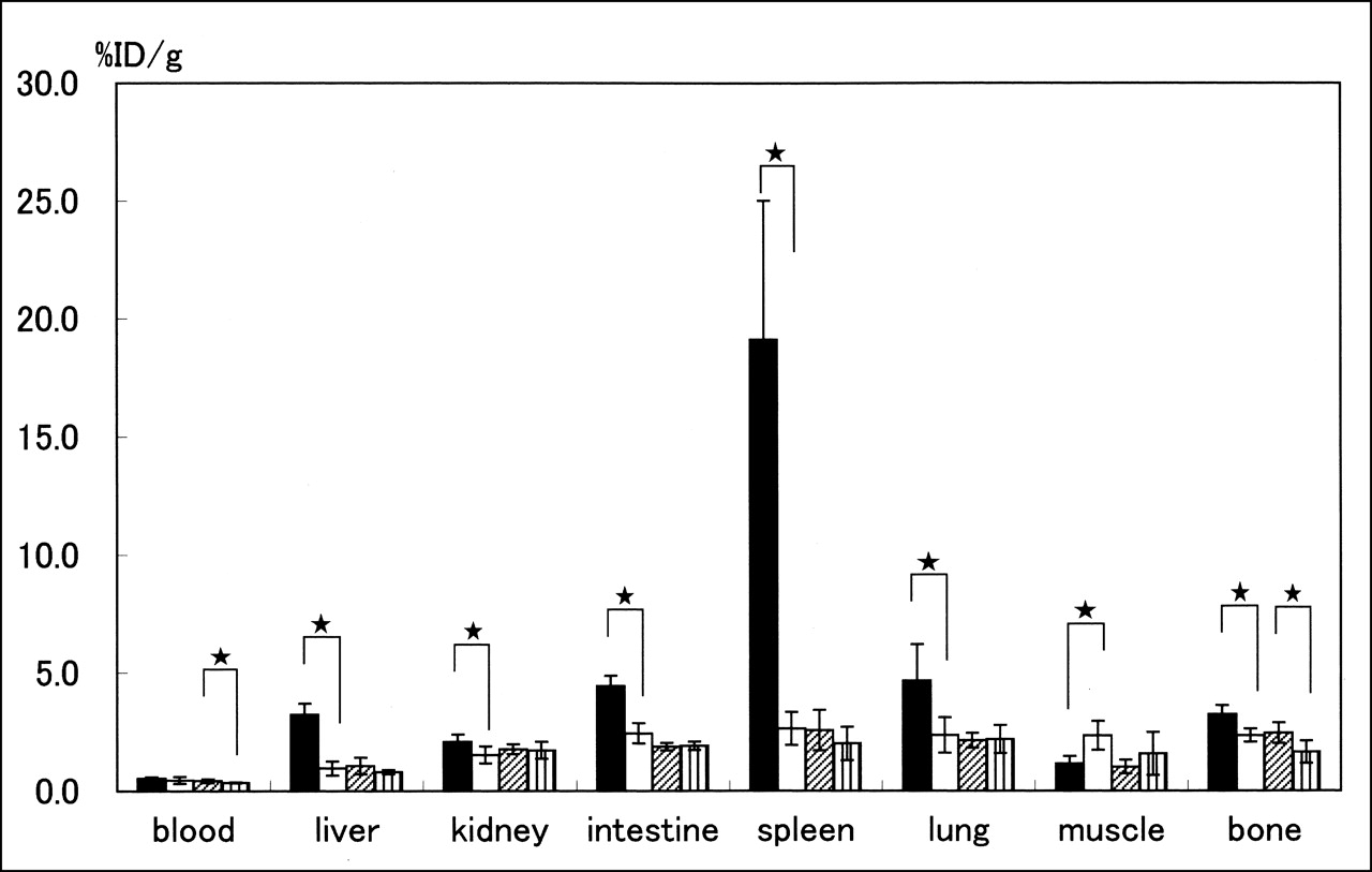
Biodistribution of 18F-FDG at 1 h after injection in immunocompetent BALB/c mice with Con A pretreatment (filled bar) or without Con A pretreatment (open bar) and in nude mice with Con A pretreatment (hatched bar) or without Con A pretreatment (striped bar). Values are expressed as mean and SD for percentage injected dose (%ID) per gram of tissue (n = 5 for each group). Stars indicate significant differences (P < 0.05).
In contrast, Con A pretreatment of immunodeficient mice did not significantly change the 18F-FDG uptake, except in bone and blood.
Autoradiography and Histology of Liver
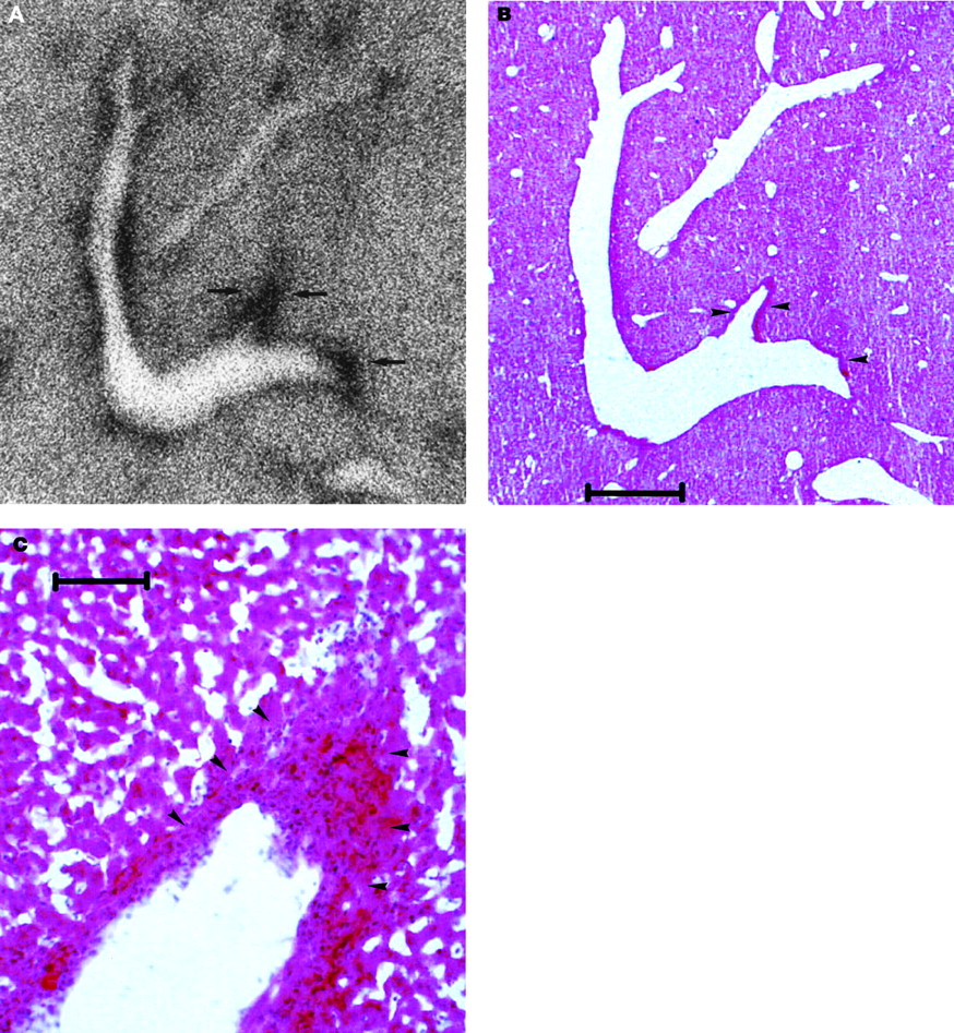
Autoradiographs of the liver of Con A-treated and untreated BALB/c mice are shown in Figures 2 and 3, along with the same section stained with H & E. Without Con A pretreatment, the livers appeared histologically normal, withno specific localization of 18F-FDG detected by autoradiography (Figs. 2A and 3A). In contrast, 24 h after injection of Con A, a specific localization of 18F-FDG was seen at periportal areas, where numerous sites of cellular infiltration were observed through H & E staining, corresponding to Con A-induced acute liver injury (Figs. 2A–2C). The infiltrated cells were mainly lymphocytes.

Autoradiography (A) and H & E staining (B and C) of liver of Con A-treated BALB/c mice. Bars indicate 1 mm (B) and 0.1 mm (C). After Con A pretreatment, 18F-FDG localizes specifically at periportal areas (arrow), where numerous sites of cellular infiltration are observed through H & E staining (arrowhead).

Autoradiography (A) and H & E staining (B and C) of liver of untreated BALB/c mice. Bars indicate 1 mm (B) and 0.1 mm (C). Without Con A pretreatment, no specific 18F-FDG localization or cellular infiltration is observed.
In Vitro 18F-FDG Uptake to In Vivo Activated Splenocytes
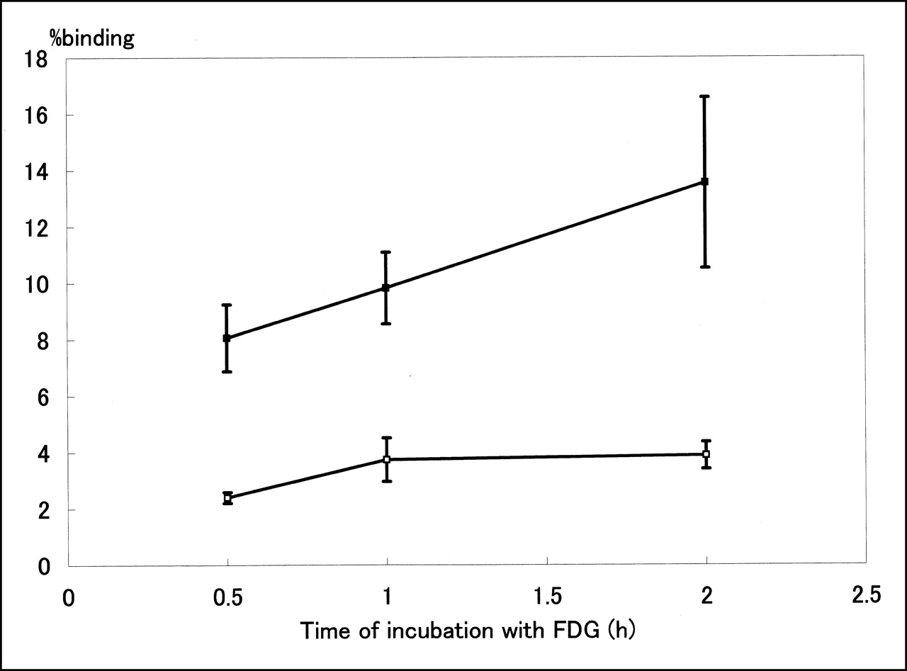
In vitro binding of 18F-FDG to the splenocytes from BALB/c mice was significantly higher when the mice were pretreated with Con A (Fig. 4). The higher uptake was first observed after 30 min of incubation and increased thereafter (from 8.06% ± 2.38% at 30 min to 13.54% ± 6.06% at 2 h).

In vitro binding of 18F-FDG to splenocytes from Con A-treated (▪) and untreated (□) BALB/c mice. Values are expressed as mean and SD for percentage binding (n = 4 for each group).
DISCUSSION
18F-FDG is widely used for the evaluation of malignant tumors in clinical oncology. At first, 18F-FDG PET was expected to play an important role in the differentiation of malignant from benign diseases (1–3). However, it gave numerous false-positive results (13–19). In addition to accumulating in malignant tissues, 18F-FDG has been shown to accumulate in nontumorous tissues, such as tissues with acute inflammation, granulomatous tissues, and tissues containing autoimmune lesions (4–6,20).
A full understanding of the mechanism of this nontumorous 18F-FDG uptake requires basic investigations of 18F-FDG uptake into the cells responsive to inflammation, granulomatous disease, and autoimmunity. However, only a few studies have investigated 18F-FDG uptake and distribution in inflammation that was induced experimentally in animal models. Yamada et al. (7) reported that subcutaneous injection of turpentine oil could produce inflammation in rats, and high 18F-FDG uptake in the inflammatory tissue was observed. Increased 18F-FDG uptake in inflammation was also reported in animal models of bacterial infection (21,22), in which high 18F-FDG uptake in the inflammatory cell infiltration was shown. In addition, Scharko et al. (21) studied 18F-FDG distribution in activated lymphoid tissues after viral infection and showed high uptake in B cells. In this study, we selected Con A to activate murine lymphocytes and used this as a model to investigate 18F-FDG uptake in inflammatory tissue and in activated lymphocytes. Con A is widely regarded as a model for investigating inflammation but has not yet been applied to 18F-FDG-related experiments.
In this study, Con A pretreatment in immunocompetent mice caused a marked increase in 18F-FDG uptake in the spleen, which is actually rich in lymphoid tissues. Increased 18F-FDG uptake in the lung and intestine may also reflect the activation of lymphocytes in the lymphoid tissue of both organs. The effect of Con A was observed only with immunocompetent mice and not with T cell-deficient nude mice, indicating that Con A-mediated cellular activation takes place only in T lymphocytes and that activated T lymphocytes took up more 18F-FDG than did nonactivated T lymphocytes. This finding was also confirmed by an in vitro binding study of 18F-FDG to activated and nonactivated splenocytes—a study in which activated splenocytes showed more than 3-fold higher binding.
As reported by Tiegs et al. (8), Con A pretreatment induced acute liver injury that caused prominent cellular infiltration at periportal areas only in immunocompetent mice. As shown by autoradiography, 18F-FDG specifically accumulated in these areas, confirming that these infiltrating cells were activated T lymphocytes that actively took up 18F-FDG.
This situation caused by Con A treatment did not exactly correspond to the situation of natural inflammation, because inflammatory cells other than T cells are also involved and may contribute to the high 18F-FDG uptake in inflammatory tissues. In experimental inflammatory tissue, young fibroblasts, endothelial cells of vessels, and phagocytes of neutrophils and macrophages were reported to show high 18F-FDG uptake (7), and high 18F-FDG uptake into B cells was also reported (23). However, in addition to acute inflammation, the processes of T cell activation are also involved in the formation of autoimmunity, in graft and tumor rejection, and in other such events, and this murine model of Con A activation can be used for evaluating the mechanism of various diseases and also for developing specific methods to reduce unfavorable 18F-FDG uptake in nontumorous tissues.
CONCLUSION
This investigation showed that Con A activation of lymphocytes, a model of intense inflammation, markedly increased 18F-FDG uptake by lymphocytes both in vitro and in vivo and that 18F-FDG specifically accumulated in Con A-mediated acute inflammatory tissues. This experimental model would be applicable to further investigations to evaluate mechanisms of 18F-FDG uptake in inflammatory cells.
Footnotes
Received May 29, 2001; revision accepted Jan. 16, 2002.
For correspondence or reprints contact: Takayoshi Ishimori, MD, Department of Nuclear Medicine and Diagnostic Imaging, Graduate School of Medicine, Kyoto University, 54 Kawahara-cho, Shogoin, Sakyo-ku, Kyoto, 606-8507 Japan.
E-mail: ishimori{at}kuhp.kyoto-u.ac.jp
REFERENCES
In this issue


Jump to section
Related Articles
Cited By...
- 18F-FAC PET Selectively Images Liver-Infiltrating CD4 and CD8 T Cells in a Mouse Model of Autoimmune Hepatitis
- Diagnostic Accuracy of 18F-FDG PET/CT in Infective Endocarditis and Implantable Cardiac Electronic Device Infection: A Cross-Sectional Study
- Prognostic Value of Dual-Time-Point 18F-FDG PET for Idiopathic Pulmonary Fibrosis
- Targeted noninvasive imaging of the innate immune response
- Metabolic Activity of the Spleen and Bone Marrow in Patients With Acute Myocardial Infarction Evaluated by 18F-Fluorodeoxyglucose Positron Emission Tomograpic Imaging
- Radiologic Responses in Cynomolgus Macaques for Assessing Tuberculosis Chemotherapy Regimens
- Inflammation Imaging in Atherosclerosis
- Quantification of Inflammation Within Rabbit Atherosclerotic Plaques Using the Macrophage-Specific CT Contrast Agent N1177: A Comparison with 18F-FDG PET/CT and Histology
- 18F-FDG PET for Semiquantitative Evaluation of Acute Allograft Rejection and Immunosuppressive Therapy Efficacy in Rat Models of Liver Transplantation
- Simvastatin Attenuates Plaque Inflammation: Evaluation by Fluorodeoxyglucose Positron Emission Tomography
- ENHANCEMENT OF SPLENIC GLUCOSE METABOLISM DURING ACUTE MALARIAL INFECTION: CORRELATION OF FINDINGS OF FDG-PET IMAGING WITH PATHOLOGICAL CHANGES IN A PRIMATE MODEL OF SEVERE HUMAN MALARIA.
- Comparison of 18F-FLT PET and 18F-FDG PET in Esophageal Cancer
- Tetraphenylphosphonium as a Novel Molecular Probe for Imaging Tumors
- Characterization of 18F-FDG Uptake in Human Endothelial Cells In Vitro