
Main menu

  • Home
  • Content
    • Current
    • Ahead of print
    • Past Issues
    • JNM Supplement
    • SNMMI Annual Meeting Abstracts
    • Continuing Education
    • JNM Podcasts
  • Subscriptions
    • Subscribers
    • Institutional and Non-member
    • Rates
    • Journal Claims
    • Corporate & Special Sales
  • Authors
    • Submit to JNM
    • Information for Authors
    • Assignment of Copyright
    • AQARA requirements
  • Info
    • Reviewers
    • Permissions
    • Advertisers
  • About
    • About Us
    • Editorial Board
    • Contact Information
  • More
    • Alerts
    • Feedback
    • Help
    • SNMMI Journals
  • SNMMI
    • JNM
    • JNMT
    • SNMMI Journals
    • SNMMI

User menu

  • Subscribe
  • My alerts
  • Log in
  • Log out
  • My Cart

Search

  • Advanced search
Journal of Nuclear Medicine
  • SNMMI
    • JNM
    • JNMT
    • SNMMI Journals
    • SNMMI
  • Subscribe
  • My alerts
  • Log in
  • Log out
  • My Cart
Journal of Nuclear Medicine

Advanced Search

  • Home
  • Content
    • Current
    • Ahead of print
    • Past Issues
    • JNM Supplement
    • SNMMI Annual Meeting Abstracts
    • Continuing Education
    • JNM Podcasts
  • Subscriptions
    • Subscribers
    • Institutional and Non-member
    • Rates
    • Journal Claims
    • Corporate & Special Sales
  • Authors
    • Submit to JNM
    • Information for Authors
    • Assignment of Copyright
    • AQARA requirements
  • Info
    • Reviewers
    • Permissions
    • Advertisers
  • About
    • About Us
    • Editorial Board
    • Contact Information
  • More
    • Alerts
    • Feedback
    • Help
    • SNMMI Journals
  • View or Listen to JNM Podcast
  • Visit JNM on Facebook
  • Join JNM on LinkedIn
  • Follow JNM on Twitter
  • Subscribe to our RSS feeds
Meeting ReportPhysics, Instrumentation & Data Sciences

Radiomics Analysis Based on18F-FDG and18F-FLT PET/CT Images may Facilitate the Differentiation of Benign and Malignant Pulmonary Nodules.

Jing Ning, Peng Yu, Jingjing Cui, Yan Jia, Panli Zuo, JiaHe Tian and Baixuan Xu
Journal of Nuclear Medicine May 2020, 61 (supplement 1) 273;
Jing Ning
1Nuclear Medicine Chinese PLA Medical School Beijing China
  • Find this author on Google Scholar
  • Find this author on PubMed
  • Search for this author on this site
Peng Yu
2BeiJing China
  • Find this author on Google Scholar
  • Find this author on PubMed
  • Search for this author on this site
Jingjing Cui
3Huiying Medical Technology Co., Ltd. Beijing China
  • Find this author on Google Scholar
  • Find this author on PubMed
  • Search for this author on this site
Yan Jia
4Huiying Medical Technology Co., Ltd Beijing China
  • Find this author on Google Scholar
  • Find this author on PubMed
  • Search for this author on this site
Panli Zuo
4Huiying Medical Technology Co., Ltd Beijing China
  • Find this author on Google Scholar
  • Find this author on PubMed
  • Search for this author on this site
JiaHe Tian
2BeiJing China
  • Find this author on Google Scholar
  • Find this author on PubMed
  • Search for this author on this site
Baixuan Xu
5Chinese PLA General Hospital Beijing China
  • Find this author on Google Scholar
  • Find this author on PubMed
  • Search for this author on this site
  • Article
  • Figures & Data
  • Info & Metrics
Loading

Abstract

273

Objectives: explore the value of radiomics analysis based on 18F-FDG and 18F-FLT PET/CT images in the differentiation of benign and malignant pulmonary nodules (PNs).

Methods: We retrospectively analyzed 113 patients getting clinicians into trouble of differential diagnosis between benign and malignant PNs on 18F-FDG PET/CT images, all of whom were followed by additional preoperative 18F-FLT PET/CT scans within a week. Three analysis methods including visual analysis, radiomics analysis based on 18F-FDG PET/CT images alone and radiomics analysis based on dual tracer PET/CT images were evaluated in the differential diagnostic value of benign and malignant PNs. Radiomics analysis went in details like dividing the dataset into training set and test set in a ratio of 7:3 (The distribution of the dataset was shown in Table 1), extracting specific quantitative features via analysis of variance (ANOVA) and least absolute shrinkage and selection operator (LASSO), constructing the logistic regression (LR) model, and evaluating their performances via receiver operating characteristic (ROC), sensitivity and specificity.

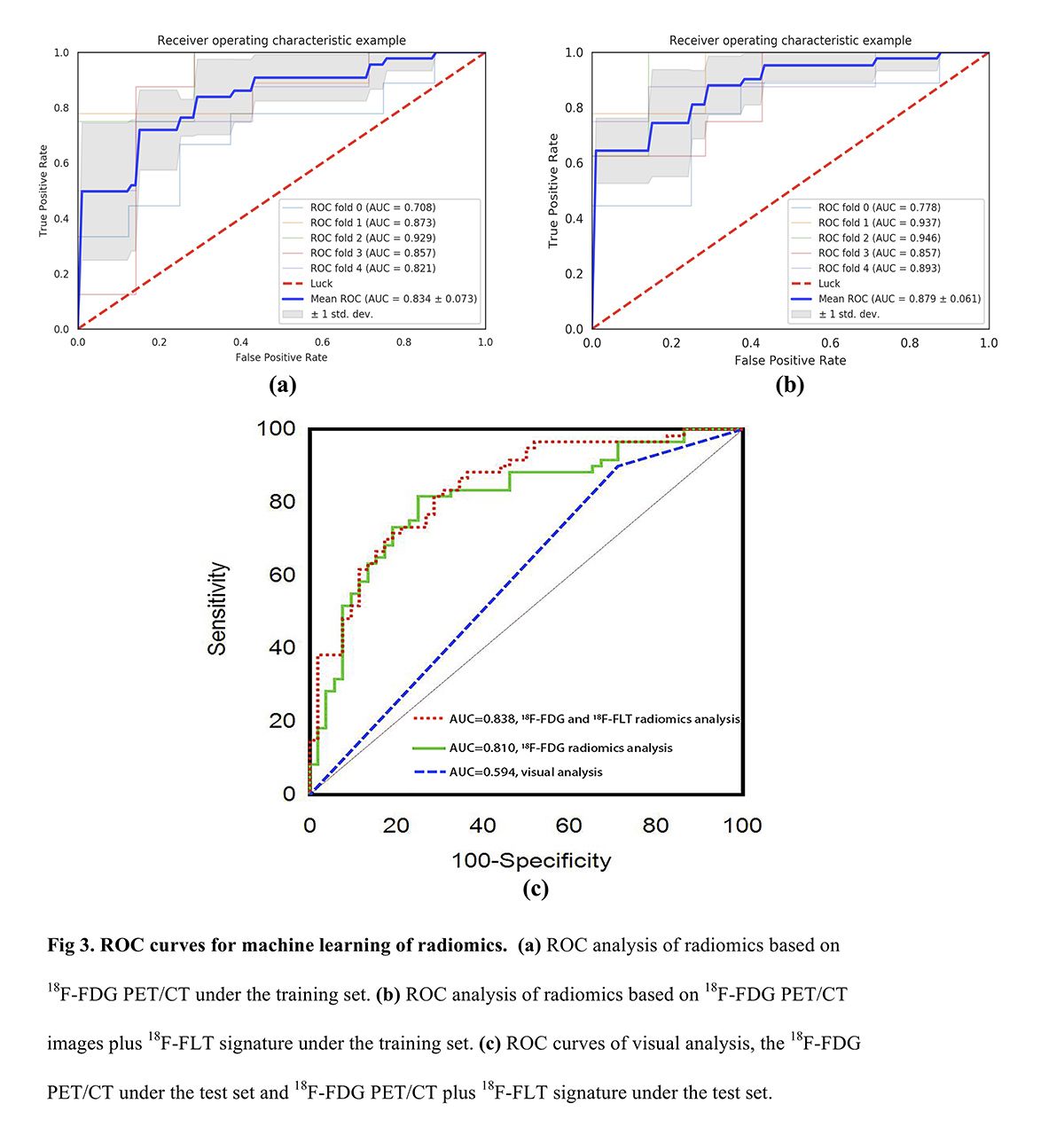
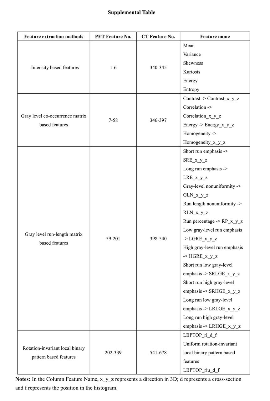
Results: A total of 123 lesions were acquired from 113 patients (74 men, 39 women; mean age 56.4 years; age range, 27-83 years). To be more detailed, 63 benign lesions were respectively diagnosed as inflammation (n=12), tuberculosis (n=29), or other diseases confirmed by follow-up (n=22) while 60 malignant lesions included adenocarcinoma (n=30), squamous cell carcinoma (n=16), small cell carcinoma (n=1), bronchioloalveolar carcinoma (n=6) and others (n=7), as presented in Table 2. Based on visual analysis of 18F-FDG PET/CT images,the diagnostic accuracy, sensitivity and specificity were 61.6%, 90% and 28.8% respectively. 678 radiomics features (see Supplemental Table) were extracted from Volumes of Interest (VOIs) of PNs (The process was revealed in Fig. 1). And 19 features were selected by ANOVA. After LASSO optimization, 14 valuable features were screened out (Fig. 2 shows the above two steps). As vividly indicated in Fig. 3, based on 18F-FDG PET/CT plus 18F-FLT signature, the area under the curve (AUC), sensitivity and specificity under the test set are 0.838, 0.778, and 0.750 respectively while under the training set, they are 0.879, 0.810, and 0.750 successively. And based on 18F-FDG PET/CT, the AUC, sensitivity and specificity under the test set are 0.810, 0.778, and 0.688 respectively while under the training set, they are 0.834, 0.786, and 0.778 successively (The results were listed in Table 3).

Conclusions: Radiomics analysis based on dual-tracer PET/CT images may be clinically potential and feasible for the discernable evaluation of benign and malignant PNs.

Figure
  • Download figure
  • Open in new tab
  • Download powerpoint
Figure
  • Download figure
  • Open in new tab
  • Download powerpoint
Figure
  • Download figure
  • Open in new tab
  • Download powerpoint
Figure
  • Download figure
  • Open in new tab
  • Download powerpoint
View this table:
  • View inline
  • View popup

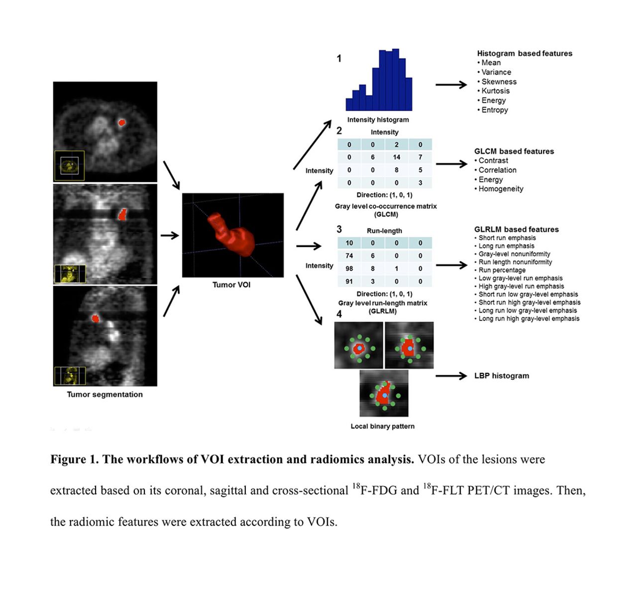
Table 1. The distribution of the dataset between benign and malignant PNs.

View this table:
  • View inline
  • View popup

Table 2. The pathological diagnosis of 123 lesions

View this table:
  • View inline
  • View popup

Table 3. The AUC, sensitivity, specificity of three analysis methods under the test set.

Previous
Back to top

In this issue

Journal of Nuclear Medicine
Vol. 61, Issue supplement 1
May 1, 2020
  • Table of Contents
  • Index by author
Article Alerts
Sign In to Email Alerts with your Email Address
Email Article

Thank you for your interest in spreading the word on Journal of Nuclear Medicine.

NOTE: We only request your email address so that the person you are recommending the page to knows that you wanted them to see it, and that it is not junk mail. We do not capture any email address.

Enter multiple addresses on separate lines or separate them with commas.
Radiomics Analysis Based on18F-FDG and18F-FLT PET/CT Images may Facilitate the Differentiation of Benign and Malignant Pulmonary Nodules.
(Your Name) has sent you a message from Journal of Nuclear Medicine
(Your Name) thought you would like to see the Journal of Nuclear Medicine web site.
Citation Tools
Radiomics Analysis Based on18F-FDG and18F-FLT PET/CT Images may Facilitate the Differentiation of Benign and Malignant Pulmonary Nodules.
Jing Ning, Peng Yu, Jingjing Cui, Yan Jia, Panli Zuo, JiaHe Tian, Baixuan Xu
Journal of Nuclear Medicine May 2020, 61 (supplement 1) 273;

Citation Manager Formats

  • BibTeX
  • Bookends
  • EasyBib
  • EndNote (tagged)
  • EndNote 8 (xml)
  • Medlars
  • Mendeley
  • Papers
  • RefWorks Tagged
  • Ref Manager
  • RIS
  • Zotero
Share
Radiomics Analysis Based on18F-FDG and18F-FLT PET/CT Images may Facilitate the Differentiation of Benign and Malignant Pulmonary Nodules.
Jing Ning, Peng Yu, Jingjing Cui, Yan Jia, Panli Zuo, JiaHe Tian, Baixuan Xu
Journal of Nuclear Medicine May 2020, 61 (supplement 1) 273;
Twitter logo Facebook logo LinkedIn logo Mendeley logo
  • Tweet Widget
  • Facebook Like
  • Google Plus One
Bookmark this article

Jump to section

  • Article
  • Figures & Data
  • Info & Metrics

Related Articles

  • No related articles found.
  • Google Scholar

Cited By...

  • No citing articles found.
  • Google Scholar

More in this TOC Section

Physics, Instrumentation & Data Sciences

  • Exploration of Multi-objective Optimization with Genetic Algorithms for PET Image Reconstruction
  • AI-based methods for nuclear-medicine imaging: Need for objective task-specific evaluation
  • Keel-Edge Height Selection for Improved Multi-Pinhole 123I Brain SPECT Imaging
Show more Physics, Instrumentation & Data Sciences

Radiomics (Data Analysis & Management)

  • Performance comparison of modified ComBat for harmonization of radiomic features in multicentric studies
  • Voxel-based analysis facilitates the identification of robust machine-learning based models for tumor classification
  • Can we identify “twin patients” to predict response to neoadjuvant chemotherapy in breast cancer?
Show more Radiomics (Data Analysis & Management)

Similar Articles

SNMMI

© 2025 SNMMI

Powered by HighWire