Main menu

  • Home
  • Content
    • Current
    • Ahead of print
    • Past Issues
    • JNM Supplement
    • SNMMI Annual Meeting Abstracts
    • Continuing Education
    • JNM Podcasts
  • Subscriptions
    • Subscribers
    • Institutional and Non-member
    • Rates
    • Journal Claims
    • Corporate & Special Sales
  • Authors
    • Submit to JNM
    • Information for Authors
    • Assignment of Copyright
    • AQARA requirements
  • Info
    • Reviewers
    • Permissions
    • Advertisers
  • About
    • About Us
    • Editorial Board
    • Contact Information
  • More
    • Alerts
    • Feedback
    • Help
    • SNMMI Journals
  • SNMMI
    • JNM
    • JNMT
    • SNMMI Journals
    • SNMMI

User menu

  • Subscribe
  • My alerts
  • Log in
  • Log out
  • My Cart

Search

  • Advanced search
Journal of Nuclear Medicine
  • SNMMI
    • JNM
    • JNMT
    • SNMMI Journals
    • SNMMI
  • Subscribe
  • My alerts
  • Log in
  • Log out
  • My Cart
Journal of Nuclear Medicine

Advanced Search

  • Home
  • Content
    • Current
    • Ahead of print
    • Past Issues
    • JNM Supplement
    • SNMMI Annual Meeting Abstracts
    • Continuing Education
    • JNM Podcasts
  • Subscriptions
    • Subscribers
    • Institutional and Non-member
    • Rates
    • Journal Claims
    • Corporate & Special Sales
  • Authors
    • Submit to JNM
    • Information for Authors
    • Assignment of Copyright
    • AQARA requirements
  • Info
    • Reviewers
    • Permissions
    • Advertisers
  • About
    • About Us
    • Editorial Board
    • Contact Information
  • More
    • Alerts
    • Feedback
    • Help
    • SNMMI Journals
  • View or Listen to JNM Podcast
  • Visit JNM on Facebook
  • Join JNM on LinkedIn
  • Follow JNM on Twitter
  • Subscribe to our RSS feeds
OtherThis Month in JNM

THIS MONTH IN JNM

Journal of Nuclear Medicine August 2004, 45 (8) 8A-9A;
  • Article
  • Info & Metrics
  • PDF
Loading

Buck and colleagues assess published reports suggesting that, in addition to its utility in staging and restaging lung cancer, 18F-FDG PET provides significant prognostic information on patients with non-small cell lung cancer.

Page 1274

Scheffel and Pomper look at the promise and potential limitations of current tracers used in PET imaging of gastrin-releasing peptide receptor expression in prostate cancer.

Page 1277

Lemke and colleagues report on incremental diagnostic information provided by retrospective PET/CT image fusion on data from patients with suspected pancreatic lesions.

Page 1279

Figure

Erdi and colleagues evaluate the effects of motion and respiratory phase on standardized uptake values in PET/CT of patients with lung cancer.

Page 1287

Pirotte and colleagues compare the merits of 18F-FDG and 11C-methionine as tracers for PET-guided neurosurgical procedures on gliomas.

Page 1293

Knappen and colleagues investigate the advantages of the perfusable tissue index in noninvasive assessment of myocardial fibrosis in patients with idiopathic dilated cardiomyopathy.

Page 1299

Tanabe and colleagues apply 123I-MIBG myocardial scintigraphy to the physiologic assessment of patients with panic disorder and find evidence of impaired cardiac sympathetic function.

Page 1305

Figure

Wells and colleagues present results evaluating the utility of dSPECT, an iterative algorithm, in removing bladder artifacts from clinically acquired pelvic bone SPECT images.

Page 1309

Jani and colleagues assess the role of radioimmunoscintigraphy directed against prostate-specific membrane antigen in influencing radiotherapy toxicity and biochemical control after prostatectomy.

Page 1315

Fuster and colleagues compare the accuracy of 18F-FDG PET with that of standard diagnostic clinical procedures in detecting locoregional disease and distant metastases in patients referred for suspected recurrent melanoma.

Page 1323

Sundaram and colleagues describe a simplified kinetic analysis method for dynamic assessment of 18F-FDG uptake in tumor.

Page 1328

Guo and colleagues investigate correlations between 18F-FDG uptake in PET imaging, lactate and choline concentrations measured by in vitro 1H magnetic resonance spectroscopy, and survival in human lung adenocarcinoma.

Page 1334

Bagheri and colleagues describe the additional value of 18F-FDG PET/CT over PET alone in reliable visualization of the normal adrenal gland.

Page 1340

Weber and colleagues evaluate a continuous-infusion H215O PET and acetazolamide challenge protocol as an alternative to single-bolus injection for assessment of cerebral perfusion status in patients with cerebrovascular disease.

Page 1344

Figure

Oturai and colleagues compare the diagnostic accuracies of γ-camera PET and dedicated PET in patients with suspected lung cancer.

Page 1351

Pandit-Taskar and colleagues provide a comprehensive review of radiopharmaceuticals currently used for palliation in patients with osseous metastases, including approved dosages, methods of administration, indications for use, and recommended guidelines.

Page 1358

Sgouros and colleagues describe and evaluate 124I PET-based, patient-specific, 3-dimensional dosimetry for 131I therapy in thyroid cancer.

Page 1366

Blankenberg and colleagues propose a novel imaging complex with a radiolabeled adapter protein bound to a docking tag and fused to a targeting protein as a model system for targeted imaging with a variety of protein-based radiotracers.

Page 1373

Thakur and colleagues assess the feasibility of 64Cu-TP3982 PET imaging of oncogene VPAC1 receptors overexpressed in human breast cancer cells.

Page 1381

Chen and colleagues use small-animal PET and autoradiographic imaging in prostate cancer-xenografted mice to evaluate 64Cu-radiolabeled gastrin-releasing peptide analogs for imaging peptide receptor expression.

Page 1390

Toyama and colleagues report on the use of 18F-FDG and small-animal PET for investigating the feasibility of absolute quantification of regional cerebral glucose use in a mouse model.

Page 1398

Koort and colleagues describe a laboratory study comparing the 18F-FDG PET characteristics of normal bone healing with those of local osteomyelitis and evaluating the ability of PET to differentiate bone healing from local infection.

Page 1406

Divgi and colleagues determine the maximum tolerated whole-body radiation-absorbed dose for a 131I-labeled fractionated radioimmunotherapy used for metastatic renal cancer.

Page 1412

ON THE COVER

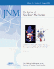
This image depicts localization of 64Cu-DOTA-Lys3-bombesin in PC-3 and CWR22 tumor-bearing mice as determined by microPET imaging (Concorde Microsystems, Inc.) followed by whole-body autoradiography. The microPET image is concordant with the whole-body autoradiographic section. Both the PC-3 tumor and the CWR22 tumor are visible, with clear contrast from the adjacent background. Prominent uptake is also seen in the liver and kidneys, and clearance of activity through the urinary bladder is evident.

Figure
PreviousNext
Back to top

In this issue

Journal of Nuclear Medicine: 45 (8)
Journal of Nuclear Medicine
Vol. 45, Issue 8
August 1, 2004
  • Table of Contents
  • Table of Contents (PDF)
  • About the Cover
  • Index by author
Print
Download PDF
Article Alerts
Sign In to Email Alerts with your Email Address
Email Article

Thank you for your interest in spreading the word on Journal of Nuclear Medicine.

NOTE: We only request your email address so that the person you are recommending the page to knows that you wanted them to see it, and that it is not junk mail. We do not capture any email address.

Enter multiple addresses on separate lines or separate them with commas.
THIS MONTH IN JNM
(Your Name) has sent you a message from Journal of Nuclear Medicine
(Your Name) thought you would like to see the Journal of Nuclear Medicine web site.
Citation Tools
THIS MONTH IN JNM
Journal of Nuclear Medicine Aug 2004, 45 (8) 8A-9A;

Citation Manager Formats

  • BibTeX
  • Bookends
  • EasyBib
  • EndNote (tagged)
  • EndNote 8 (xml)
  • Medlars
  • Mendeley
  • Papers
  • RefWorks Tagged
  • Ref Manager
  • RIS
  • Zotero
Share
THIS MONTH IN JNM
Journal of Nuclear Medicine Aug 2004, 45 (8) 8A-9A;
Twitter logo Facebook logo LinkedIn logo Mendeley logo
  • Tweet Widget
  • Facebook Like
  • Google Plus One
Bookmark this article

Jump to section

  • Article
  • Info & Metrics
  • PDF

Related Articles

  • Quantification of 18F-FDG Uptake in Non–Small Cell Lung Cancer: A Feasible Prognostic Marker?
  • PET Imaging of GRP Receptor Expression in Prostate Cancer
  • Retrospective Digital Image Fusion of Multidetector CT and 18F-FDG PET: Clinical Value in Pancreatic Lesions—A Prospective Study with 104 Patients
  • The CT Motion Quantitation of Lung Lesions and Its Impact on PET-Measured SUVs
  • Comparison of 18F-FDG and 11C-Methionine for PET-Guided Stereotactic Brain Biopsy of Gliomas
  • Perfusable Tissue Index as a Potential Marker of Fibrosis in Patients with Idiopathic Dilated Cardiomyopathy
  • 123I-Metaiodobenzylguanidine Myocardial Scintigraphy in Panic Disorder
  • Reducing Bladder Artifacts in Clinical Pelvic SPECT Images
  • Radioimmunoscintigraphy for Postprostatectomy Radiotherapy: Analysis of Toxicity and Biochemical Control
  • Is 18F-FDG PET More Accurate Than Standard Diagnostic Procedures in the Detection of Suspected Recurrent Melanoma?
  • Simplified Kinetic Analysis of Tumor 18F-FDG Uptake: A Dynamic Approach
  • In Vitro Proton Magnetic Resonance Spectroscopic Lactate and Choline Measurements, 18F-FDG Uptake, and Prognosis in Patients with Lung Adenocarcinoma
  • Characterization of the Normal Adrenal Gland with 18F-FDG PET/CT
  • Constant-Infusion H215O PET and Acetazolamide Challenge in the Assessment of Cerebral Perfusion Status
  • γ-Camera 18F-FDG PET in Diagnosis and Staging of Patients Presenting with Suspected Lung Cancer and Comparison with Dedicated PET
  • Radiopharmaceutical Therapy for Palliation of Bone Pain from Osseous Metastases
  • Patient-Specific Dosimetry for 131I Thyroid Cancer Therapy Using 124I PET and 3-Dimensional-Internal Dosimetry (3D-ID) Software
  • Tumor Imaging Using a Standardized Radiolabeled Adapter Protein Docked to Vascular Endothelial Growth Factor
  • PET Imaging of Oncogene Overexpression Using 64Cu-Vasoactive Intestinal Peptide (VIP) Analog: Comparison with 99mTc-VIP Analog
  • microPET and Autoradiographic Imaging of GRP Receptor Expression with 64Cu-DOTA-[Lys3]Bombesin in Human Prostate Adenocarcinoma Xenografts
  • Absolute Quantification of Regional Cerebral Glucose Utilization in Mice by 18F-FDG Small Animal PET Scanning and 2-14C-DG Autoradiography
  • Comparative 18F-FDG PET of Experimental Staphylococcus aureus Osteomyelitis and Normal Bone Healing
  • Phase I Clinical Trial with Fractionated Radioimmunotherapy Using 131I-Labeled Chimeric G250 in Metastatic Renal Cancer
  • Google Scholar

Cited By...

  • No citing articles found.
  • Google Scholar

More in this TOC Section

  • This Month in JNM
  • This Month in JNM
  • This Month in JNM
Show more This Month in JNM

Similar Articles

SNMMI

© 2025 SNMMI

Powered by HighWire