


Visual Abstract

Abstract
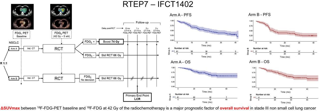
The purpose of this study was to assess the prognostic value of 18F-FDG PET parameter variation between baseline and 42 Gy (PET2) of radiochemotherapy at 6 mo and 1 y of evaluation in patients with stage III inoperable nonsmall cell lung cancer based on RECIST 1.1. Methods: In total, 158 patients in a prospective multicenter phase II/III study were analyzed. Patients were randomized into 2 groups: an experimental arm (group A) and a standard arm (group B). Patients from group A with residual metabolism on PET2 (group A+) at 42 Gy received a radiation boost (74 Gy). Patients without residual uptake on 18F-FDG PET at 42 Gy (group A−) and patients in group B received a standard radiotherapy dose (66 Gy). We compared group A with group B. The 18F-FDG PET parameters SUVmax, SUVmean, SUVpeak, peak SUV normalized on lean body mass, mean SUV normalized on lean body mass, total lesion glycolysis, total metabolic tumor volume (MTV) (tumor and nodes), and tumor MTV were measured. All patients were evaluated with RECIST 1.1 using CT at 6 mo and 1 y after radiochemotherapy. Progression-free survival and overall survival were evaluated. Results: Except for the radiotherapy dose (P < 0.001), patient demographic characteristics were similar between the 2 groups (A vs. B). All 18F-FDG PET uptake and volume parameter measurements were correlated. Therefore, only the change in SUVmax (ΔSUVmax) and total MTV were selected for the analysis. There was no significant difference in any variable between the 2 groups. In the multivariate analysis, ΔSUVmax appeared to be the most important prognostic factor for overall survival, and SUVmax of PET2 appeared to be the most important prognostic factor for progression-free survival. Conclusion: 18F-FDG PET at 42 Gy can be used to identify good responders to radiochemotherapy in patients with inoperable stage III nonsmall cell lung cancer. The SUVmax of PET2 and ΔSUVmax are independent prognostic factors.
Footnotes
Published online Feb. 27, 2025.
- © 2025 by the Society of Nuclear Medicine and Molecular Imaging.
This article requires a subscription to view the full text. If you have a subscription you may use the login form below to view the article. Access to this article can also be purchased.
SNMMI members
Login to the site using your SNMMI member credentials
Individuals
Login as an individual user
In this issue


Jump to section
Related Articles
Cited By...
- No citing articles found.