
Main menu

  • Home
  • Content
    • Current
    • Ahead of print
    • Past Issues
    • JNM Supplement
    • SNMMI Annual Meeting Abstracts
    • Continuing Education
    • JNM Podcasts
  • Subscriptions
    • Subscribers
    • Institutional and Non-member
    • Rates
    • Journal Claims
    • Corporate & Special Sales
  • Authors
    • Submit to JNM
    • Information for Authors
    • Assignment of Copyright
    • AQARA requirements
  • Info
    • Reviewers
    • Permissions
    • Advertisers
  • About
    • About Us
    • Editorial Board
    • Contact Information
  • More
    • Alerts
    • Feedback
    • Help
    • SNMMI Journals
  • SNMMI
    • JNM
    • JNMT
    • SNMMI Journals
    • SNMMI

User menu

  • Subscribe
  • My alerts
  • Log in
  • My Cart

Search

  • Advanced search
Journal of Nuclear Medicine
  • SNMMI
    • JNM
    • JNMT
    • SNMMI Journals
    • SNMMI
  • Subscribe
  • My alerts
  • Log in
  • My Cart
Journal of Nuclear Medicine

Advanced Search

  • Home
  • Content
    • Current
    • Ahead of print
    • Past Issues
    • JNM Supplement
    • SNMMI Annual Meeting Abstracts
    • Continuing Education
    • JNM Podcasts
  • Subscriptions
    • Subscribers
    • Institutional and Non-member
    • Rates
    • Journal Claims
    • Corporate & Special Sales
  • Authors
    • Submit to JNM
    • Information for Authors
    • Assignment of Copyright
    • AQARA requirements
  • Info
    • Reviewers
    • Permissions
    • Advertisers
  • About
    • About Us
    • Editorial Board
    • Contact Information
  • More
    • Alerts
    • Feedback
    • Help
    • SNMMI Journals
  • View or Listen to JNM Podcast
  • Visit JNM on Facebook
  • Join JNM on LinkedIn
  • Follow JNM on Twitter
  • Subscribe to our RSS feeds
Meeting ReportData Analysis & Management

Objective task-based evaluation of a deep-learning-based segmentation method for quantitative SPECT

Ziping Liu, Zekun Li and Abhinav Jha
Journal of Nuclear Medicine August 2022, 63 (supplement 2) 3200;
Ziping Liu
1Washington University in St. Louis
  • Find this author on Google Scholar
  • Find this author on PubMed
  • Search for this author on this site
Zekun Li
1Washington University in St. Louis
  • Find this author on Google Scholar
  • Find this author on PubMed
  • Search for this author on this site
Abhinav Jha
1Washington University in St. Louis
  • Find this author on Google Scholar
  • Find this author on PubMed
  • Search for this author on this site
  • Article
  • Figures & Data
  • Info & Metrics
Loading

Abstract

3200

Introduction: Quantitative SPECT (QSPECT) is showing significant promise in multiple clinical applications. In many of these applications, the task is quantifying the mean uptake within region(s) of interest. Reliable quantification requires accurate segmentation of these regions on the SPECT images. However, SPECT segmentation is challenging, a major reason being partial-volume effects. To address this challenge, we recently proposed a deep-learning (DL)-based segmentation method that estimates the fractional volume occupied by each considered region within each image-voxel (Liu et al. 2021a). The method was evaluated in the context of segmenting DaTscan SPECT images (Liu et al. 2021b), and was observed to yield accurate segmentation performance with high Dice scores and significantly outperform conventional methods. However, for clinical translation of this method for QSPECT, it is important that the method is evaluated on the clinical task of quantifying regional uptake. The aim of our study was to conduct this objective task-based evaluation.

Methods: To objectively evaluate segmentation methods for QSPECT, it is important that the procedure to quantify regional uptake should be optimal when the true boundaries of considered regions are known. The typical process of quantifying regional uptake from OSEM-reconstructed SPECT images is sub-optimal since these estimates are known to be biased and/or imprecise. Thus, such procedure is not suitable to objectively evaluate segmentation methods. In this context, an optimal procedure is projection-domain quantification (PDQ). The PDQ is a maximum-likelihood technique that estimates regional uptake directly from projection data by accurately modeling the imaging-system physics. Given the true segmentations, the PDQ approach yields an unbiased estimate of regional uptake with a variance close to Cramér-Rao bound (Li et al. 2021). Thus, when using the PDQ approach to evaluate a segmentation method, any error in the measured regional uptake would be due to the error in segmentation. Thus, we used the PDQ approach to objectively evaluate the proposed method.

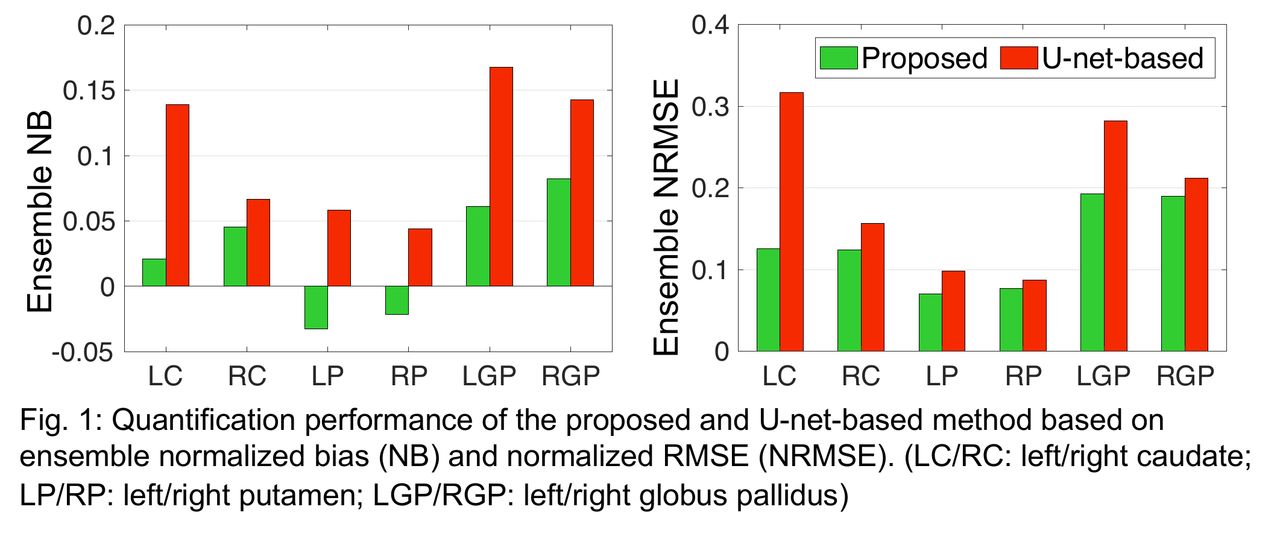
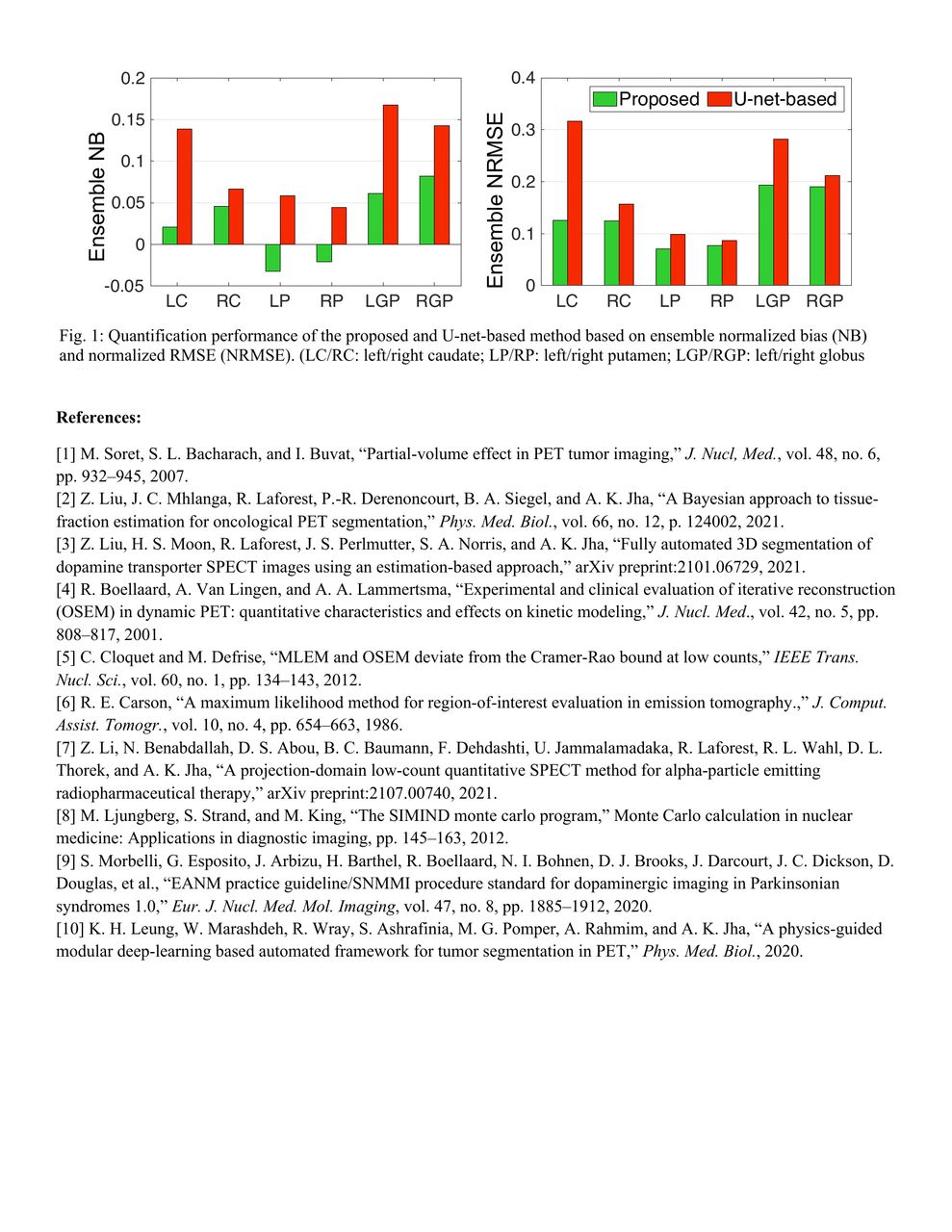
The proposed method was evaluated on the clinical task of quantifying regional uptake in caudate, putamen and globus pallidus from DaTscan SPECT. The evaluation was conducted using clinically realistic simulations with the details presented in Liu et al. (2021b). We generated 100 test digital brain phantoms with clinically relevant DaT activity distributions. From these activity distributions, realistic projection data were simulated using SIMIND by modeling all the relevant image-degrading processes and by following the practice guideline for DaTscan SPECT acquisition. The projection data were reconstructed using an OSEM algorithm that compensated for attenuation, scatter, and collimator-detector response. The proposed method was applied to segment the considered regions from the reconstructed SPECT images. We then used the PDQ approach to estimate the uptake within these segmented regions directly from the projection data. The performance of the proposed method was assessed using ensemble normalized bias (NB) and normalized RMSE (NRMSE) between the estimated and true uptake. The proposed method was also compared to a state-of-the-art U-net-based method based on these metrics.

Results: Fig. 1 shows the ensemble NB and NRMSE of the estimates yielded by the proposed and U-net-based methods. The proposed method yielded low NB < 10% and NRMSE < 20% for all regions, demonstrating both accurate and precise quantification. The proposed method also outperformed the U-net-based method based on ensemble NB and NRMSE.

Conclusions: The proposed deep-learning-based segmentation method yielded accurate and precise quantification of regional uptake from DaTscan SPECT. These results motivate further clinical evaluation of the method. Further, we outlined a framework to objectively evaluate segmentation methods for quantitative SPECT on the task of regional uptake quantification.

Figure
  • Download figure
  • Open in new tab
  • Download powerpoint
Figure
  • Download figure
  • Open in new tab
  • Download powerpoint
Figure
  • Download figure
  • Open in new tab
  • Download powerpoint
Previous
Back to top

In this issue

Journal of Nuclear Medicine
Vol. 63, Issue supplement 2
August 1, 2022
  • Table of Contents
  • Index by author
Article Alerts
Sign In to Email Alerts with your Email Address
Email Article

Thank you for your interest in spreading the word on Journal of Nuclear Medicine.

NOTE: We only request your email address so that the person you are recommending the page to knows that you wanted them to see it, and that it is not junk mail. We do not capture any email address.

Enter multiple addresses on separate lines or separate them with commas.
Objective task-based evaluation of a deep-learning-based segmentation method for quantitative SPECT
(Your Name) has sent you a message from Journal of Nuclear Medicine
(Your Name) thought you would like to see the Journal of Nuclear Medicine web site.
Citation Tools
Objective task-based evaluation of a deep-learning-based segmentation method for quantitative SPECT
Ziping Liu, Zekun Li, Abhinav Jha
Journal of Nuclear Medicine Aug 2022, 63 (supplement 2) 3200;

Citation Manager Formats

  • BibTeX
  • Bookends
  • EasyBib
  • EndNote (tagged)
  • EndNote 8 (xml)
  • Medlars
  • Mendeley
  • Papers
  • RefWorks Tagged
  • Ref Manager
  • RIS
  • Zotero
Share
Objective task-based evaluation of a deep-learning-based segmentation method for quantitative SPECT
Ziping Liu, Zekun Li, Abhinav Jha
Journal of Nuclear Medicine Aug 2022, 63 (supplement 2) 3200;
Twitter logo Facebook logo LinkedIn logo Mendeley logo
  • Tweet Widget
  • Facebook Like
  • Google Plus One
Bookmark this article

Jump to section

  • Article
  • Figures & Data
  • Info & Metrics

Related Articles

  • No related articles found.
  • Google Scholar

Cited By...

  • No citing articles found.
  • Google Scholar

More in this TOC Section

  • Assessment and optimization of CT radiation dose for body PET/CT across different scanner systems and different anatomical ranges.
  • Shortened whole body dynamic PET 18F-FDG Patlak imaging using a population input function and the Biograph Vision Quadra PET/CT
  • Lymphovascular Invasion prediction in Non-Small Cell Lung Carcinoma Using Multimodality PET/CT Fusion Radiomics
Show more Data Analysis & Management

Similar Articles

SNMMI

© 2025 SNMMI

Powered by HighWire