


A 29-y-old woman with relapsed Hodgkin lymphoma was being assessed for response to ifosfamide, carboplatin, and etoposide therapy. 18F-FDG PET/CT showed progressive hypermetabolic mediastinal lymphadenopathy (Fig. 1A) with diffuse hypermetabolism in the bone marrow, likely secondary to marrow stimulation by granulocyte–colony stimulating factor therapy. Treatment was escalated to gemcitabine, vinorelbine, and doxorubicin and to pembrolizumab. 18F-FDG PET/CT performed 6 wk later showed nearly complete metabolic resolution of the prior mediastinal lymphadenopathy. However, interval development of hepatomegaly with multifocal hypermetabolic lesions throughout the liver was noted (Fig. 1B). In view of the favorable response at the primary site, the new hepatic lesions were favored to represent immune-related hepatitis over progressive lymphomatous involvement of the liver. The patient’s laboratory results obtained within 4 d of the PET/CT showed elevated levels of aspartate transaminase (115 U/L) and alanine transaminase (151 U/L) (11 and 15 U/L, respectively, 1 mo beforehand). On the basis of the suggestion of immune-related hepatitis, pembrolizumab was discontinued and prednisone 40 mg daily was started, tapering by 10 mg weekly over 4 wk. The regimen of gemcitabine, vinorelbine, and doxorubicin was continued. Reassessment 18F-FDG PET/CT performed 5 wk later showed continued remission of the mediastinal disease, with marked anatomic and metabolic improvement of the liver lesions (Fig. 1C). The patient received an autologous stem cell transplantation 2 wk later, and reassessment 18F-FDG PET/CT (Fig. 1D) showed continued response by the mediastinal disease and complete resolution of the liver lesions.

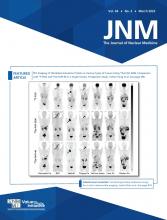
(A) 18F-FDG PET/CT maximum-intensity projection (top) and PET/CT transaxial images (bottom) at baseline show hypermetabolic mediastinal lymphadenopathy (dashed arrows). (B) 18F-FDG PET/CT performed 6 wk after treatment with pembrolizumab shows metabolic resolution of mediastinal lymphadenopathy and interval development of multiple discrete hypermetabolic lesions in liver (solid arrows). (C and D) 18F-FDG PET/CT performed 5 wk after discontinuation of pembrolizumab and treatment with prednisone shows significant improvement in liver lesions (C), which resolved completely at 8 wk (D).
Immune-related adverse events, such as hepatitis, are known to occur in patients treated with immune-checkpoint inhibitors. However, the presentation of immune-related hepatitis as multifocal involvement of the liver is uncommon and can mimic the appearance of progressive disease. In such cases, a reassessment 18F-FDG PET/CT performed at least 4 wk after discontinuation of immunotherapy can help improve the diagnostic accuracy. The detection of immune-related adverse events on PET/CT is critical, as patients with severe immune-related adverse events require cessation of immunotherapy and initiation of corticosteroids.
DISCLOSURE
No potential conflict of interest relevant to this article was reported.
Footnotes
Published online Oct. 13, 2022.
- © 2023 by the Society of Nuclear Medicine and Molecular Imaging.


