


Abstract
The purpose of this study was to examine the profound effect of the ring size of the radiolabeled lactam bridge–cyclized α-melanocyte–stimulating hormone (α-MSH) peptide on its melanoma-targeting properties. Methods: A novel cyclic α-MSH peptide, 1,4,7,10-tetraazacyclododecane-1,4,7,10-tetraacetic acid-Nle-c[Asp-His-d-Phe-Arg-Trp-Lys]-CONH2 (DOTA-Nle-CycMSHhex), was synthesized and radiolabeled with 111In. The melanocortin-1 receptor–binding affinity of DOTA-Nle-CycMSHhex was determined in B16/F1 melanoma cells. The internalization and efflux of 111In-DOTA-Nle-CycMSHhex were examined in B16/F1 cells. The melanoma-targeting properties and SPECT/CT characteristics of 111In-DOTA-Nle-CycMSHhex were determined in B16/F1 melanoma–bearing C57 mice. Results: DOTA-Nle-CycMSHhex displayed 1.77 nM receptor-binding affinity. 111In-DOTA-Nle-CycMSHhex exhibited rapid internalization and extended retention in B16/F1 cells. The tumor uptake of 111In-DOTA-Nle-CycMSHhex was 24.94% ± 4.58% and 10.53% ± 1.11% injected dose per gram at 0.5 and 24 h after injection, respectively. Greater than 82% of the injected radioactivity was cleared through the urinary system by 2 h after injection. The tumor-to-kidney uptake ratios reached 2.04 and 1.70 at 2 and 4 h after injection, respectively. Flank melanoma tumors were clearly visualized by SPECT/CT using 111In-DOTA-Nle-CycMSHhex as an imaging probe at 2 and 24 h after injection. The radioactivity accumulation in normal organs, except for the kidneys, was low at 2, 4, and 24 h after injection. Conclusion: The reduction of the peptide ring size dramatically increased the melanoma uptake and decreased the renal uptake of 111In-DOTA-Nle-CycMSHhex, providing a new insight into the design of a novel radiolabeled lactam bridge–cyclized α-MSH peptide for melanoma imaging and treatment.
Skin cancer is the most commonly diagnosed cancer in the United States. Melanoma accounts for less than 5% of skin cancer cases but causes greater than 75% of deaths from skin cancer. It is predicted that 68,720 new cases will be diagnosed and 8,650 deaths will occur in 2009 (1). Early diagnosis and prompt surgical removal are the best opportunities for a patient's cure, because no curative treatment exists for metastatic melanoma. Despite the clinical use of 18F-FDG for PET diagnosis and staging of melanoma, 18F-FDG is not a melanoma-specific imaging agent and is also not effective in imaging small melanoma metastases (<5 mm) and melanomas that have primary energy sources other than glucose (2–4). Alternatively, melanocortin-1 (MC1) receptor is a distinct molecular target because of its overexpression on both human and mouse melanoma cells (5–9). Radiolabeled α-melanocyte–stimulating hormone (α-MSH) peptides can bind the MC1 receptors with nanomolar binding affinities (10–20) and represent a class of promising melanoma-specific radiopharmaceuticals for melanoma imaging and therapy.
Recently, we have developed a novel class of 111In-labeled lactam bridge–cyclized 1,4,7,10-tetraazacyclododecane-1,4,7,10-tetraacetic acid (DOTA)-conjugated α-MSH peptides for melanoma detection (21,22). Lactam bridge cyclization was used to improve the stabilities of the α-MSH peptides against the proteolytic degradations in vivo and enhance the binding affinities of the α-MSH peptides through stabilization of their secondary structures such as β-turns (23–26). The radiometal chelator DOTA was attached to the N terminus of the lactam bridge–cyclized α-MSH peptide (12 amino acids in the peptide ring) for 111In radiolabeling. For instance, 111In-DOTA-GlyGlu-CycMSH (111In-DOTA-Gly-Glu-c[Lys-Nle-Glu-His-d-Phe-Arg-Trp-Gly-Arg-Pro-Val-Asp]) exhibited high MC1 receptor–mediated tumor uptake (10.40 ± 1.40 percentage injected dose per gram [%ID/g] at 2 h after injection) in flank B16/F1 melanoma–bearing C57 mice (21). Both primary and pulmonary metastatic melanoma flank lesions were clearly visualized by small-animal SPECT/CT using 111In-DOTA-GlyGlu-CycMSH as an imaging probe (21,22), highlighting its potential as an effective imaging probe for melanoma detection.
One advantage of the lactam bridge–cyclized α-MSH peptide is that the peptide ring size can be finely modified by either adding or deleting amino acids without sacrificing the binding affinity of the peptide (21,22). The studies on the α-MSH peptide agonists for the MC1 receptor revealed that the lactam bridge–cyclized α-MSH peptide with a 6-amino acid peptide ring (Ac-Nle-c[Asp-His-d-Phe-Arg-Trp-Lys(CONH2)]) displayed not only higher MC1 receptor–binding affinity but also a slower MC1 receptor dissociation rate than the native α-MSH peptide (Ac-Ser-Tyr-Ser-Met-Glu-His-Phe-Arg-Trp-Gly-Lys-Pro-Val-NH2) (27,28). A slow MC1 receptor dissociation rate might contribute to the prolonged biologic activity of Ac-Nle-c[Asp-His-d-Phe-Arg-Trp-Lys(CONH2) in vitro and in vivo (27). In this study, we conjugated the radiometal chelator DOTA to the N terminus of the Ac-Nle-c[Asp-His-d-Phe-Arg-Trp-Lys(CONH2) peptide to generate a novel DOTA-conjugated lactam bridge–cyclized α-MSH peptide with a 6-amino acid peptide ring (DOTA-Nle-CycMSHhex) to examine the effect of peptide ring size on its melanoma-targeting and pharmacokinetic properties. The MC1 receptor–binding affinity of DOTA-Nle-CycMSHhex was determined in B16/F1 melanoma cells. DOTA-Nle-CycMSHhex was radiolabeled with 111In, a commercially available diagnostic radionuclide with a half-life of 2.8 d. The melanoma-targeting and pharmacokinetic properties and SPECT/CT of 111In-labeled DOTA-Nle-CycMSHhex were determined in B16/F1 melanoma–bearing C57 mice.
MATERIALS AND METHODS
Chemicals and Reagents
Amino acid and resin were purchased from Advanced ChemTech Inc. and Novabiochem. DOTA-tri-t-butyl ester was purchased from Macrocyclics Inc. 111InCl3 was purchased from Trace Life Sciences, Inc. 125I-Tyr2-[Nle4, d-Phe7]-α-MSH (125I-(Tyr2)-NDP-MSH) was obtained from PerkinElmer, Inc. All other chemicals used in this study were purchased from Thermo Fischer Scientific and used without further purification. B16/F1 melanoma cells were obtained from American Type Culture Collection.
Peptide Synthesis
DOTA-Nle-CycMSHhex was synthesized using standard fluorenylmethyloxycarbonyl chemistry. Briefly, an intermediate scaffold of (tBu)3DOTA-Nle-Asp(O-2-PhiPr)-His(Trt)-d-Phe-Arg(Pbf)-Trp(Boc)-Lys(Dde) was synthesized on H2N-Sieber amide resin by an Advanced ChemTech multiple-peptide synthesizer. The protecting group of Dde was removed by 2% hydrazine for peptide cyclization. The protecting group of 2-phenylisopropyl from the Asp residue was removed, and the protected peptide was cleaved from the resin by treatment with a mixture of 2.5% of trifluoroacetic acid (TFA) and 5% of triisopropylsilane for 1 h. After precipitation with ice-cold ether and characterization by liquid chromatography–mass spectroscopy (LC-MS), the protected peptide was dissolved in H2O:CH3CN (30:70) and lyophilized to remove the reagents such as TFA and triisopropylsilane. The protected peptide was further cyclized by coupling the carboxylic group from the Asp with the ε-amino group from the Lys. The cyclization reaction was achieved by an overnight reaction in dimethylformamide using benzotriazole-1-yl-oxy-tris-pyrrolidino-phosphonium-hexafluorophosphate as a coupling agent in the presence of N,N-diisopropylethylamine. After characterization by LC-MS, the cyclized protected peptide was dissolved in H2O:CH3CN (30:70) and lyophilized to remove the reagents such as benzotriazole-1-yl-oxy-tris-pyrrolidino-phosphonium-hexafluorophosphate and N,N-diisopropylethylamine. The protecting groups were totally removed by treatment with a mixture of TFA, thioanisole, phenol, water, ethanedithiol, and triisopropylsilane (87.5:2.5:2.5:2.5:2.5:2.5) for 4 h at room temperature (25°C). The peptide was precipitated and washed with ice-cold ether 4 times, purified by reversed-phase (RP) high-performance liquid chromatography (HPLC), and characterized by LC-MS.
In Vitro Competitive Binding Assay
The inhibitory concentration of 50% (IC50) value of DOTA-Nle-CycMSHhex was determined by an in vitro competitive binding assay according to our previously published procedure (21). B16/F1 cells were harvested and seeded into a 24-well cell culture plate (5 × 10⁵ cells per well) and incubated at 37°C overnight. After the cells were washed twice with binding medium, Dulbecco's modified Eagle's medium with 25 mM N-(2-hydroxyethyl)-piperazine-N′-(2-ethanesulfonic acid) (pH 7.4, 0.2% bovine serum albumin [BSA], and 0.3 mM 1,10-phenathroline), they were incubated at room temperature (25°C) for 2 h with approximately 60,000 counts per minute of 125I-Tyr2-NDP-MSH in the presence of increasing concentrations (10−12 to 10−5 M) of DOTA-Nle-CycMSHhex in 0.3 mL of binding medium. The reaction medium was aspirated after the incubation. The cells were rinsed twice with 0.5 mL of ice-cold, pH 7.4, 0.2% BSA/0.01 M phosphate-buffered saline and lysed in 0.5 mL of 1N NaOH for 5 min. The activities associated with cells were measured in a Wallac 1480 automated γ-counter (PerkinElmer). The IC50 value of the peptide was calculated using Prism software (GraphPad Software).
Peptide Radiolabeling with 111In
111In-DOTA-Nle-CycMSHhex was prepared in a 0.5 M NH4OAc-buffered solution at pH 4.5 according to our published procedure (21). Briefly, 50 μL of 111InCl3 (37–74 MBq [1–2 mCi] in 0.05 M HCl aqueous solution), 10 μL of a 1 mg/mL aqueous solution of DOTA-Nle-CycMSHhex, and 400 μL of 0.5 M NH4OAc (pH 4.5) were added into a reaction vial and incubated at 75°C for 45 min. After the incubation, 10 μL of 0.5% ethylenediaminetetraacetic acid aqueous solution were added into the reaction vial to scavenge potential unbound 111In3+ ions. The radiolabeled peptide was purified to a single species by Waters RP HPLC on a Grace Vydac C-18 reversed-phase analytic column using a 20-min gradient of 18%−28% acetonitrile in 20 mM HCl aqueous solution with a flow rate of 1.0 mL/min. The purified peptide sample was purged with N2 gas for 20 min to remove the acetonitrile. The pH of the final solution was adjusted to 7.4 with 0.1N NaOH and sterile normal saline for animal studies. The in vitro serum stability of HPLC-purified 111In-DOTA-Nle-CycMSHhex was determined by incubation in mouse serum at 37°C for 24 h and monitored for degradation by RP HPLC.
Cellular Internalization and Efflux of 111In-DOTA-Nle-CycMSHhex
Cellular internalization and efflux of 111In-DOTA-Nle-CycMSHhex were evaluated in B16/F1 melanoma cells. After being washed twice with the binding medium, the B16/F1 cells seeded in cell culture plates were incubated at 25°C for 20, 40, 60, 90, and 120 min (n = 3) in the presence of approximately 200,000 counts per minute of HPLC-purified 111In-DOTA-Nle-CycMSHhex. After incubation, the reaction medium was aspirated, and the cells were rinsed with 2 × 0.5 mL of ice-cold, pH 7.4, 0.2% BSA/0.01 M phosphate-buffered saline. Cellular internalization of 111In-DOTA-Nle-CycMSHhex was assessed by washing the cells with acidic buffer (40 mM sodium acetate [pH 4.5] containing 0.9% NaCl and 0.2% BSA) to remove the membrane-bound radioactivity. The remaining internalized radioactivity was obtained by lysing the cells with 0.5 mL of 1N NaOH for 5 min. Membrane-bound and internalized 111In activities were counted in a γ-counter. Cellular efflux of 111In-DOTA-Nle-CycMSHhex was determined by incubating the B16/F1 cells with 111In-DOTA-Nle-CycMSHhex for 2 h at 25°C, removing nonspecifically bound activity with 2 × 0.5 mL of ice-cold phosphate-buffered saline rinse, and monitoring radioactivity released into the cell culture medium. At 20, 40, 60, 90, and 120 min, the radioactivities on the cell surface and inside the cells were separately collected and counted in a γ-counter.
Biodistribution Studies
All the animal studies were conducted in compliance with Institutional Animal Care and Use Committee approval. The mice were housed 5 animals per cage in sterile microisolator cages in a temperature- and humidity-controlled room with a 12-h light/12-h dark schedule. The pharmacokinetics of 111In-DOTA-Nle-CycMSHhex were determined in B16/F1 melanoma–bearing C57 female mice (Harlan). C57 mice were subcutaneously inoculated on the right flank with 1 × 10⁶ B16/F1 cells. The weight of tumors reached approximately 0.2 g 10 d after cell inoculation. Each melanoma-bearing mouse was injected with 0.037 MBq (1 μCi) of 111In-DOTA-Nle-CycMSHhex via the tail vein. Groups of 5 mice were sacrificed at 0.5, 2, 4, and 24 h after injection, and tumors and organs of interest were harvested, weighed, and counted. Blood values were taken as 6.5% of the whole-body weight. The tumor uptake specificity of 111In-DOTA-Nle-CycMSHhex was determined by coinjecting 10 μg of unlabeled NDP-MSH, a linear α-MSH peptide analog with picomolar affinity for the MC1 receptor present on the melanoma cells. To examine whether L-lysine coinjection can reduce the renal uptake, a group of 5 mice was injected with a mixture of 12 mg of L-lysine and 0.037 MBq (1 μCi) of 111In-DOTA-Nle-CycMSHhex. The mice were sacrificed at 2 h after injection. The tumor and organs of interest were harvested, weighed, and counted.
Melanoma Imaging with 111In-DOTA-Nle-CycMSHhex
Two B16/F1 melanoma–bearing C57 mice (10 d after the cell inoculation) were injected with 37 MBq (1 mCi) of 111In-DOTA-Nle-CycMSHhex via the tail vein. The mice were sacrificed for small-animal SPECT/CT (Nano-SPECT/CT; Bioscan) at 2 and 24 h after injection. The 9-min CT scan was immediately followed by the whole-body SPECT scan. The SPECT scans of 24 projections were acquired, and total acquisition time was approximately 60 min. Reconstructed data from SPECT and CT were visualized and coregistered using InVivoScope (Bioscan).
Urinary Metabolites of 111In-DOTA-Nle-CycMSHhex
The mouse used for the imaging study (2 h after injection) was euthanized, and the urine was collected for identification of the metabolites. The urinary sample was centrifuged at 16,000g for 5 min before the HPLC analysis. The radioactive metabolite in the urine was analyzed by injecting aliquots of urine into the chomatograph. A 20-min gradient of 18%−28% acetonitrile/20 mM HCl was used for the urine analysis.
Statistical Analysis
Statistical analysis was performed using the Student t test for unpaired data. A 95% confidence level was chosen to determine the significance between the tumor uptake of 111In-DOTA-Nle-CycMSHhex with or without NDP-MSH coinjection and the renal uptake of 111In-DOTA-Nle-CycMSHhex with or without L-lysine coinjection in the biodistribution studies described earlier. Differences at the 95% confidence level (P < 0.05) were considered significant.
RESULTS
To examine the profound effect of peptide ring size on melanoma and kidney uptake of the 111In-labeled lactam bridge–cyclized α-MSH peptide, a novel peptide of DOTA-Nle-CycMSHhex was synthesized and purified by RP HPLC. The identity of the peptide was confirmed by electrospray ionization mass spectrometry (molecular weight, 1,368.5; calculated molecular weight, 1,368.2). DOTA-Nle-CycMSHhex displayed greater than 95% purity, with 30% overall synthetic yield. The schematic structures of DOTA-Nle-CycMSHhex and DOTA-GlyGlu-CycMSH are shown in Figure 1. Figure 2 illustrates the synthetic scheme of DOTA-Nle-CycMSHhex. The competitive binding curve of DOTA-Nle-CycMSHhex is presented in Figure 3A. The IC50 value of DOTA-Nle-CycMSHhex was 1.77 nM in B16/F1 cells.

Structures of DOTA-GlyGlu-CycMSH (A), DOTA-Re(Arg11)CCMSH (B), and DOTA-Nle-CycMSHhex (C).
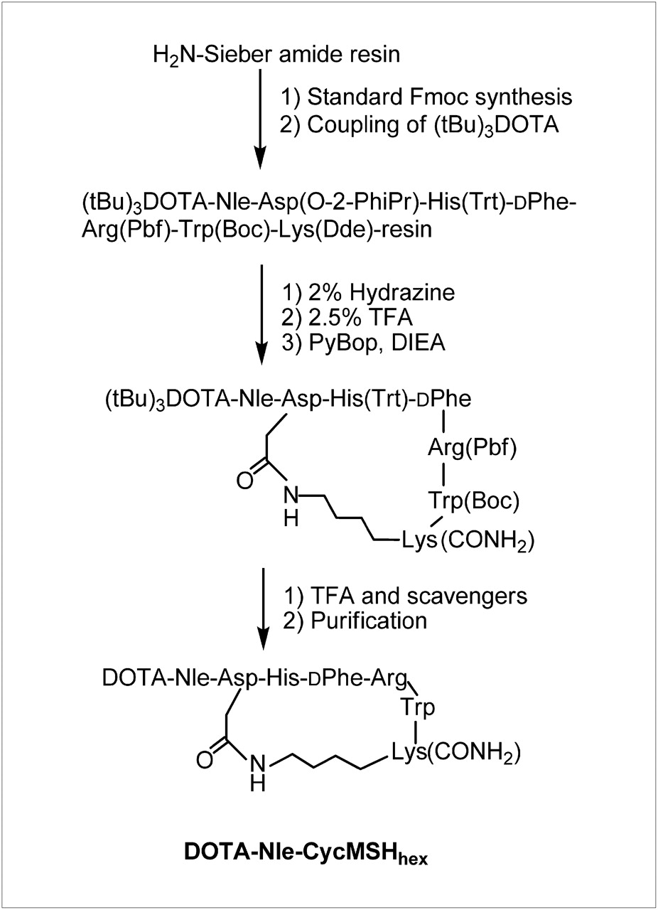
Synthetic scheme of DOTA-Nle-CycMSHhex.

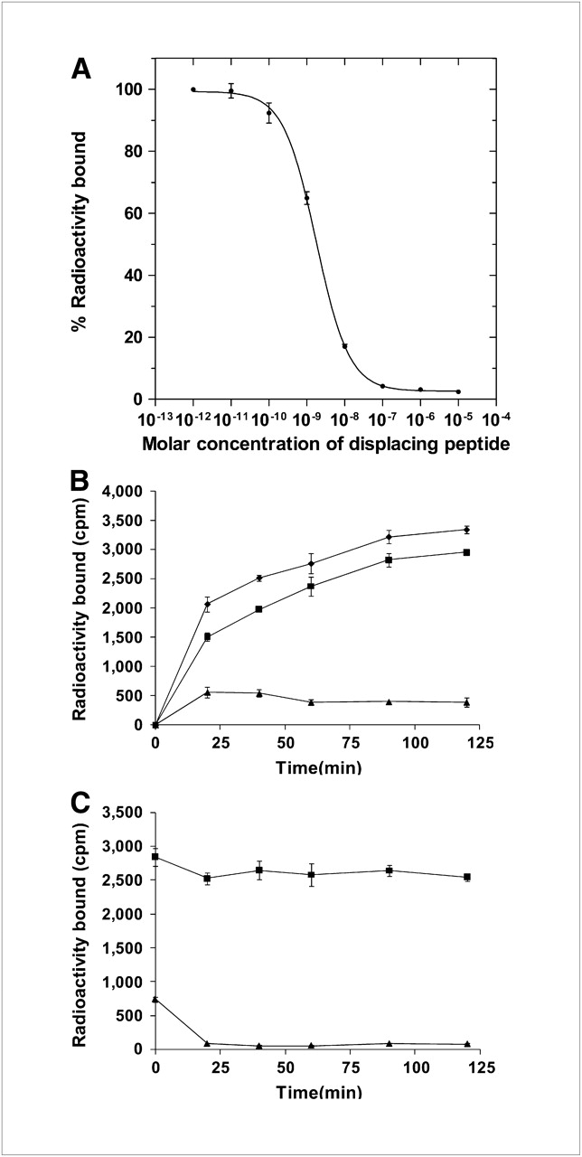
Competitive binding curve (A) of DOTA-Nle-CycMSHhex in B16/F1 melanoma cells. IC50 value of DOTA-Nle-CycMSHhex was 1.77 nM. Cellular internalization (B) and efflux (C) of 111In-DOTA-Nle-CycMSHhex in B16/F1 melanoma cells at 25°C. Total bound radioactivity (♦), internalized activity (▪), and cell membrane activity (▴) were presented as counts per minute (cpm).
The peptide was readily labeled with 111In in 0.5 M ammonium acetate at pH 4.5 with greater than 95% radiolabeling yield. 111In-DOTA-Nle-CycMSHhex was completely separated from its excess nonlabeled peptide by RP HPLC. The retention time of 111In-DOTA-Nle-CycMSHhex was 10.7 min. 111In-DOTA-Nle-CycMSHhex showed greater than 98% radiochemical purity after the HPLC purification. 111In-DOTA-Nle-CycMSHhex was stable in mouse serum at 37°C for 24 h. Only the 111In-DOTA-Nle-CycMSHhex was detected by RP HPLC after 24 h of incubation.
Cellular internalization and efflux of 111In-DOTA-Nle-CycMSHhex were evaluated in B16/F1 cells. Figures 3B and 3C illustrate the cellular internalization and efflux of 111In-DOTA-Nle-CycMSHhex, respectively. 111In-DOTA-Nle-CycMSHhex exhibited rapid cellular internalization and extended cellular retention. At 20 and 120 min after incubation, 72.9% ± 3.5% and 88.3% ± 0.7%, respectively, of the cellular uptake of 111In-DOTA-Nle-CycMSHhex activity had internalized in the B16/F1 cells. Cellular efflux results demonstrated that 89.5% ± 1.9% of internalized 111In-DOTA-Nle-CycMSHhex activity remained inside the cells 2 h after the cells were incubated in culture medium.
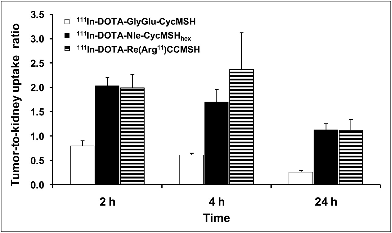
The melanoma-targeting and pharmacokinetic properties of 111In-DOTA-Nle-CycMSHhex were determined in B16/F1 melanoma–bearing C57 mice. The biodistribution results of 111In-DOTA-Nle-CycMSHhex are shown in Table 1. 111In-DOTA-Nle-CycMSHhex exhibited rapid high melanoma uptake and prolonged tumor retention in melanoma-bearing mice. At 0.5 h after injection, 111In-DOTA-Nle-CycMSHhex reached its peak tumor uptake of 24.94 ± 4.58 %ID/g. At 4 and 24 h after injection, 17.01 ± 2.54 %ID/g and 10.53 ± 1.11 %ID/g of the 111In-DOTA-Nle-CycMSHhex activity, respectively, remained in the tumors. In the melanoma uptake blocking study, the tumor uptake of 111In-DOTA-Nle-CycMSHhex with 10 μg of nonradiolabeled NDP-MSH coinjection was only 4.2% of the tumor uptake without NDP-MSH coinjection at 2 h after dose administration (P < 0.05), demonstrating that the tumor uptake was specific and MC1 receptor–mediated. Whole-body clearance of 111In-DOTA-Nle-CycMSHhex was rapid, with approximately 82% of the injected radioactivity cleared through the urinary system by 2 h after injection (Table 1). Normal-organ uptake of 111In-DOTA-Nle-CycMSHhex was low (<1.89 %ID/g), except for the kidneys, at 2, 4, and 24 h after injection. High tumor-to-blood and high tumor–to–normal-organ uptake ratios were achieved as early as 0.5 h after injection (Table 1). As the major excretion pathway of 111In-DOTA-Nle-CycMSHhex, the kidney uptake was 16.20 ± 4.32 %ID/g at 0.5 h after injection and decreased to 9.31 ± 0.91 %ID/g at 24 h after injection. The tumor-to-kidney uptake ratios of 111In-DOTA-Nle-CycMSHhex are presented in Figure 4. The tumor-to-kidney uptake ratios of 111In-DOTA-Nle-CycMSHhex were 2.04, 1.70, and 1.13 at 2, 4, and 24 h, respectively, after injection. Coinjection of NDP-MSH did not reduce renal uptake of 111In-DOTA-Nle-CycMSHhex activity at 2 h after injection, indicating that renal uptake was not MC1 receptor–mediated. Coinjection of L-lysine significantly (P < 0.05) reduced kidney uptake by 30% at 2 h after injection (Table 1).

Tumor-to-kidney uptake ratios of 111In-DOTA-GlyGlu-CycMSH, 111In-DOTA-Nle-CycMSHhex, and 111In-DOTA-Re(Arg11)CCMSH at 2, 4, and 24 h after injection. Calculated tumor-to-kidney uptake ratios of 111In-DOTA-GlyGlu-CycMSH and 111In-DOTA-Re(Arg11)CCMSH were based on results published by Cheng et al. (17) and Miao et al. (21).
Biodistribution of 111In-DOTA-Nle-CycMSHhex in B16/F1 Melanoma–Bearing C57 Mice
Two B16/F1 melanoma–bearing C57 mice were separately injected with 37 MBq (1 mCi) of 111In-DOTA-Nle-CycMSHhex through the tail vein to visualize the tumors at 2 and 24 h after dose administration. The whole-body SPECT/CT images are presented in Figures 5A and 5B. Flank melanoma tumors were clearly visualized by SPECT/CT at 2 and 24 h after injection of 111In-DOTA-Nle-CycMSHhex. In agreement with the biodistribution results, both images showed high tumor–to–normal-organ uptake ratios except for the kidneys. The urinary metabolite of 111In-DOTA-Nle-CycMSHhex was analyzed by RP HPLC at 2 h after injection. Figure 5C illustrates the urinary HPLC profile of 111In-DOTA-Nle-CycMSHhex. 111In-DOTA-Nle-CycMSHhex remained intact in the urine 2 h after injection.
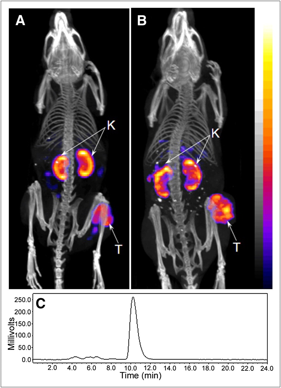
Whole-body SPECT/CT images of B16/F1 flank melanoma–bearing C57 mice at 2 (A) and 24 h (B) after injection of 37 MBq (1 mCi) of 111In-DOTA-Nle-CycMSHhex. Tumor (T) and kidneys (K) are highlighted with arrows on images. HPLC profile (C) of radioactive urine sample of B16/F1 melanoma–bearing C57 mouse at 2 h after injection of 111In-DOTA-Nle-CycMSHhex. 111In-DOTA-Nle-CycMSHhex remained intact in urine at 2 h after injection.
DISCUSSION
Cyclization strategies using a disulfide bridge, a lactam bridge, and metal coordination have been successfully applied to cyclize the α-MSH peptides to enhance the binding affinities and in vivo stabilities of the peptides (23–26). Both 111In-labeled metal-cyclized and lactam bridge–cyclized α-MSH peptides exhibited greater melanoma uptake and lower renal uptake than 111In-labeled disulfide bridge–cyclized α-MSH peptide (21,29). We have reported a novel class of melanoma-specific 111In-labeled lactam bridge–cyclized α-MSH peptides for both primary and metastatic melanoma imaging (21,22). 111In-DOTA-GlyGlu-CycMSH (Fig. 1), with a 12-amino acid peptide ring, exhibited great potential as a melanoma-specific imaging probe in detecting both primary and metastatic melanoma lesions (21,22). However, tumor uptake of 111In-DOTA-GlyGlu-CycMSH was 60.15% that of 111In-DOTA-Re(Arg11)CCMSH, whereas kidney uptake of 111In-DOTA-GlyGlu-CycMSH was 1.5 times renal uptake of 111In-DOTA-Re(Arg11)CCMSH at 2 h after injection in B16/F1 melanoma–bearing C57 mice (17,21). The structural differences between 111In-DOTA-GlyGlu-CycMSH and 111In-DOTA-Re(Arg11)CCMSH (Fig. 1) indicated that the smaller size of the peptide ring might contribute to the more favorable melanoma targeting and pharmacokinetic properties of 111In-DOTA-Re(Arg11)CCMSH because there was an 8-amino acid peptide ring in 111In-DOTA-Re(Arg11)CCMSH, whereas there was a 12-amino acid peptide ring in 111In-DOTA-GlyGlu-CycMSH. Moreover, it was reported that the lactam bridge–cyclized α-MSH peptide with a 6-amino acid peptide ring (Ac-Nle-c[Asp-His-d-Phe-Arg-Trp-Lys(CONH2)]) displayed not only higher MC1 receptor–binding affinity but also a slower MC1 receptor dissociation rate than the native α-MSH peptide (27). Therefore, we synthesized a novel DOTA-conjugated lactam bridge–cyclized peptide with a 6-amino acid peptide ring (DOTA-Nle-CycMSHhex) to examine the profound effect of the peptide ring size on the tumor and kidney uptake in this study.
The conjugation of DOTA to the N terminus of the peptide and the reduction of the peptide ring size did not sacrifice the MC1 receptor–binding affinity of DOTA-Nle-CycMSHhex. DOTA-Nle-CycMSHhex exhibited 1.77 nM MC1 receptor–binding affinity in B16/F1 melanoma cells (Fig. 3A), whereas DOTA-GlyGlu-CycMSH and DOTA-Re(Arg11)CCMSH displayed 0.90 and 2.10 nM MC1 receptor–binding affinities in B16/F1 cells (17,21). 111In-DOTA-Nle-CycMSHhex displayed rapid internalization and prolonged retention in B16/F1 melanoma cells, highlighting its potential as an effective imaging probe for melanoma detection and a therapeutic agent for melanoma treatment when labeled with a therapeutic radionuclide. As we anticipated, the strategy of reducing the ring size of the lactam bridge–cyclized α-MSH peptide resulted in improved tumor uptake and prolonged tumor retention. Compared with 111In-DOTA-GlyGlu-CycMSH with a 12-amino acid peptide ring, 111In-DOTA-Nle-CycMSHhex (Fig. 1) had only a 6-amino acid peptide ring. The tumor uptake (19.39 ± 2.72 %ID/g) of 111In-DOTA-Nle-CycMSHhex was 1.86 times that of 111In-DOTA-GlyGlu-CycMSH at 2 h after injection in B16/F1 melanoma–bearing C57 mice. 111In-DOTA-Nle-CycMSHhex also exhibited more prolonged tumor retention than 111In-DOTA-GlyGlu-CycMSH. At 24 h after injection, 54.3% of the 111In-DOTA-Nle-CycMSHhex activity present at 2 h after injection (10.53 ± 1.11 %ID/g) remained in the tumors (Table 1), whereas only 22.8% of the 111In-DOTA-GlyGlu-CycMSH radioactivity present at 2 h after injection (2.37 ± 0.28 %ID/g) remained in the tumors. Urinary analysis demonstrated that the 111In-DOTA-Nle-CycMSHhex remained intact 2 h after injection (Fig. 5C). It is likely that both high in vivo stability of 111In-DOTA-Nle-CycMSHhex and low MC1 receptor dissociation rate (27) contributed to the rapid high melanoma uptake (24.94 ± 4.58 %ID/g at 0.5 h after injection) and prolonged tumor retention (10.53 ± 1.11 %ID/g at 24 h after injection) of 111In-DOTA-Nle-CycMSHhex in B16/F1 melanoma–bearing C57 mice.
The reduction of the peptide ring size also decreased the nonspecific kidney uptake of 111In-DOTA-Nle-CycMSHhex, compared with 111In-DOTA-GlyGlu-CycMSH (21), at 2 and 4 h after injection. Renal uptake of 111In-DOTA-Nle-CycMSHhex was only 72.8% and 82.4% that of 111In-DOTA-GlyGlu-CycMSH at 2 and 4 h after injection, respectively. Renal uptake of 111In-DOTA-Nle-CycMSHhex was further reduced with L-lysine coinjection by 30% at 2 h after injection, demonstrating that the electrostatic interaction between 111In-DOTA-Nle-CycMSHhex and kidney cells played an important role in renal uptake of 111In-DOTA-Nle-CycMSHhex. The synergistic effects of an increase in tumor uptake and a decrease in renal uptake dramatically improved the tumor-to-kidney uptake ratios of 111In-DOTA-Nle-CycMSHhex at all time points investigated in this study. Improved tumor uptake and decreased kidney uptake resulted in tumor-to-kidney uptake ratios of 111In-DOTA-Nle-CycMSHhex that were superior to those of 111In-DOTA-GlyGlu-CycMSH at 2, 4, and 24 h after injection. The tumor-to-kidney uptake ratios of 111In-DOTA-Nle-CycMSHhex were 2.55, 2.79, and 4.35 times those of 111In-DOTA-GlyGlu-CycMSH at 2, 4, and 24 h after injection, respectively (Fig. 4). 111In-DOTA-Nle-CycMSHhex remained intact in the urine (Fig. 5C), whereas all 111In-DOTA-GlyGlu-CycMSH transformed into 2 polar metabolites in the urine at 2 h after injection (21), possibly contributing to the decreased renal uptake of 111In-DOTA-Nle-CycMSHhex.
Recently, 99mTc-labeled lactam bridge–cyclized α-MSH peptides ([Ac-Nle4,Asp5,d-Phe7,Lys11(pz-99mTc(CO)3)]α-MSH4–11 and 99mTc(CO)3-pz-βAla-Nle-cyclo[Asp-His-d-Phe-Arg-Trp-Lys]-NH2) have been reported for melanoma targeting (30,31). 99mTc(CO)3-pz-βAla-Nle-cyclo[Asp-His-d-Phe-Arg-Trp-Lys]-NH2 exhibited melanoma uptake (11.31 ± 1.81 %ID/g) superior to that of [Ac-Nle4,Asp5, d-Phe7,Lys11(pz-99mTc(CO)3)]α-MSH4–11 (4.24 ± 0.94 %ID/g) at 4 h after injection in B16/F1 melanoma–bearing C57 mice. However, 99mTc(CO)3-pz-βAla-Nle-cyclo[Asp-His-d-Phe-Arg-Trp-Lys]-NH2 displayed high accumulation and prolonged retention in both liver (22.86 ± 1.17 %ID/g) and kidneys (32.12 ± 1.57 %ID/g) at 4 h after injection, possibly limiting its potential application in metastatic melanoma imaging. In this study, tumor uptake of 111In-DOTA-Nle-CycMSHhex was 1.5 times that of 99mTc(CO)3-pz-βAla-Nle-cyclo[Asp-His-d-Phe-Arg-Trp-Lys]-NH2 at 4 h after injection, whereas liver and kidney uptake of 111In-DOTA-Nle-CycMSHhex were only 7.5% and 31.1% that of 99mTc(CO)3-pz-βAla-Nle-cyclo[Asp-His-d-Phe-Arg-Trp-Lys]-NH2 at 4 h after injection. The dramatic increase in tumor uptake and decrease in liver and kidney uptake of 111In-DOTA-Nle-CycMSHhex were likely due to the structural differences between 99mTc(CO)3-pz-βAla-Nle-cyclo[Asp-His-d-Phe-Arg-Trp-Lys]-NH2 and 111In-DOTA-Nle-CycMSHhex.
Currently, metal-cyclized 111In-DOTA-Re(Arg11)CCMSH showed the highest melanoma uptake among all reported 111In-labeled linear and cyclic α-MSH peptides (17). The tumor uptake of 111In-DOTA-Re(Arg11)CCMSH was 17.29 ± 2.49 %ID/g, 17.41 ± 5.63 %ID/g, and 8.19 ± 1.63 %ID/g at 2, 4, and 24 h after injection, respectively (17). Remarkably, 111In-DOTA-Nle-CycMSHhex exhibited 1.12, 0.98, and 1.29 times the tumor uptake of 111In-DOTA-Re(Arg11)CCMSH at 2, 4, and 24 h after injection, respectively. Meanwhile, 111In-DOTA-Nle-CycMSHhex showed renal uptake that was slightly higher than but similar to 111In-DOTA-Re(Arg11)CCMSH at 2 and 4 h after injection. 111In-DOTA-Nle-CycMSHhex exhibited tumor-to-kidney ratios comparable to 111In-DOTA-Re(Arg11)CCMSH at 2 and 24 h after injection despite the fact that the tumor-to-kidney uptake ratio of 111In-DOTA-Nle-CycMSHhex was 28% less than that of 111In-DOTA-Re(Arg11)CCMSH at 4 h after injection. It was reported that a single-dose treatment of 7.4 MBq (200 μCi) of 212Pb-labeled DOTA-Re(Arg11)CCMSH resulted in 44% cures in B16/F1 melanoma–bearing mice (11). Accordingly, it would be likely that treatment with 212Pb-labeled DOTA-Nle-CycMSHhex would yield similar quantitative therapeutic effect for melanoma in the future because 111In-DOTA-Nle-CycMSHhex displayed tumor-to-kidney ratios comparable to 111In-DOTA-Re(Arg11)CCMSH at 2 and 24 h after injection.
CONCLUSION
The ring size of the 111In-labeled lactam bridge–cyclized α-MSH peptide exhibited a profound effect on its melanoma-targeting and pharmacokinetic properties. The reduction of the peptide ring size dramatically increased the melanoma uptake and decreased the renal uptake of 111In-DOTA-Nle-CycMSHhex, providing a new insight into the design of novel radiolabeled lactam bridge–cyclized α-MSH peptide for melanoma imaging and treatment.
Acknowledgments
We thank Benjamin M. Gershman for his technical assistance. This work was supported in part by the Southwest Melanoma SPORE Developmental Research Program, the DOD grant W81XWH-09-1-0105, and the NIH grant NM-INBRE P20RR016480. The images in Figure 5 were generated by the Keck-UNM Small Animal Imaging Resource established with funding from the W.M. Keck Foundation and the University of New Mexico Cancer Research and Treatment Center (NIH P30 CA118100).
Footnotes
-
COPYRIGHT © 2010 by the Society of Nuclear Medicine, Inc.
References
- Received for publication October 20, 2009.
- Accepted for publication December 3, 2009.


Jump to section
Related Articles
Cited By...
- 68Ga-DOTA-GGNle-CycMSHhex targets the melanocortin-1 receptor for melanoma imaging
- Introduction of an 8-Aminooctanoic Acid Linker Enhances Uptake of 99mTc-Labeled Lactam Bridge-Cyclized {alpha}-MSH Peptide in Melanoma
- Effects of the Amino Acid Linkers on the Melanoma-Targeting and Pharmacokinetic Properties of 111In-Labeled Lactam Bridge-Cyclized {alpha}-MSH Peptides