


Abstract
In recent years, implementation of 68Ga-radiometalated peptides for PET imaging of cancer has attracted the attention of clinicians. Herein, we propose the use of 44Sc (half-life = 3.97 h, average β+ energy [Eβ+av] = 632 keV) as a valuable alternative to 68Ga (half-life = 68 min, Eβ+av = 830 keV) for imaging and dosimetry before 177Lu-based radionuclide therapy. The aim of the study was the preclinical evaluation of a folate conjugate labeled with cyclotron-produced 44Sc and its in vitro and in vivo comparison with the 177Lu-labeled pendant. Methods: 44Sc was produced via the 44Ca(p,n)44Sc nuclear reaction at a cyclotron (17.6 ± 1.8 MeV, 50 μA, 30 min) using an enriched 44Ca target (10 mg 44CaCO3, 97.00%). Separation from the target material was performed by a semiautomated process using extraction chromatography and cation exchange chromatography. Radiolabeling of a DOTA-folate conjugate (cm09) was performed at 95°C within 10 min. The stability of 44Sc-cm09 was tested in human plasma. 44Sc-cm09 was investigated in vitro using folate receptor–positive KB tumor cells and in vivo by PET/CT imaging of tumor-bearing mice Results: Under the given irradiation conditions, 44Sc was obtained in a maximum yield of 350 MBq at high radionuclide purity (>99%). Semiautomated isolation of 44Sc from 44Ca targets allowed formulation of up to 300 MBq of 44Sc in a volume of 200–400 μL of ammonium acetate/HCl solution (1 M, pH 3.5–4.0) within 10 min. Radiolabeling of cm09 was achieved with a radiochemical yield of greater than 96% at a specific activity of 5.2 MBq/nmol. In vitro, 44Sc-cm09 was stable in human plasma over the whole time of investigation and showed folate receptor–specific binding to KB tumor cells. PET/CT images of mice injected with 44Sc-cm09 allowed excellent visualization of tumor xenografts. Comparison of cm09 labeled with 44Sc and 177Lu revealed almost identical pharmacokinetics. Conclusion: This study presents a high-yield production and efficient separation method of 44Sc at a quality suitable for radiolabeling of DOTA-functionalized biomolecules. An in vivo proof-of-concept study using a DOTA-folate conjugate demonstrated the excellent features of 44Sc for PET imaging. Thus, 44Sc is a valid alternative to 68Ga for imaging and dosimetry before 177Lu-radionuclide tumor therapy.
In the past decade, applications of radiometal-based PET have rapidly increased, particularly for oncologic imaging purposes (1). Clinical implementation of 68Ga-labeled somatostatin analogs (e.g., 68Ga-DOTATATE, 68Ga-DOTATOC) for imaging of neuroendocrine tumors has raised attention because of the excellent imaging quality that can be achieved and the on-site availability of 68Ga (half-life [T1/2] = 68 min, average β+ energy [Eβ+av] = 830 keV, intensity = 89%) by the 68Ge/68Ga generator (2–6). Somatostatin receptor–targeted PET is currently used for dosimetry before application of 177Lu-based radionuclide therapy (7). These facts have established the basis of a new era of PET applications using radiometal-based radiopharmaceuticals. An advantage of 68Ga is its easy availability by the 68Ge/68Ga generator system. However, because of their short half-lives, 68Ga-based radiopharmaceuticals are predominantly used in-house. Also, in most countries radiochemical laboratories in compliance with good-manufacturing practice and qualified personnel are required (8). These circumstances lead to high overall cost and render 68Ga radiopharmaceuticals of limited interest for centralized production and commercial distribution. An alternative is the cyclotron-produced 64Cu (T1/2 = 12.7 h, Eβ+av = 278 keV, intensity = 17.6%), which has been used in a large number of preclinical and clinical PET studies (9–13). The half-life of 12.7 h provides the flexibility to use it in combination with small molecules and with slow-clearing targeting agents and for transportation of 64Cu radiopharmaceuticals to hospitals without cyclotrons and radiopharmacies (14). However, 64Cu possesses a complex redox chemistry, which has to be taken into consideration when choosing the chelating agents. In addition, coemission of β−-particles (∼39% branch ratio) contribute to additional dose burden to the patient without diagnostic benefit. Recently, 44Sc was proposed as an alternative radionuclide for PET imaging using radiometalated peptides and other small-molecular-weight biomolecules (15). 44Sc is a rare earth metal, which decays by emission of positrons (Eβ+av = 632 keV, intensity = 94.3%) with a half-life of 3.97 h. This almost 4-fold-longer half-life of 44Sc, compared with 68Ga, potentially allows centralized production and cost-efficient distribution of 44Sc radiopharmaceuticals to even remote PET centers. Moreover, the application of 44Sc may be appealing if applied with biomolecules of longer biologic half-lives and for applications, which should cover several hours after administration of the radiopharmaceutical. These advantages have to be carefully traded off against the coemission of high-energy γ-rays by 44Sc (Eγ = 1,157 keV, intensity = 99.9% and 1,499 keV, intensity = 0.9%).
44Sc can be produced via a 44Ti/44Sc generator systems (15–17). However, 44Ti (T1/2 = 60 y) can be produced only at a small number of facilities in the world, with limited yields and at high costs (18). Therefore, the accessibility of cyclotron-produced 44Sc by proton irradiation of natural calcium targets has recently been proposed as an inexpensive alternative by Severin et al. (19). Also, the idea of using enriched calcium targets to optimize radionuclide purity of the produced 44Sc was reported in the literature (20–22).
The present study is the first report, to our knowledge, on the cyclotron production of 44Sc at a quality and quantity sufficient for radiolabeling of a biomolecule, including preclinical evaluation. We demonstrate the feasibility of the production of 44Sc via the 44Ca(p,n)44Sc nuclear reaction. Moreover, we present a semiautomated process, which allowed isolation of 44Sc in a short time and at high radionuclide purity for direct radiolabeling of biomolecules. For the in vivo proof-of-concept study, we chose a recently published folic acid conjugate with a DOTA chelator (cm09) (23). The 177Lu-labeled version (177Lu-cm09) proved to have excellent characteristics with regard to the uptake in folate receptor (FR)–positive tumors in a well-established tumor mouse model (23). In vivo imaging of tumor-bearing mice after the administration of 44Sc-cm09 demonstrated the promising features of 44Sc for PET imaging purposes.
MATERIALS AND METHODS
Production of 44Sc
44Sc was produced via the 44Ca(p,n)44Sc nuclear reaction at the PSI Injector 2 cyclotron facility. Targets for irradiation were prepared with enriched 44CaCO3 (5–10 mg; 97.00% 44Ca; 2.89% 40Ca; 0.06% 42Ca; 0.03% 43Ca; 0.02% 48Ca; and <0.002% 46Ca [Trace Sciences International]). Graphite powder (150 mg, 99.9999%; Alfa Aesar) was used as matrix material. The enriched 44CaCO3 powder was pressed on the surface of the prepressed graphite disk, followed by encapsulation of the target in aluminum. The targets were irradiated with protons of 17.6 ± 1.8 MeV (degraded from an initial beam of 72 MeV) at a beam current of 50 μA for 30–40 min. For the chemical separation of 44Sc(III) from Ca(II), a semiautomated separation system was developed. The irradiated target (with typical activities of ∼350 MBq) was transferred into a conical glass vial and dissolved in HCl (3 M, prepared from 30% HCl and Suprapur and MilliQ water; Merck KGaA). A small aliquot of dissolved 44CaCO3 was analyzed by γ-ray spectrometry. The unsolved graphite remained in the conical glass vial, and the radioactive solution mixture was pumped onto the first column (1-mL column of 55 mm × 5.65 mm, fitted with a 20-μm frit; ISOLUTE SPE Accessories) filled with 50–70 mg of N,N,N′,N′-tetra-n-octyldiglycolamide (DGA) (50–100 μm; Triskem International) resin. The adsorbed 44Sc(III) was eluted from the DGA resin with HCl (0.1 M, 2–3 mL). Afterward, the acidic 44Sc(III) solution was loaded on a second column (1-mL column of 55 mm × 5.65 mm, fitted with 20-μm frit; ISOLUTE SPE Accessories) filled with 100–140 μL of a cation exchange resin in water (DOWEX 50, hydrogen form, 200–400 mesh; Fluka Analytic). 44Sc(III) was eluted in ammonium acetate (CH3COONH4/HCl 1 M, 200–400 μL, pH 3.5–4.0; CH3COONH4: Trace SELECT, ≥99.9999% [Fluka Analytic]) at a radioactivity concentration of up to 980 MBq/mL. For the adjustment of the pH to 3.5–4.0, HCl (0.1 M, 10–30 μL) was added before the use of the 44Sc(III) solution for the radiolabeling procedure.
Radiosynthesis
A stock solution of cm09 (25–35 μL, 10−3 M) was added to 200–400 μL of the 44Sc solution (130–180 MBq, pH 3.5–4.0) and incubated at 95°C for 10 min. Then, Na-diethylenetriaminepentaacetic acid (Na-DTPA, 10 μL, 5 mM, pH 5) was added to the reaction mixture for complexation of traces of free 44Sc(III). Quality control was performed by high-performance liquid chromatography (HPLC) using a C-18 reversed-phase column (Xterra MS C18, 5 μm, 15 × 4.6 cm; Waters). The mobile phase consisted of MilliQ water with 0.1% trifluoracetic acid (A) and methanol (B) with a linear gradient from 95% A and 5% B to 20% A and 80% B over 25 min with a flow rate of 1 mL/min. For in vitro and in vivo application, the labeling mixture containing 44Sc-cm09 was diluted with water to reduce the osmolarity.
177Lu-cm09 was prepared for in vitro cell experiments, which were performed in parallel with 44Sc-cm09. The radiosynthesis and quality control of 177Lu-cm09 was performed as previously reported (23). The specific activity of 177Lu-cm09 was adapted to allow its application at the same molar amount, which was used for 44Sc-cm09.
In Vitro Stability
For the determination of the stability of 44Sc-cm09 in human plasma, a solution of the radiotracer (50 µL, ∼1.0 MBq) was mixed with a sample of human plasma (250 μL) and incubated at 37°C. After 1, 2, and 4 h, aliquots (40 µL) were taken, and the plasma proteins were precipitated by the addition of methanol (200 μL). The suspension was centrifuged (8,000 rcf, 3 min) twice and the supernatants analyzed by HPLC. To investigate potential radiolysis, 44Sc-cm09 was incubated at a high-radioactivity concentration (150–160 MBq in a volume of 500 μL) over several hours at room temperature. After 1, 2, 3, and 4 h, aliquots (5 µL) were taken and analyzed by HPLC.
Determination of Octanol/Phosphate-Buffered Saline (PBS) Distribution Coefficient (LogD)
For determination of the distribution coefficient (logD), 44Sc-cm09 was purified via HPLC using the same system as described above for analytic purposes. The logD value of 44Sc-cm09 was determined by the shake-flask method as previously published in the literature (23,24). In brief, a sample of 44Sc-cm09 (25 µL, ∼0.25 MBq) was added to a mixture of n-octanol (1,500 μL) and PBS (pH 7.4, 1,475 μL). The resulting biphasic system was mixed again by vortex for 1 min at room temperature, followed by centrifugation (2,500 rpm, 6 min). Aliquots of each layer were measured for radioactivity in a γ-counter. The logD value was expressed as the ratio of counts per minute measured in a defined volume of the n-octanol phase to the counts per minute measured in an equal volume of the PBS phase. The results represent the average ± SD of quintuplets of 2 independent experiments.
Cell Culture
KB cells (human cervical carcinoma cell line, HeLa subclone; ACC-136) were purchased from the German Collection of Microorganisms and Cell Cultures (DSMZ). The cells were cultured as monolayers at 37°C in a humidified atmosphere containing 5% CO2. Importantly, KB cells were cultured in a folate-free cell culture medium, FFRPMI (modified RPMI, without folic acid, vitamin B12, and phenol red; Cell Culture Technologies GmbH). The FFRPMI medium was supplemented with 10% heat-inactivated fetal calf serum, l-glutamine, and antibiotics (penicillin/streptomycin/fungizone). Routine culture treatment was performed twice a week using 0.25% trypsin-ethylenediamine tetraacetic acid (1×) (Gibco by Life Technologies) to release cells from cell culture flasks.
Cell Experiments
KB cells were seeded in 12-well plates (∼700,000 cells in 2 mL of FFRPMI medium per well), allowing cell adhesion and growth overnight at 37°C. After removal of the supernatant, cells were washed once with PBS before the addition of FFRPMI medium (975 μL/well) without supplements. 44Sc-cm09 (25 μL, ∼38 kBq, ∼8 pmol) or 177Lu-cm09 (25 μL, ∼38 kBq, ∼8 pmol) was added to each well. In some cases, cells were incubated with an excess of folic acid (100 μM) to block FRs on the surface of KB cells. After incubation of the well plates for different time periods (0, 5, 15, 30, 60, 120, and 240 min) at 37°C, the cells were washed twice with ice-cold PBS to determine total uptake of 44Sc-cm09. To assess the fraction of internalized 44Sc-cm09, KB cells were additionally washed with a stripping buffer (aqueous solution of 0.1 M acetic acid and 0.15 M NaCl, pH 3 (25)) to release FR-bound radiofolates from the cell surface (26). Cell samples were lysed by the addition of NaOH (1 M, 1 mL) to each well. The cell suspensions were then transferred to 4-mL tubes, and each sample was counted for radioactivity in a γ-counter. After homogenization by vortex, the concentration of proteins was determined for each sample by a Micro BCA Protein Assay kit (Pierce, Thermo Scientific) to standardize measured radioactivity to the average content of 0.2 mg of protein in a single well.
Biodistribution Studies
In vivo experiments were approved by the local veterinarian department and conducted in accordance with the Swiss law of animal protection. Six- to 8-wk-old female, athymic nude mice (CD-1 Foxn-1/nu) were purchased from Charles River Laboratories. The animals were fed with a folate-deficient rodent diet (ssniff Spezialdiäten GmbH) starting 5 d before tumor cell inoculation (27). Mice were inoculated with KB cells (5 × 10⁶ cells in 100 μL of PBS) into the subcutis of each shoulder. Biodistribution studies were performed in triplicate approximately 14 d after cell inoculation. 44Sc-cm09 (2 MBq/mouse in a volume of 100 μL) at a specific activity of approximately 4 MBq/nmol was injected into a lateral tail vein. Importantly, the same molar amount of cm09 (0.5 nmol/mouse) was used as previously used for the tissue distribution of 177Lu-cm09 (23) because alteration of the molar amount of the biomolecule may critically affect its tissue distribution. Blocking studies were performed by the injection of excess folic acid (100 μg in a volume of 100 μL of PBS, pH 7.4) immediately before administration of 44Sc-cm09. The animals were sacrificed at predetermined time points after administration of 44Sc-cm09. Selected tissues and organs were collected, weighed, and counted for radioactivity in a γ-counter. The results were listed as a percentage of the injected dose per gram of tissue weight (%ID/g), using counts of defined volume of the original injection solution counted at the same time.
PET/CT Imaging
PET/CT scans were obtained with a dedicated small-animal PET/CT camera (Vista eXplore, Sedecal Spain; GE Healthcare). Reconstruction of PET and CT data was performed using the instrument’s software. For PET reconstruction, the 2-dimensional ordered-subset expectation maximization algorithm was used. PET and CT data were fused using PMOD software (version 3.3; PMOD Technologies Ltd.).
Imaging studies were performed with nude mice approximately 14 d after KB tumor cell inoculation. 44Sc-cm09 (25 MBq, 6 nmol per mouse) was intravenously injected. During the PET scans, mice were anesthetized by inhalation of isoflurane in an air–oxygen mixture approximately 5 min before PET data acquisition. Static PET scans were obtained with each of the mice from 240 to 270 min after injection of 44Sc-cm09. All PET scans were followed by a CT scan.
RESULTS
Production of 44Sc
44Sc was produced via the 44Ca(p,n)44Sc nuclear reaction (Fig. 1A) in typical yields of approximately 350 MBq under the given irradiation conditions (10 mg of 44CaCO3 on the surface of a graphite disk). This resulted in an apparent production cross section of about 300 millibarn at an initial proton beam energy of 17.6 ± 1.8 MeV. Significantly higher quantities of 44Sc can readily be obtained by optimization of the beam energy, by increasing the amount of 44Ca, and by extending the time of irradiation. This procedure requires a fully automated separation process in shielded hot cells to avoid high radiation doses to the technical personnel. (The high-energy γ-radiation [Eγ = 1,157 keV, 99.9% and 1,499 keV, 0.9%] coemitted with the β+-particles have to be taken into consideration for radiation protection precautions.) These optimizations are the topic of ongoing investigations in our laboratories.
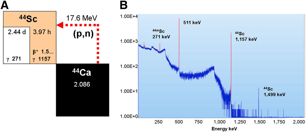
(A) Nuclear reaction for production of 44Sc from enriched 44Ca targets. (B) γ-ray spectrum of 44Sc obtained from irradiated 44Ca targets 5 h after end of beam.
The γ-ray spectrum of the dissolved target material showed almost exclusively the γ-lines of 44Sc (511, 1,157, and 1,499 keV), proving the high radionuclide purity of greater than 99%, which is achievable by this method (Fig. 1B). As the only coproduced scandium isotope of enriched 44Ca targets, the longer-lived 44mSc (T1/2 = 2.4 d) was identified by its 271-keV γ-line as a radioisotopic impurity (<1%) (supplemental data, Supplemental Fig. 1 [supplemental materials are available at http://jnm.snmjournals.org]). A schematic illustration of the separation process of 44Sc from the 44Ca/graphite target is given in the supplemental data (Supplemental Fig. 2). DGA-based extraction chromatography resulted in adsorption of 44Sc(III) on the column and hence separation from Ca(II), which was not retained on the column. 44Sc was almost quantitatively eluted from the DGA resin by 0.1 M HCl. Cation-based exchange chromatography was used to concentrate the 44Sc solution to 200–400 μL (1 M ammonium acetate). The purification process from target dissolution to the elution of 44Sc was accomplished within 10 min. Before use, the pH value of the 44Sc solution was adjusted to 3.5–4.0.
Radiolabeling and In Vitro Characterization
44Sc-cm09 was prepared by radiolabeling of cm09 (25 nmol) with 44Sc(III) (130 MBq, 200 μL) in an ammonium acetate/HCl solution at a pH of 3.5–4.0 (Fig. 2). Quality control performed by HPLC showed the product peak of 44Sc-cm09 with a retention time of 19.6 min, which was largely identical to 177Lu-cm09 (retention time = 19.7 min) (supplemental data, Supplemental Fig. 3). The radiochemical yield was greater than 96% at a specific activity of up to 5.2 MBq/nmol, representing a 44Sc–to–ligand molar ratio of 1:5,600. If the molar amount of cm09 was reduced, the labeling yield dropped rapidly. Neither varying the pH value of the labeling solution nor increasing the incubation time improved these results. For comparison, we prepared 177Lu-cm09 at the same specific activity (∼5.2 MBq/nmol) and calculated a metal-to-ligand ratio of about 1:138.

Chemical structure of DOTA-folate conjugate (cm09) and preparation of 44Sc-cm09.
Investigation of the stability of 44Sc-cm09 in blood plasma showed no decomposition product or free 44Sc(III) over the time of investigation. Also, radiolysis of 44Sc-cm09 was not observed over at least 4 h, even at a radioactivity concentration of up to 400 MBq/mL. The distribution coefficient (logD) of 44Sc-cm09 between n-octanol and PBS pH 7.4 revealed a value of −4.49 ± 0.12. This value was comparable to the logD value obtained for 177Lu-cm09 (−4.25 ± 0.41) (23).
Cell Experiments
Uptake and internalization of 44Sc-cm09 into FR-positive KB tumor cells was determined in vitro over a period of 4 h. The internalized fraction of 44Sc-cm09 accounted for 30%–50% of the total FR-bound fraction of 44Sc-cm09. Analysis of cell samples, which were coincubated with excess folic acid to block FRs, showed a decline of 44Sc-cm09 uptake to less than 1%. These results were comparable to those of other folate radioconjugates previously developed in our group (28). Direct comparison of 44Sc-cm09 and 177Lu-cm09 revealed slight variations of the values for the total cell uptake (Fig. 3A) but almost identical results for the internalized fraction over 4 h (Fig. 3B).
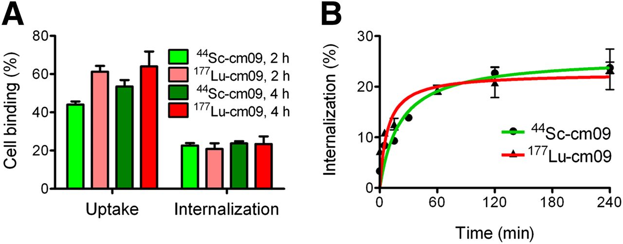
(A) Cell uptake and internalization of 44Sc-cm09 (green) and 177Lu-cm09 (red) after incubation time of 2 or 4 h at 37°C, respectively. (B) Time-dependent internalization of 44Sc-cm09 (green) and 177Lu-cm09 (red) over 4 h.
Biodistribution Studies
The tissue distribution of 44Sc-cm09 in athymic nude mice showed an excellent accumulation of radioactivity in KB tumor xenografts (Table 1). Already 2 h after injection of 44Sc-cm09, the tumor uptake was high (8.37 ± 0.41 %ID/g) and reached a value that was almost doubled at 20 h after injection (14.05 ± 2.29 %ID/g). The enhanced blood circulation time of 44Sc-cm09 as a consequence of the albumin-binding entity integrated in the structure of cm09 was reflected by a relatively high amount of radioactivity in the blood pool (3.74 ± 0.88 %ID/g) at 2 h after injection. In nontargeted tissues and organs such as liver, stomach lung, muscle, and bones, accumulation of 44Sc-cm09 was low and no higher than background levels 20 h after injection. Specific uptake of 44Sc-cm09 was found in the salivary glands and in the kidneys, which are known to express the FR. Renal retention of 44Sc-cm09 was in the range of 19–23 %ID/g at all investigated time points. The excellent accumulation of 44Sc-cm09 in the tumor tissue resulted in high tumor-to-kidney ratios, which reached values between 0.45 and 0.65.
Biodistribution of 44Sc-cm09 in KB Tumor–Bearing Female Nude Mice
The tissue distribution data of 44Sc-cm09 were also compared with our previous data obtained with 177Lu-cm09 (23). Retention of 44Sc-cm09 in the blood circulation was somewhat lower than found for the 177Lu-labeled radioconjugate (supplemental data, Supplemental Table 1) (23). However, the tumor-to-background ratios were comparable for both 44Sc-cm09 and 177Lu-cm09 at early time points after injection and almost identical at 1 d after injection (Figs. 4A and 4B). Also, the relative clearances from blood and nontargeted organs such as liver, lung, and spleen (with the 2-h-after-injection time point set to 100%) revealed equivalent values for 44Sc-cm09 and 177Lu-cm09 (Figs. 4C–4F).
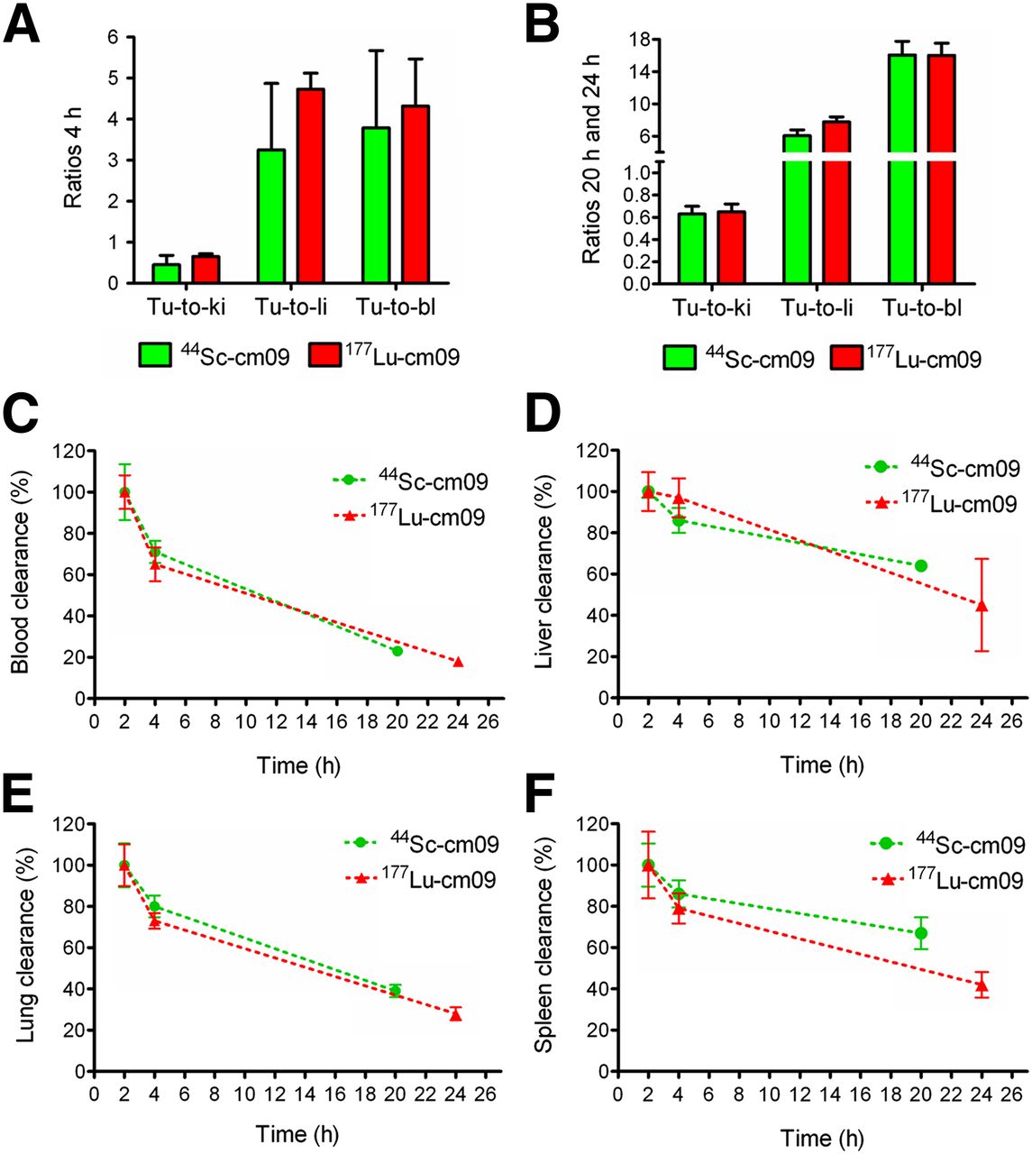
(A) Tumor-to-background ratios 4 h after injection of 44Sc-cm09 (green) and 177Lu-cm09 (red). (B) Tumor-to-background ratios 20 h after injection of 44Sc-cm09 (green) and 24 h 177Lu-cm09 (red). Blood clearance (C), liver clearance (D), lung clearance (E), and spleen clearance (F) of 44Sc-cm09, compared with 177Lu-cm09 (the %ID/g values at 2 h after injection were set to 100%).
In Vivo PET Imaging Studies Using 44Sc-cm09
PET/CT imaging studies were performed 4 h after injection of approximately 25 MBq of 44Sc-cm09. The enhanced blood circulation time of 44Sc-cm09 led to a high tumor uptake and a reduced renal retention, resulting in an excellent tumor-to-kidney ratio of about 1. On the other hand, the tumor-to-nontarget ratio was somewhat lower than what is usually observed for fast clearing small-molecular-weight molecules. However, the PET image allowed excellent visualization of tumor xenografts located on each shoulder of the mouse and in the kidneys, which were the 2 major sites of accumulated radioactivity (Fig. 5A). Besides the uptake of 44Sc-cm09 in these FR-positive organs and tissue, radioactivity was found only in the urinary bladder. Studies with a KB tumor–bearing mouse injected with excess folic acid immediately before injection of 44Sc-cm09 resulted in a significant reduction of accumulated radioactivity in tumor xenografts and in the kidneys (supplemental data, Supplemental Fig. 4). The PET scans obtained 4 h after injection of 44Sc-cm09 showed the same distribution profile of radioactivity, which was previously observed on SPECT/CT images of a mouse 4 h after injection of 177Lu-cm09 (Fig. 5B).

(A) PET/CT image of KB tumor–bearing mouse 4 h after injection of approximately 25 MBq of 44Sc-cm09. (B) SPECT/CT image of KB tumor–bearing mouse 4 h after injection of approximately 35 MBq of 177Lu-cm09 (23). Tumors (Tu) are indicated with red arrows, kidneys (Ki) are indicated with yellow arrows, and urinary bladder (Bl) is indicated with white arrow.
DISCUSSION
Proton irradiation of enriched 44Ca targets resulted in the production of 44Sc in radiochemical purity higher than 99%. As the only radioactive side product, traces of the longer-lived isotope 44mSc were determined by γ-ray spectrometry. In contrast, the quality of 44Sc obtained from irradiation of natural calcium targets was much lower because of several coproduced scandium radioisotopes as reported by Severin et al. (supplemental data, Supplemental Fig. 1) (19). The semiautomated separation system allowed isolation of more than 85% of the initial activity of 44Sc(III) at high radioactivity concentrations (>900 MBq/mL) within only 10 min. For the in vivo assessment of 44Sc, we have chosen a folate derivative (cm09) comprising an albumin-binding entity. This novel folate conjugate reveals an enhanced blood circulation time and, hence, slower pharmacokinetic properties, making it suitable for combining with a longer-lived PET and therapeutic radiometal. 44Sc radiolabeling of cm09 was achieved at a specific activity of up to 5.2 MBq/nmol, which is an unprecedentedly high value, compared with other 44Sc-labeling results reported in the literature (29–31).
In vitro 44Sc-cm09 was tested in parallel to 177Lu-cm09 on FR-positive KB tumor cells. The results were largely in agreement with the results previously obtained with 177Lu-cm09 (23). In vivo, we found an excellent tissue distribution of 44Sc-cm09, with increasing accumulation of radioactivity in FR-positive tumor xenografts over time (8.37 ± 0.41 %ID/g at 1 h after injection to 14.05 ± 2.29 %ID/g at 20 h after injection). Accumulation in nontargeted tissue and organs such as liver, lung, intestinal tract, muscle, and bone was largely absent.
Although the radius of Sc(III) is similar to that of Ga(III), its chemical properties resemble more closely those of Y(III) (pseudolanthanide) and lanthanides such as 177Lu(III) with common coordination numbers of 8 and 9 (32). This allows the formation of stable 44Sc complexes with DOTA chelators. Recently, it was shown that DOTA was the most preferred chelator for 44Sc(III) from a series of macrocyclic ligands (33). All 4 amine and carboxyl groups of a DOTA chelator contribute to the complexation of 44Sc(III), resulting in a stability constant comparable to those of Y(III) and lanthanides (33). In theory, comparable results for 44Sc-labeled and 177Lu-labeled biomolecules would be expected.
The reason why the values for uptake in the tumor tissue and in the kidneys were somewhat lower for 44Sc-cm09 than previously published values for 177Lu-cm09 can presumably be attributed to interexperimental variability (Table 1; supplemental data, Supplemental Table 1) (23). However, the tumor-to-background ratios of 44Sc-cm09 and 177Lu-cm09 were almost the same, particularly at late time points (20 and 24 h, respectively) after application.
Similar to what has been published for 44Sc- and 177Lu- DOTATATE complexes (33), we determined largely the same hydrophilicity for 44Sc-cm09 (logD, −4.49 ± 0.12) and 177Lu-cm09 (logD, −4.25 ± 0.41) (23). Our findings and those of recent literature reports (30,33) corroborated the hypothesis of a similar in vivo behavior for 44Sc- and 177Lu-labeled biomolecules rather than for a 68Ga-labeled pendant. Moreover, because of the reduced β+-energy of 44Sc (Eβ+av = 632 keV), compared with 68Ga (Eβ+av = 830 keV), the resolution of PET images obtained with 44Sc is expected to be equal or even superior to that of 68Ga. This has been demonstrated by PET imaging studies using Derenzo phantoms (supplemental data, Supplemental Fig. 5). Moreover, the longer half-life makes 44Sc an attractive alternative to 68Ga, allowing distribution of 44Sc-labeled biomolecules over long distances.
It should also be recognized that 47Sc exists as a chemically absolute identical therapeutic match to 44Sc (34). 47Sc decays by the emission of low-energy β−-particles (average β− energy [Eβ−av] = 162 keV) similar to 177Lu (Eβ−av = 134 keV). The much shorter half-life of 47Sc (T1/2 = 3.35 d), compared with 177Lu (T1/2 = 6.65 d), may result in a lower dose burden to the patient. Moreover, it is important to recognize that 47Sc also emits γ-rays of an ideal energy (Eγ = 159 keV) for SPECT imaging. Direct monitoring of a therapeutic application of 47Sc radiopharmaceuticals would be accessible as it is currently performed with 177Lu-labeled compounds.
CONCLUSION
In this study, we demonstrated the feasibility to use cyclotron-produced 44Sc of a high quality for the preparation of a 44Sc-labeled folate conjugate, which was evaluated in a proof-of-concept in vivo study. Optimizations of the production process of 44Sc to obtain higher quantities of activity is the topic of ongoing investigations. On the basis of the excellent features of 44Sc for PET, it is likely that this radioisotope will find application in clinical nuclear imaging and pretherapeutic dosimetry of cancer patients, who would subsequently be treated with the 177Lu-labeled pendant. Moreover, clinical implementation of 47Sc as a therapeutic match would allow application of identical radiopharmaceuticals with equal pharmacokinetics. Future prospects with the matched pair 44Sc/47Sc for PET imaging and radionuclide therapy are dependent on the realization of the theranostic principle and routine availability of these novel radioisotopes.
DISCLOSURE
The costs of publication of this article were defrayed in part by the payment of page charges. Therefore, and solely to indicate this fact, this article is hereby marked “advertisement” in accordance with 18 USC section 1734. The project was financially supported by the Swiss National Science Foundation (Ambizione PZ00P3_121772 & PZ00P3_138834), COST-BM0607 (C08.0026), and the Swiss Cancer League (KLS-02762-02-2011). No other potential conflict of interest relevant to this article was reported.
Acknowledgments
We thank Walter Hirzel (PSI), Ernst Schaub (PSI), Yvonne Eichholzer (PSI), Alexander Sommerhalder (PSI), Holger Dorrer (PSI), Stephanie Haller (PSI), Nadja Romano (PSI), Claudia Keller (ETH), and Martin Hungerbühler (ETH) for technical assistance.
Footnotes
↵* Contributed equally to this work.
Published online Nov. 6, 2013.
- © 2013 by the Society of Nuclear Medicine and Molecular Imaging, Inc.
REFERENCES
- Received for publication April 3, 2013.
- Accepted for publication July 31, 2013.


Jump to section
Related Articles
Cited By...
- First positronium image of the human brain in vivo
- Production and Use of the First-Row Transition Metal PET Radionuclides 43,44Sc, 52Mn, and 45Ti
- Therapeutic Radiometals Beyond 177Lu and 90Y: Production and Application of Promising {alpha}-Particle, {beta}--Particle, and Auger Electron Emitters
- Promising Prospects for 44Sc-/47Sc-Based Theragnostics: Application of 47Sc for Radionuclide Tumor Therapy in Mice