
Main menu

  • Home
  • Content
    • Current
    • Ahead of print
    • Past Issues
    • JNM Supplement
    • SNMMI Annual Meeting Abstracts
    • Continuing Education
    • JNM Podcasts
  • Subscriptions
    • Subscribers
    • Institutional and Non-member
    • Rates
    • Journal Claims
    • Corporate & Special Sales
  • Authors
    • Submit to JNM
    • Information for Authors
    • Assignment of Copyright
    • AQARA requirements
  • Info
    • Reviewers
    • Permissions
    • Advertisers
  • About
    • About Us
    • Editorial Board
    • Contact Information
  • More
    • Alerts
    • Feedback
    • Help
    • SNMMI Journals
  • SNMMI
    • JNM
    • JNMT
    • SNMMI Journals
    • SNMMI

User menu

  • Subscribe
  • My alerts
  • Log in
  • My Cart

Search

  • Advanced search
Journal of Nuclear Medicine
  • SNMMI
    • JNM
    • JNMT
    • SNMMI Journals
    • SNMMI
  • Subscribe
  • My alerts
  • Log in
  • My Cart
Journal of Nuclear Medicine

Advanced Search

  • Home
  • Content
    • Current
    • Ahead of print
    • Past Issues
    • JNM Supplement
    • SNMMI Annual Meeting Abstracts
    • Continuing Education
    • JNM Podcasts
  • Subscriptions
    • Subscribers
    • Institutional and Non-member
    • Rates
    • Journal Claims
    • Corporate & Special Sales
  • Authors
    • Submit to JNM
    • Information for Authors
    • Assignment of Copyright
    • AQARA requirements
  • Info
    • Reviewers
    • Permissions
    • Advertisers
  • About
    • About Us
    • Editorial Board
    • Contact Information
  • More
    • Alerts
    • Feedback
    • Help
    • SNMMI Journals
  • View or Listen to JNM Podcast
  • Visit JNM on Facebook
  • Join JNM on LinkedIn
  • Follow JNM on Twitter
  • Subscribe to our RSS feeds
Research ArticleFocus on Molecular Imaging

Production and Use of the First-Row Transition Metal PET Radionuclides 43,44Sc, 52Mn, and 45Ti

Ivis F. Chaple and Suzanne E. Lapi
Journal of Nuclear Medicine November 2018, 59 (11) 1655-1659; DOI: https://doi.org/10.2967/jnumed.118.213264
Ivis F. Chaple
Department of Radiology, University of Alabama at Birmingham, Birmingham, Alabama
  • Find this author on Google Scholar
  • Find this author on PubMed
  • Search for this author on this site
Suzanne E. Lapi
Department of Radiology, University of Alabama at Birmingham, Birmingham, Alabama
  • Find this author on Google Scholar
  • Find this author on PubMed
  • Search for this author on this site
  • Article
  • Figures & Data
  • Info & Metrics
  • PDF
Loading

Abstract

With the increasing focus on a more personalized approach to medicine using imaging techniques to select patients for targeted treatment or theranostic strategies, interest in the development of new radionuclides is expanding. Through the development of production and radiochemistry techniques, several new radiometals are being added to the toolbox of the nuclear imaging community. 43,44g,mSc, 52gMn, and 45Ti are all emerging transition metal radionuclides and will be discussed in this short review. Each of these nuclides has unique imaging characteristics and shows promise for the development of new molecular imaging agents.

  • scandium-43
  • scandium-44
  • manganese-52
  • titanium-45
  • isotope production
  • positron emission tomography

The field of PET imaging has changed considerably in the last 5 y, shifting toward production and use of metal radionuclides in addition to continued research for 18F and 11C compounds. Transition metal PET research is a rapidly growing area of interest. Specifically, this review will focus on work over the last 5 y involving 43,44g,mSc, 52gMn, and 45Ti.

Although scandium itself is not a true transition metal, its chemical properties are similar enough to that of transition metals to earn it the title of “transition metallike element.” Because of the imaging and therapeutic characteristics of the scandium radionuclides 43,44g,mSc and 47Sc, respectively, scandium is a potential candidate for the development of theranostic strategies that use complementary imaging and therapeutic compounds labeled with matching isotope pairs.

Interest in using manganese as a contrast agent for manganese-enhanced MRI has increased in recent years as an alternative to gadolinium-based contrast agents because of the toxicity issues of the latter (1). During the development of new agents for this technique, PET imaging with 52Mn can also help quantitatively determine the distribution, uptake, and excretory patterns of manganese-based MRI agents.

45Ti is a promising candidate for radiolabeling peptides. Its half-life of 3.08 h, although slightly shorter than that of the scandium radionuclides discussed, is longer than that of 68Ga, making it favorable for longer imaging times and centralized distribution. Additionally, its economical production makes implementation in many centers possible if chemistry challenges can be overcome.

PRODUCTION OF 43,44G,MSC

Recently, scandium nuclides have been of interest because of the potential therapeutic properties of 47Sc, which can be combined with 43,44gSc for imaging. An excellent review article describes the production routes of these 3 scandium isotopes using proton beams, α-particles, neutrons, and various target materials (2). 43Sc (β+ = 88.1%, Eβ+ = 476 keV) and 44gSc (β+ = 94.3%, Eβ+ = 632 keV) have half-lives of 3.89 and 3.97 h, respectively, (3). Although the 2 isotopes are similar, the main difference is the high-energy γ-ray (1.157 MeV, emitted 99.9% of the time) associated with 44gSc. Additionally, 44mSc, which is coproduced with 44gSc, has a half-life of 58.6 h and has been suggested for use as an in vivo PET generator for 44gSc (4). However, the longer half-life coupled with the high-energy γ-ray emitted from 44gSc, may lead to unfavorable dosimetry for shorter studies.

Production routes for 43,44g,m,47Sc use either calcium or titanium targets, typically enriched for the desired isotope. 43Sc can be produced via proton irradiation of 43Ca or 46Ti, as well as α-irradiation of 40Ca or deuteron irradiation of 44Ca (5–7). 44g,mSc can be produced via proton irradiation of 44Ca, 47Ti, or 45Sc or deuteron irradiation of 44Ca (2,4–20). Enriched targets are preferred to avoid production of contaminants such as 46,48Sc (18). Production studies of 44g,44mSc using deuterons yielded up to 90 MBq (4 h after the end of bombardment) with a 3-h bombardment (21). In a similar study, 44g,44mSc was produced via deuteron irradiation of enriched 44CaCO3 targets, and the authors concluded that production of 44g,mSc via calcium targets is preferable to 43Sc because of the higher natural abundance of 44Ca than of 43Ca (7,22). Researchers have also studied the 46Ti(p,α)43Sc reaction and showed that this pathway produced 43Sc of higher radionuclidic purity than when using the 43Ca(p,n)43Sc production route; however, the latter route produced 43Sc in higher yields (18). Another source for 44gSc is the 44Ti/44Sc generator (8,23). Although the generator method has been investigated, only a small number of facilities in the world have these generators in use.

Purification of 43Sc is detailed in a recent report that included various methods by several researchers (9). This article described separation methods summarized in Table 1, all of which had favorable separation efficiency and recovery yields of more than 80% (9). Finally, another report detailed the separation of 44gSc, showing recovery of up to 80% using H2O as the final eluent on a UTEVA resin (Eichrom Technologies, LLC) (11). These methods for production and separation of 43,44gSc enhance the availability of these nuclides for further development.

View this table:
  • View inline
  • View popup
TABLE 1

43Sc Production Routes Highlighted by Walczak et al. (9)

RADIOCHEMISTRY AND IMAGING OF 43,44SC

43,44gSc has been proposed as a matched-pair theranostic radioisotope that is a longer-lived alternative to 68Ga for imaging analogs of 177Lu. Scandium exists in the +3 oxidation state and is suitable for labeling of DOTA-derivatized biomolecules (24). In studies investigating 44gSc as an imaging surrogate for 177Lu, 44gSc-labeled cm09 (DOTA-folate conjugate) was determined to have comparable properties, with pharmacokinetics nearly identical to the same compound labeled with 177Lu (25).

However, using 2 different elements may create problems if they show different biodistributions. Radioscandium allows for the development of true matched-pair theranostics using chemically identical constructs as opposed to chemically similar constructs. In this case, the pair would be 43,44gSc as the diagnostic agent and 47Sc as the therapeutic agent. Studies have compared the labeling efficiencies of 68Ga, 177Lu, and 44gSc for DOTA-HPMA (N-(2-hydroxypropyl) methacrylamide) conjugates and showed that the 44gSc-labeled polymer was highly stable for up to 24 h, showing promising characteristics for in vivo imaging (12). A similar study determined that reaction rates were increased when buffers including EtOH were used, allowing for lower temperatures and a reduced time to achieve labeling yields above 90% for DOTA complexes with these 3 radioisotopes (26).

44gSc was used to radiolabel DOTA-NAPamide for targeting of the melanocortin-1 receptor, with higher uptake in melanocortin-1 receptor–positive B16-F10 xenografts than in melanocortin-1 receptor–negative A375 xenografts (27). In another study, 44gSc-DOTANOC was synthesized with high radiochemical yields and used for dynamic PET imaging, showing continuous uptake and high-quality images in AR42J tumor xenografts, encouraging the transition into clinical applications (24). A similar study optimized radiolabeling conditions for peptides such as DOTANOC and NODAGANOC with 44gSc. After this, the authors determined that compounds labeled with 44gSc had similar biodistribution to the same compounds labeled with 68Ga except for a higher liver uptake seen in the latter (28). As these studies have indicated, 43,44gSc shows promise for the development of matched-pair theranostic agents with 47Sc.

PRODUCTION OF 52MN

With a longer half-life of 5.6 d, a β+ branching ratio of 29.4%, and Eβ+ of 242 keV, 52gMn has characteristics complementary to the shorter-lived nuclides discussed and is a promising candidate for imaging of longer biologic processes or compounds that intrinsically contain manganese. However, 52gMn emits multiple high-energy γ-rays, making work less favorable with 52gMn than with some other radioisotopes (29). 52gMn can be produced via the 52Cr(p,x)52gMn reaction, but radioisotope contaminants, including the long-lived 54Mn, can be produced via the 54Cr(p,n) reaction when natural chromium targets are used (27–29). Several groups have studied cross sections of these reactions to attain the highest yields with the fewest impurities (30,31). Various natural chromium target configurations have been studied, including metal targets, electrodeposition, and pressed powder targets (29,32,33). Targets have been irradiated with 16-MeV protons or less, because of the desirable cross section between 6 and 20 MeV, illustrating production of 52Mn with low-energy cyclotrons (29,30,32–34).

Several separation methods have been developed for the purification of 52gMn and are summarized in Table 2 (35,36). Although further optimization of the purification chemistry is required, these purification strategies have enabled the availability of this nuclide for research use.

View this table:
  • View inline
  • View popup
TABLE 2

52Mn Separation Methods

RADIOCHEMISTRY AND IMAGING OF 52MN

The radiochemistry of manganese has been deemed challenging because manganese and chromium have multiple oxidation states and because Mn(II) and Cr(III) behave similarly (32). Recent studies have noted that 52gMn can be chelated with EDTA, DTPA, and DOTA, with straightforward labeling chemistry similar to radionuclides of scandium previously discussed (36).52gMn has been used in several imaging applications. For example, recent studies showed that Mn2+ can enter neurons, and thus, 52gMn has been studied as a neural imaging agent (37). Because manganese is also used as a contrast agent in manganese-enhanced MRI, MRI tools can benefit from the use of 52gMn to enhance the understanding of the pharmacokinetics of these agents (38). An excellent example of using 52gMn for dual PET/MRI made use of Mn2+ for manganese-enhanced MRI and 52gMn for PET imaging studies on neural progenitor cells that overexpress divalent metal transporter 1 (32). Researchers found that the effectiveness of cell therapy could be measured using the dual-modality PET/MRI method by applying divalent metal transporter 1 as a reporter gene (32). In another study, the use of 52gMn coupled with a DOTA antibody conjugate (DOTA-TRC105) for PET imaging of mice bearing 4T1 xenografts was reported (34). That study showed that 52gMn can be used as a PET agent for imaging time points of up to 120 h and resulted in high-quality images as shown in Figure 1. Although DOTA-coupled compounds can be labeled with both 52gMn and 43,44gSc, the application is important for radionuclide selection, with the former having longer imaging times and the latter being used for the development of true theranostics (28).

FIGURE 1.
  • Download figure
  • Open in new tab
  • Download powerpoint
FIGURE 1.

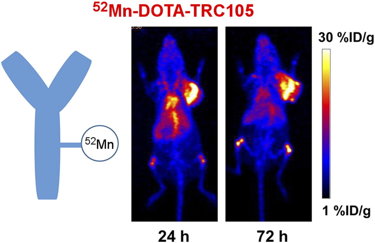
Results of 52Mn-DOTA-TRC105 injection into 4T1 xenograft tumor–bearing mice. %ID = percentage injected dose. (Reprinted with permission from (34).)

Another interesting study showed the uptake of 52gMn-Mn2+ in monkeys after nostril administration of the radiotracer (37). Brain uptake in rhesus macaque monkeys was determined over 8 sessions with at least 6 mo between each session. After 7 d, activity was seen in parts of the brain after following the olfactory tract, which demonstrated that 52gMn-Mn2+ can be used to for neuronal imaging (39). 52gMn-Mn(oxinate)2 was synthesized and determined to have suitable in vitro stability to study extravasation in vivo when conjugated with a liposomal nanomedicine (40). In another study, which leveraged the biologic properties of Mn2+, 52gMn2+-Mn2+ was successfully used to image functional β-cell mass in mice with type 1 and type 2 diabetes (41). Finally, 52gMn2+-Mn2+ was used to investigate the biodistribution of manganese when administered intravenously or through inhalation (38). That study found most organs to have higher uptake when the dose was administered intravenously, with the highest uptake being in the kidney and liver (38). In summary, the implementation of 52gMn could provide a new way to image long-circulating compounds and to enhance the results from manganese MRI tools.

PRODUCTION OF 45TI

45Ti has many favorable characteristics for PET imaging, including a naturally monoisotopic target material, a half-life of 3.08 h, low maximum positron energy (Eβ+ = 439 keV), and a high positron branching ratio (β+ = 84.8%). Compared with the previous nuclides discussed, 45Ti production may be the most economical. Although not heavily studied to date, recent articles showed the utility of 45Ti. 45Ti is produced via proton bombardment of natural scandium foils through the 45Sc(p,n)45Ti reaction. Promising results were achieved using 11.8-MeV protons to avoid production of the long-lived 44Ti (42). An excellent review article has described the historical interest and significance of 45Ti, as well as some of the most recent advances in its production and separation methods (43). For the purification of 45Ti, studies have used hydroxamate resin with reported yields of up to 42% ± 6%, with a separation time of an hour (42). Production yields of 4.6 ± 0.4 GBq (20 μA,11.8-MeV protons, 1 h) have been reported, compared with previously calculated theoretic yields of 2.165 GBq (5 μA, 14.5-MeV protons, 1 h) (42,44–46). A novel separation method achieved 93% ± 3% recovery via use of a HypoGel 200 (Rapp Polymere GmbH) 1,3-diol resin to retain 45Ti on the column and then subsequently introduced salan followed by dipic in pyridine for radiolabeling on the same column, to produce 45Ti-(salan)Ti(dipic) (47). These production parameters indicate that 45Ti can be readily produced in significant quantities using low-cost target material.

RADIOCHEMISTRY AND IMAGING OF 45TI

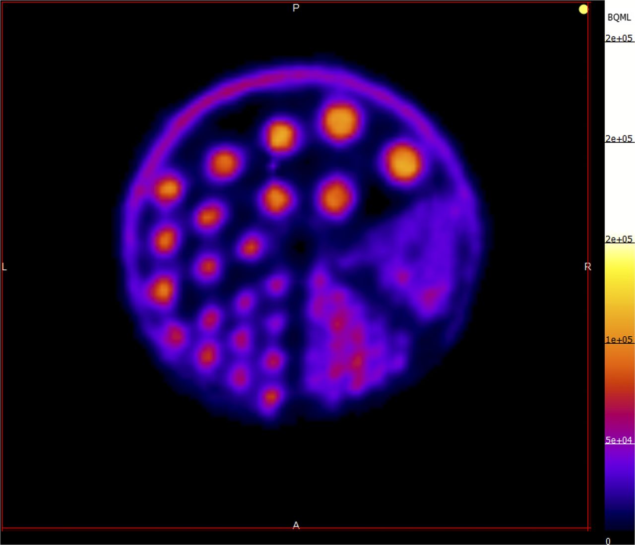
Radiochemistry with 45Ti, which exists in the +3 and +4 oxidation states, is challenging because of the potential oxidation of Ti to TiO2, its instability in an aqueous environment, and the challenge of finding stable titanium chelators (47). Researchers illustrated that 45Ti could be used to radiolabel mesoporous silica nanoparticles using a chelator-free radiolabeling technique (42). In another study using an on-column radiolabeling technique, the radiolabeled antitumor agent 45Ti-(salan)Ti(dipic) was applied for PET/CT imaging and biodistribution studies and showed rapid clearance from blood and liver, whereas the uptake in the tumor remained constant for 400 min (47). Previous studies showed that 45Ti could be used to radiolabel apotransferrin, which forms a complex that remains intact in vivo, demonstrating transport of 45Ti to tumors for 24 h after injection (48). 45Ti can yield high-resolution images as illustrated with recent phantom imaging studies in our group. Figure 2 shows a miniature Derenzo phantom imaged with 3.7 MBq of 45Ti on the small-animal PET scanner at the University of Alabama at Birmingham. When isotopes with moderate half-lives are compared, 45Ti and 43,44gSc are comparable; however, the straightforward production may make 45Ti more appealing as a diagnostic radionuclide if chemistry challenges can be overcome.

FIGURE 2.
  • Download figure
  • Open in new tab
  • Download powerpoint
FIGURE 2.

Miniature Derenzo phantom image (3.7 MBq of 45Ti; image taken after 60-min scan time).

CONCLUSION

The field of nuclear imaging is growing rapidly, with many recent advancements being made in the area of radioisotope development and implementation. Each of these isotopes possesses unique properties. The characteristics of 43,44g,mSc may allow for implementation as theranostic agents when combined with 47Sc. The long half-life of 52gMn is promising for longer imaging studies and could be used for the development of PET/MRI agents. 45Ti is an interesting isotope that has much to offer, with its favorable half-life and potential ease of production. With additional development, 43,44g,mSc, 52gMn, and 45Ti, may prove to be useful additions to the PET isotope toolbox.

DISCLOSURE

No potential conflict of interest relevant to this article was reported.

Footnotes

  • Published online Sep. 27, 2018.

  • © 2018 by the Society of Nuclear Medicine and Molecular Imaging.

REFERENCES

  1. 1.↵
    1. Rogosnitzky M,
    2. Branch S
    . Gadolinium-based contrast agent toxicity: a review of known and proposed mechanisms. Biometals. 2016;29:365–376.
    OpenUrlCrossRefPubMed
  2. 2.↵
    1. Müller C,
    2. Domnanich KA,
    3. Umbricht CA,
    4. van der Meulen NP
    . Scandium and terbium radionuclides for radiotheranostics: current state of development towards clinical application. Br J Radiol. June 15, 2018 [Epub ahead of print].
  3. 3.↵
    1. Singh B,
    2. Chen J
    . Nuclear data sheets for A = 43. Nucl Data Sheets. 2015;126:1–150.
    OpenUrl
  4. 4.↵
    1. Alliot C,
    2. Kerdjoudj R,
    3. Michel N,
    4. Haddad F,
    5. Huclier-Markai S
    . Cyclotron production of high purity 44m,44Sc with deuterons from 44CaCO3 targets. Nucl Med Biol. 2015;42:524–529.
    OpenUrl
  5. 5.↵
    1. Szkliniarz K,
    2. Sitarz M,
    3. Walczak R,
    4. et al
    . Production of medical Sc radioisotopes with an alpha particle beam. Appl Radiat Isot. 2016;118:182–189.
    OpenUrl
  6. 6.
    1. Minegishi K,
    2. Nagatsu K,
    3. Fukada M,
    4. Suzuki H,
    5. Ohya T,
    6. Zhang M-R
    . Production of scandium-43 and -47 from a powdery calcium oxide target via the nat/44Ca(α,x)-channel. Appl Radiat Isot. 2016;116:8–12.
    OpenUrl
  7. 7.↵
    1. Duchemin C,
    2. Guertin A,
    3. Haddad F,
    4. Michel N,
    5. Metivier V
    . Production of scandium-44 m and scandium-44 g with deuterons on calcium-44: cross section measurements and production yield calculations. Phys Med Biol. 2015;60:6847–6864.
    OpenUrl
  8. 8.↵
    1. Radchenko V,
    2. Meyer CAL,
    3. Engle JW,
    4. et al
    . Separation of 44Ti from proton irradiated scandium by using solid-phase extraction chromatography and design of 44Ti/44Sc generator system. 2016;1477:39–46.
  9. 9.↵
    1. Walczak R,
    2. Krajewski S,
    3. Szkliniarz K,
    4. et al
    . Cyclotron production of 43Sc for PET imaging. EJNMMI Phys. 2015;2:33.
    OpenUrl
  10. 10.
    1. Carzaniga TS,
    2. Auger M,
    3. Braccini S,
    4. et al
    . Measurement of 43Sc and 44Sc production cross-section with an 18MeV medical PET cyclotron. Appl Radiat Isot. 2017;129:96–102.
    OpenUrl
  11. 11.↵
    1. Hernandez R,
    2. Valdovinos HF,
    3. Yang Y,
    4. et al
    . 44Sc: an attractive isotope for peptide-based PET imaging. Mol Pharm. 2014;11:2954–2961.
    OpenUrl
  12. 12.↵
    1. Eppard E,
    2. de la Fuente A,
    3. Mohr N,
    4. et al
    . Labeling of DOTA-conjugated HPMA-based polymers with trivalent metallic radionuclides for molecular imaging. EJNMMI Res. 2018;8:16.
    OpenUrl
  13. 13.
    1. Müller C,
    2. Bunka M,
    3. Haller S,
    4. et al
    . Promising prospects for 44Sc-/47Sc-based theragnostics: application of 47Sc for radionuclide tumor therapy in mice. J Nucl Med. 2014;55:1658–1664.
    OpenUrlAbstract/FREE Full Text
  14. 14.
    1. Valdovinos HF,
    2. Hernandez R,
    3. Barnhart TE,
    4. Graves S,
    5. Cai W,
    6. Nickles RJ
    . Separation of cyclotron-produced 44Sc from a natural calcium target using a dipentyl pentylphosphonate functionalized extraction resin. Appl Radiat Isot. 2015;95c:23–29.
    OpenUrl
  15. 15.
    1. Chakravarty R,
    2. Goel S,
    3. Valdovinos HF,
    4. et al
    . Matching the decay half-life with the biological half-life: immunoPET imaging with 44Sc-labeled cetuximab Fab fragment. Bioconjug Chem. 2014;25:2197–2204.
    OpenUrlCrossRefPubMed
  16. 16.
    1. Hoehr C,
    2. Oehlke E,
    3. Benard F,
    4. et al
    . 44gSc production using a water target on a 13MeV cyclotron. Nucl Med Biol. 2014;41:401–406.
    OpenUrl
  17. 17.
    1. Synowiecki MA,
    2. Perk LR,
    3. Nijsen JFW
    . Production of novel diagnostic radionuclides in small medical cyclotrons. EJNMMI Radiopharm Chem. 2018;3:3.
    OpenUrl
  18. 18.↵
    1. Domnanich KA,
    2. Eichler R,
    3. Müller C,
    4. et al
    . Production and separation of 43Sc for radiopharmaceutical purposes. EJNMMI Radiopharm Chem. 2017;2:14.
    OpenUrl
  19. 19.
    1. Eppard E,
    2. De La Fuente A,
    3. Benešová M,
    4. et al
    . Clinical translation and first in-human use of [44Sc]Sc-PSMA-617 for PET imaging of metastasized castrate-resistant prostate cancer. Theranostics. 2017;7:4359–4369.
    OpenUrl
  20. 20.↵
    1. Severin GW,
    2. Engle JW,
    3. Valdovinos HF,
    4. Barnhart TE,
    5. Nickles RJ
    . Cyclotron produced 44gSc from natural calcium. Appl Radiat Isot. 2012;70:1526–1530.
    OpenUrlCrossRefPubMed
  21. 21.↵
    1. Alliot C,
    2. Audouin N,
    3. Barbet J,
    4. et al
    . Is there an interest to use deuteron beams to produce non-conventional radionuclides? Front Med (Lausanne). 2015;2:31.
    OpenUrl
  22. 22.↵
    1. Duchemin C,
    2. Guertin A,
    3. Haddad F,
    4. Michel N,
    5. Métivier V
    . Corrigendum: production of scandium-44m and scandium-44g with deuterons on calcium-44—cross section measurements and production yield calculations (2015 Phys. Med. Biol. 60 6847–64). Phys Med Biol. 2016;61:3637.
    OpenUrl
  23. 23.↵
    1. Roesch F
    . Scandium-44: benefits of a long-lived PET radionuclide available from the 44Ti/44Sc generator system. Curr Radiopharm. 2012;5:187–201.
    OpenUrlCrossRefPubMed
  24. 24.↵
    1. van der Meulen NP,
    2. Bunka M,
    3. Domnanich KA,
    4. et al
    . Cyclotron production of 44Sc: from bench to bedside. Nucl Med Biol. 2015;42:745–751.
    OpenUrl
  25. 25.↵
    1. Müller C,
    2. Bunka M,
    3. Reber J,
    4. et al
    . Promises of cyclotron-produced 44Sc as a diagnostic match for trivalent β−-emitters: in vitro and in vivo study of a 44Sc-DOTA-folate conjugate. J Nucl Med. 2013;54:2168–2174.
    OpenUrlAbstract/FREE Full Text
  26. 26.↵
    1. Pérez-Malo M,
    2. Szabó G,
    3. Eppard E,
    4. et al
    . Improved efficacy of synthesizing *MIII-labeled DOTA complexes in binary mixtures of water and organic solvents. a combined radio- and physicochemical study. Inorg Chem. 2018;57:6107–6117.
    OpenUrl
  27. 27.↵
    1. Nagy G,
    2. Denes N,
    3. Kis A,
    4. et al
    . Preclinical evaluation of melanocortin-1 receptor (MC1-R) specific 68Ga- and 44Sc-labeled DOTA-NAPamide in melanoma imaging. Eur J Pharm Sci. 2017;106:336–344.
    OpenUrl
  28. 28.↵
    1. Domnanich KA,
    2. Müller C,
    3. Farkas R,
    4. et al
    . 44Sc for labeling of DOTA- and NODAGA-functionalized peptides: preclinical in vitro and in vivo investigations. EJNMMI Radiopharm Chem. 2016;1:8.
    OpenUrl
  29. 29.↵
    1. Fonslet J,
    2. Tietze S,
    3. Jensen AI,
    4. Graves SA,
    5. Severin GW
    . Optimized procedures for manganese-52: production, separation and radiolabeling. Appl Radiat Isot. 2017;121:38–43.
    OpenUrl
  30. 30.↵
    1. Wooten AL,
    2. Lewis BC,
    3. Lapi SE
    . Cross-sections for (p,x) reactions on natural chromium for the production of 52,52m,54Mn radioisotopes. Appl Radiat Isot. 2015;96:154–161.
    OpenUrl
  31. 31.↵
    1. Kakavand T,
    2. Mirzaii M,
    3. Eslami M,
    4. Valizadeh S
    . Cyclotron production of 52Mn and Monte Carlo benchmarking. J Radioanalytical Nucl Chem. 2015;304:669–674.
    OpenUrl
  32. 32.↵
    1. Brunnquell C,
    2. Graves S,
    3. Hernandez R,
    4. et al
    . 52Mn production for PET/MRI tracking of human stem cells expressing divalent metal transporter 1 (DMT1). Theranostics 2015;5:227–239.
    OpenUrl
  33. 33.↵
    1. Buchholz M,
    2. Spahn I,
    3. Scholten B,
    4. Coenen HH
    . Cross-section measurements for the formation of manganese-52 and its isolation with a non-hazardous eluent. Radiochimica Acta. 2013;101:491–499.
    OpenUrl
  34. 34.↵
    1. Graves SA,
    2. Hernandez R,
    3. Fonslet J,
    4. et al
    . Novel preparation methods of 52Mn for immunoPET imaging. Bioconjug Chem. 2015;26:2118–2124.
    OpenUrlCrossRefPubMed
  35. 35.↵
    1. Topping GJ,
    2. Schaffer P,
    3. Hoehr C,
    4. Ruth TJ,
    5. Sossi V
    . Manganese-52 positron emission tomography tracer characterization and initial results in phantoms and in vivo. Med Phys. 2013;40:042502.
    OpenUrl
  36. 36.↵
    1. Jensen AI,
    2. Severin GW,
    3. Hansen AE,
    4. et al
    . Remote-loading of liposomes with manganese-52 and in vivo evaluation of the stabilities of 52Mn-DOTA and 64Cu-DOTA using radiolabelled liposomes and PET imaging. J Control Release. 2018;269:100–109.
    OpenUrl
  37. 37.↵
    1. Saar G,
    2. Millo CM,
    3. Szajek LP,
    4. Bacon J,
    5. Herscovitch P,
    6. Koretsky AP
    . Anatomy, functionality, and neuronal connectivity with manganese radiotracers for positron emission tomography. Mol Imaging Biol. 2018;20:562–574.
    OpenUrl
  38. 38.↵
    1. Wooten AL,
    2. Aweda TA,
    3. Lewis BC,
    4. Gross RB,
    5. Lapi SE
    . Biodistribution and PET imaging of pharmacokinetics of manganese in mice using manganese-52. PLoS One. 2017;12:e0174351.
    OpenUrl
  39. 39.↵
    1. Napieczynska H,
    2. Severin GW,
    3. Fonslet J,
    4. et al
    . Imaging neuronal pathways with 52Mn PET: toxicity evaluation in rats. Neuroimage. 2017;158:112–125.
    OpenUrl
  40. 40.↵
    1. Gawne P,
    2. Man F,
    3. Fonslet J,
    4. et al
    . Manganese-52: applications in cell radiolabelling and liposomal nanomedicine PET imaging using oxine (8-hydroxyquinoline) as an ionophore. Dalton Trans. 2018;17:9283–9293.
    OpenUrl
  41. 41.↵
    1. Hernandez R,
    2. Graves SA,
    3. Gregg T,
    4. et al
    . Radiomanganese PET detects changes in functional beta-cell mass in mouse models of diabetes. Diabetes. 2017;66:2163–2174.
    OpenUrlAbstract/FREE Full Text
  42. 42.↵
    1. Chen F,
    2. Valdovinos HF,
    3. Hernandez R,
    4. Goel S,
    5. Barnhart TE,
    6. Cai W
    . Intrinsic radiolabeling of titanium-45 using mesoporous silica nanoparticles. Acta Pharmacol Sin. 2017;38:907–913.
    OpenUrl
  43. 43.↵
    1. Costa P,
    2. Metello L,
    3. Alves F,
    4. Duarte Naia M
    . Cyclotron production of unconventional radionuclides for PET imaging: the example of titanium-45 and its applications. Instruments. 2018;2:8.
    OpenUrl
  44. 44.↵
    1. Gagnon K,
    2. Severin GW,
    3. Barnhart TE,
    4. Engle JW,
    5. Valdovinos HF,
    6. Nickles R J
    . 45Ti extraction using hydroxamate resin [abstract]. AIP Conf Proc. 2012;1509:211.
    OpenUrl
  45. 45.
    1. Holland JP,
    2. Sheh Y,
    3. Lewis JS
    . Standardized methods for the production of high specific-activity zirconium-89. Nucl Med Biol. 2009;36:729–739.
    OpenUrlCrossRefPubMed
  46. 46.↵
    1. Vāvere AL,
    2. Laforest R,
    3. Welch MJ
    . Production, processing and small animal PET imaging of titanium-45. Nucl Med Biol. 2005;32:117–122.
    OpenUrlCrossRefPubMed
  47. 47.↵
    1. Severin GW,
    2. Nielsen CH,
    3. Jensen AI,
    4. Fonslet J,
    5. Kjær A,
    6. Zhuravlev F
    . Bringing radiotracing to titanium-based antineoplastics: solid phase radiosynthesis, PET and ex vivo evaluation of antitumor agent [45Ti](salan)Ti(dipic). J Med Chem. 2015;58:7591–7595.
    OpenUrl
  48. 48.↵
    1. Vavere AL,
    2. Welch MJ
    . Preparation, biodistribution, and small animal PET of 45Ti-transferrin. J Nucl Med. 2005;46:683–690.
    OpenUrlAbstract/FREE Full Text
  • Received for publication July 2, 2018.
  • Accepted for publication September 17, 2018.
PreviousNext
Back to top

In this issue

Journal of Nuclear Medicine: 59 (11)
Journal of Nuclear Medicine
Vol. 59, Issue 11
November 1, 2018
  • Table of Contents
  • Table of Contents (PDF)
  • About the Cover
  • Index by author
Print
Download PDF
Article Alerts
Sign In to Email Alerts with your Email Address
Email Article

Thank you for your interest in spreading the word on Journal of Nuclear Medicine.

NOTE: We only request your email address so that the person you are recommending the page to knows that you wanted them to see it, and that it is not junk mail. We do not capture any email address.

Enter multiple addresses on separate lines or separate them with commas.
Production and Use of the First-Row Transition Metal PET Radionuclides 43,44Sc, 52Mn, and 45Ti
(Your Name) has sent you a message from Journal of Nuclear Medicine
(Your Name) thought you would like to see the Journal of Nuclear Medicine web site.
Citation Tools
Production and Use of the First-Row Transition Metal PET Radionuclides 43,44Sc, 52Mn, and 45Ti
Ivis F. Chaple, Suzanne E. Lapi
Journal of Nuclear Medicine Nov 2018, 59 (11) 1655-1659; DOI: 10.2967/jnumed.118.213264

Citation Manager Formats

  • BibTeX
  • Bookends
  • EasyBib
  • EndNote (tagged)
  • EndNote 8 (xml)
  • Medlars
  • Mendeley
  • Papers
  • RefWorks Tagged
  • Ref Manager
  • RIS
  • Zotero
Share
Production and Use of the First-Row Transition Metal PET Radionuclides 43,44Sc, 52Mn, and 45Ti
Ivis F. Chaple, Suzanne E. Lapi
Journal of Nuclear Medicine Nov 2018, 59 (11) 1655-1659; DOI: 10.2967/jnumed.118.213264
Twitter logo Facebook logo LinkedIn logo Mendeley logo
  • Tweet Widget
  • Facebook Like
  • Google Plus One
Bookmark this article

Jump to section

  • Article
    • Abstract
    • PRODUCTION OF 43,44G,MSC
    • RADIOCHEMISTRY AND IMAGING OF 43,44SC
    • PRODUCTION OF 52MN
    • RADIOCHEMISTRY AND IMAGING OF 52MN
    • PRODUCTION OF 45TI
    • RADIOCHEMISTRY AND IMAGING OF 45TI
    • CONCLUSION
    • DISCLOSURE
    • Footnotes
    • REFERENCES
  • Figures & Data
  • Info & Metrics
  • PDF

Related Articles

  • This Month in JNM
  • PubMed
  • Google Scholar

Cited By...

  • No citing articles found.
  • Google Scholar

More in this TOC Section

  • From Genome to Phenome: Opportunities and Challenges of Molecular Imaging
  • Imaging the Activity of Efflux Transporters at the Blood–Brain Barrier in Neurologic Diseases: Radiotracer Selection Criteria
  • Molecular Imaging of Pulmonary Fibrosis
Show more Focus on Molecular Imaging

Similar Articles

Keywords

  • Scandium-43
  • Scandium-44
  • Manganese-52
  • Titanium-45
  • isotope production
  • Positron Emission Tomography
SNMMI

© 2025 SNMMI

Powered by HighWire