


Abstract
The aim of this study was to examine the effect of rhenium-mediated peptide cyclization on melanoma targeting, biodistribution, and clearance kinetics of the α-melanocyte-stimulating hormone (α-MSH) analog 1,4,7,10-tetraazacyclododecane-1,4,7,10-tetraacetic acid (DOTA) coupled ReO-cyclized [Cys3,4,10,d-Phe7]α-MSH3–13 (DOTA-ReCCMSH). Methods: DOTA-ReCCMSH was compared with its reduced nonmetalated linear homolog, DOTA-CCMSH, and an analog in which rhenium cyclization was replaced by disulfide bond cyclization, DOTA-[Cys4,10,d-Phe7]α-MSH4–13 (CMSH). DOTA was also conjugated to the amino terminus of one of the highest-affinity α-MSH receptor-binding peptides, [Nle4,d-Phe7]α-MSH (NDP), as a linear peptide standard. The DOTA-conjugated α-MSH analogs were radiolabeled with 111In and examined for their in vitro receptor-binding affinity with B16/F1 murine melanoma cells, and their in vivo biodistribution properties were evaluated and compared in melanoma tumor–bearing C57 mice. Results: The tumor uptake values of 111In-DOTA–ReCCMSH were significantly higher than those of the other closely related 111In-DOTA–α-MSH conjugates. Even at 24 h after injection, a comparison of the tumor uptake values for 111In-DOTA-coupled ReCCMSH (4.86 ± 1.52 percentage injected dose [%ID]/g), CCMSH (1.91 ± 0.56 %ID/g), CMSH (3.09 ± 0.32 %ID/g), and NDP (2.47 ± 0.79 %ID/g) highlighted the high tumor retention property of ReCCMSH. Rhenium-coordinated cyclization resulted in less renal radioactivity accumulation of 111In-DOTA–ReCCMSH (8.98 ± 0.82 %ID/g) than of 111In-DOTA–CCMSH (63.2 ± 15.6 %ID/g), 111In-DOTA–CMSH (38.4 ± 3.6 %ID/g), and 111In-DOTA–NDP (12.0 ± 1.96 %ID/g) at 2 h after injection and significantly increased its clearance into the urine (92 %ID at 2 h after injection). A high radioactivity uptake ratio of tumor to normal tissue was obtained for 111In-DOTA–ReCCMSH (e.g., 489, 159, 100, and 49 for blood, muscle, lung, and liver, respectively, at 4 h after injection). Conclusion: The novel ReO-coordinated cyclic structure of DOTA-ReCCMSH contributes significantly to its enhanced tumor-targeting and renal clearance properties and makes DOTAReCCMSH an excellent candidate for melanoma radiodetection and radiotherapy.
Alpha-melanocyte-stimulating hormone (α-MSH) peptide analogs have been investigated for their abilities to target radionuclides (1), toxins, and chemotherapeutic agents (2) to melanoma tumor cells through specific binding with their cognate cell surface receptor. Wild-type α-MSH (Ac-Ser1-Tyr2-Ser3-Met4-Glu5-His6-Phe7-Arg8-Trp9-Gly10-Lys11-Pro12-Val13-NH2) is a tridecapeptide that is primarily responsible for the regulation of skin pigmentation (3). α-MSH receptors are present on human (4,5) and murine (6) melanoma cell lines. Moreover, reports show that >80% of human melanoma tumor samples obtained from patients with metastatic lesions display α-MSH receptors (5). Several radiolabeled α-MSH peptide analogs have been investigated for the possibility of specific melanoma targeting. For example, [Nle4,d-Phe7]α-MSH (NDP) analogs radiohalogenated with succinimidyl 3- or 4 (125I or 18F)-benzoate or radiolabeled with 99mTc were stable and exhibited quick clearance from normal tissues in vivo (7,8). However, the radiolabeled NDP analogs did not display significant tumor uptake and retention in murine melanoma–bearing mice. 111In-labeled α-MSH derivatives containing 2 NDP fragments linked through a single diethylenetriaminepentaacetic acid (DTPA) molecule have been examined for their abilities to image melanoma lesions in patients (1,9). Although the 111In-labeled DTPA–bis-NDP fragment conjugates could image melanoma tumors in vivo, routine clinical use appears limited because of high nonspecific radioactivity accumulation in the liver and kidneys. The use of 111In-DTPA–mono-NDP decreased background in liver and kidneys, but its tumor uptake value was significantly lower than that of 111In-DTPA–bis-NDP fragments (9,10).
The results of our previous studies showed significantly higher tumor radioactivity uptake values for 99mTc- or 188Re-labeled [Cys3,4,10,d-Phe7]α-MSH3–13 (CCMSH) (11) than for other radiolabeled α-MSH analogs in the B16/F1 murine melanoma–bearing mouse model (12–14). The high tumor uptake of 99mTc-CCMSH could be specifically blocked by coinjecting 2 μg nonradiolabeled peptide (14). 99mTc/188Re-CCMSH is an 11-amino acid α-MSH peptide analog cyclized through metal coordination, with 3 Cys3,4,10 sulfhydryls and 1 Cys4 amide nitrogen positioned in the sequence of the peptide. 99mTc and 188Re cyclization of CCMSH resulted in increased uptake and retention of radioactivity by tumor and accelerated renal radioactivity clearance compared with 125I-NDP and 99mTc/188Re-NDP (12). Targeted radioactivity of 125I-NDP, 99mTc[Cys-Gly-Cys-Gly]-NDP, and 99mTc-mercaptoacetylglycylglycyl-α-aminobutyrate [MAG2]-NDP was found to be rapidly washed out of tumor tissue, and coupling of the tetrapeptide or MAG2 chelator increased the hydrophobicity of the peptide complex, causing radioactivity accumulation in the liver and gastrointestinal tract (12). Although most radiolabeled α-MSH analogs were rapidly internalized after binding to tumor cells, only 99mTc/188Re-CCMSH was resistant to intracellular degradation and washout. Therefore, it has been postulated that the superior melanoma tumor-targeting properties and whole-body clearance kinetics of 99mTc/188Re-CCMSH were intimately related to the structure of the molecule (14).
In this study, the effect of metal-coordinated peptide cyclization on the tumor uptake and clearance kinetics of ReCCMSH were investigated in a B16/F1 murine melanoma animal model. The macrocyclic chelator 1,4,7,10-tetraazacyclododecane-1,4,7,10-tetraacetic acid (DOTA) was conjugated to the amino terminus of ReCCMSH and 3 closely related α-MSH analogs so that the molecules could be radiolabeled independent of their peptide sequences. The DOTA chelator was selected for the α-MSH peptide because of its ability to chelate a wide variety of imaging and therapeutic radiometals. In addition to DOTA-ReCCMSH, a nonmetalated linear homolog (DOTA-CCMSH), a disulfide bond-cyclized analog (DOTA-[Cys4,10,d-Phe7]α-MSH4–13 [CMSH]), and a linear α-MSH standard (DOTA-NDP) were synthesized. The DOTA-conjugated α-MSH analogs were radiolabeled with 111In and examined in vitro for their receptor-binding activities and in vivo for their tumor-targeting and biodistribution properties in melanoma-bearing mice. A comparison of the tumor uptake values and clearance kinetics of 111In-DOTA–ReCCMSH and the other closely related 111In-DOTA–α-MSH analogs allowed us to address the relative contributions of peptide sequence and cyclization on its in vivo properties and its potential as a melanoma-targeting agent.
MATERIALS AND METHODS
Peptide Synthesis
DOTA-CCMSH, DOTA-CMSH, DOTA-NDP, and NDP were synthesized using Fmoc/HBTU peptide synthesis chemistry on amide resin with a Synergy 432A desktop solid-phase peptide synthesizer (Applied Biosystems, Foster City, CA). Protected amino acids were purchased from Advanced ChemTech Inc. (Louisville, KY). DOTA–tri-t-butyl ester (Macrocyclic, Inc., Richardson, TX) was coupled to the N-terminus of the peptide during the terminal round of synthesis. The synthetic peptides were cleaved from the resin and deprotected by stirring in a mixture of trifluoroacetic acid (TFA):thioanisole:ethanedithiol:H2O (at a ratio of 36:2:1:1 by volume) at room temperature for 2 h. The mixture was filtered, and the peptide was precipitated and washed 4 times with ice-cold diethyl ether. After drying, the peptides were dissolved in water or 1 mmol/L dithiothreitol (for cysteine-containing peptides). Peptides were purified by high-performance liquid chromatography (HPLC) (Isco, Inc., Lincoln, NE) on a C-18 reverse-phase (RP) column (218TP54; Vydac, Hesperia, CA), lyophilized, and stored at −20°C. Identities of the peptides were confirmed by electrospray ionization mass spectrometry (ESI MS) (Mass Consortium Corp., San Diego, CA).
Peptide Cyclization
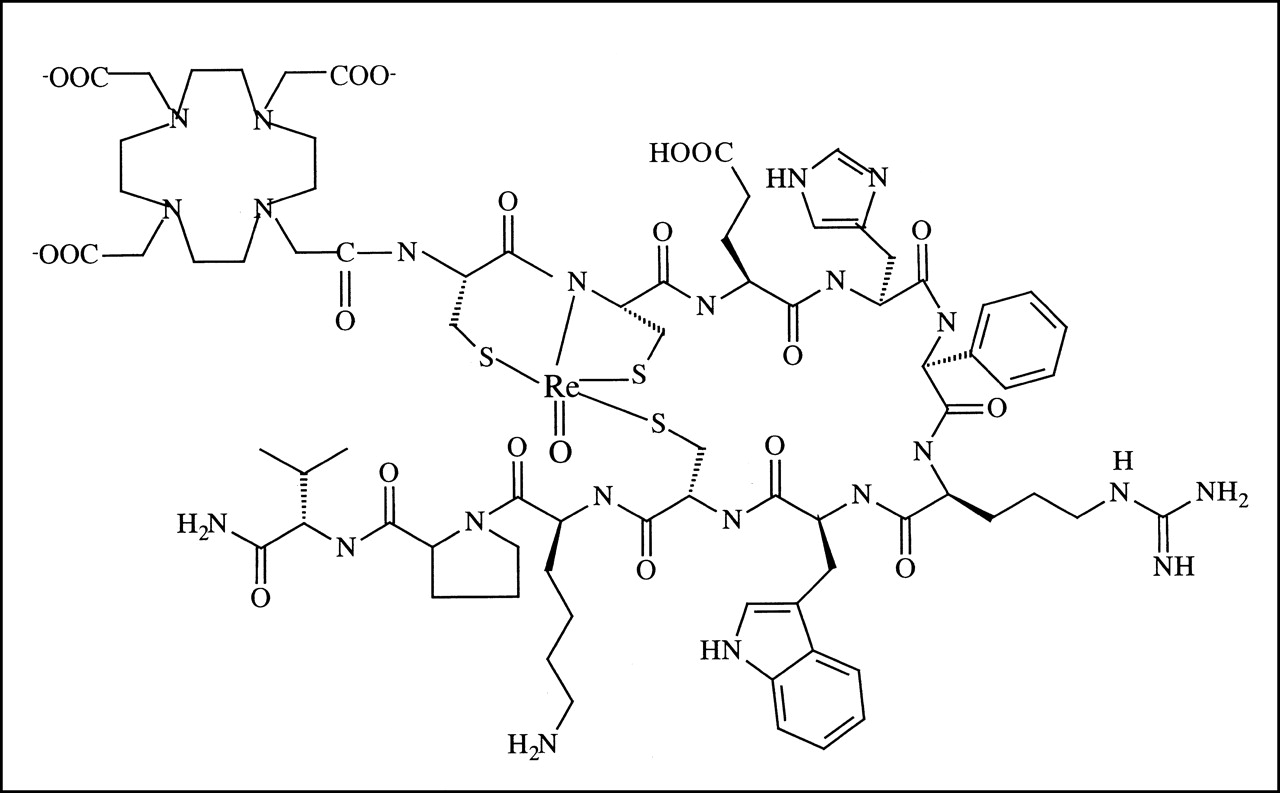
DOTA-ReCCMSH was cyclized by site-specific rhenium coordination. DOTA-CCMSH and ReOCl3(Me2S)(OPPh3) were dissolved in 60% MeOH aqueous solution with a molar ratio of 1:1.5. After the pH was adjusted to approximately 8, the reaction solution was incubated at 70°C for 1 h. The mixture was centrifuged to remove any precipitant, and the DOTA-ReCCMSH complex was purified by RP HPLC monitored at 420 nm with an inline detector. The product peak was collected, lyophilized, and confirmed by ESI MS. The structure of DOTA-ReCCMSH is shown in Figure 1.

Structure of DOTA-ReCCMSH.
DOTA-CMSH was cyclized through the formation of a disulfide bond between cysteines 4 and 10. The DOTA-CMSH peptide was dissolved in 0.1 mol/L ammonium bicarbonate to a final concentration of 0.25 mg/mL. The solution was gently bubbled with air overnight and then lyophilized. Cyclic monomer DOTA-CMSH was purified from its polymer forms by RP HPLC and confirmed by ESI MS.
Radiolabeled Complex Preparation
The 111In-labeled DOTA-ReCCMSH, DOTA-CCMSH, DOTA-CMSH, and DOTA-NDP complexes were prepared under similar conditions. Briefly, 20 μL 111InCl3 (5 mCi/500 μL in 0.04 mol/L HCl) (Mallinckrodt, St. Louis, MO); 80 μL pH 5.5, 0.1 mol/L NH4OAc; and 10 μg peptide were mixed and incubated at 70°C for 45 min. The radiolabeled complex was purified by RP HPLC with a 20-min gradient of 18%–25% acetonitrile/0.1% TFA versus H2O/0.1% TFA. Purified preparations were flushed with nitrogen gas to remove the acetonitrile, and the pH was adjusted to neutral by adding 0.2 mol/L sodium phosphate (pH 8.0)/150 mmol/L NaCl. The 111In-DOTA–CCMSH complex was prepared from fresh DOTA-CCMSH to avoid oxidation of the cysteine thiols of the peptide.
125I-(Tyr2)-NDP was prepared by the chloramine-T method (15). Ten micrograms NDP; 1.5 μL Na125I (American Radiolabeled Chemicals, Inc., St. Louis, MO); 20 μL pH 7.4, 0.2 mol/L phosphate buffer; and 10 μL freshly prepared 1 mg/mL chloramine-T aqueous solution were mixed. After incubation at room temperature for 40 s, the reaction was quenched by adding 10 μL 1 mg/mL Na2S2O4. The radioiodinated complex was purified by RP HPLC, lyophilized, and stored at −20°C.
The stability of the radiolabeled complexes was determined in pH 7.4, 0.01 mol/L phosphate-buffered saline (PBS)/0.1% bovine serum albumin (BSA) or pH 7.4, 0.01 mol/L PBS/0.1 mmol/L ethylenediaminetetraacetic acid (EDTA). Biologic activity of the radiolabeled complexes was determined by in vitro receptor-binding assays with B16/F1 melanoma cells (14).
Cell Culture, Receptor Binding, and IC50 Determinations
The B16/F1 murine melanoma cell line was obtained from American Type Culture Collection (Manassas, VA). Cells were cultured in Roswell Park Memorial Institute 1640 medium containing NaHCO3 (2 g/L), which was supplemented with 10% heat-inactivated fetal calf serum, 2 mmol/L l-glutamine, and 48 mg gentamicin. The cells were expanded in 75-cm2 tissue culture flasks and kept in a humidified atmosphere of 5% CO2 at 37°C. The media were changed every other day. Confluent monolayers were detached with 0.02% EDTA in Ca2+- and Mg2+-free, pH 7.4, 0.01 mol/L PBS and dissociated into single-cell suspensions for further cell culture.
The receptor-binding properties of the 125I- or 111In-labeled complexes were assayed on a B16/F1 murine melanoma cell line. Cells were seeded at a density of 0.2 million per well in 24-well tissue culture plates and allowed to attach overnight. After 1 washing with binding medium (modified Eagle’s medium with 25 mmol/L N-(2-hydroxyethyl)piperazine-N′-(2-ethanesulfonic acid), 0.2% BSA, and 0.3 mmol/L 1,10-phenanthroline [Sigma, St. Louis, MO]), the cells were incubated at 25°C for 3 h with approximately 50,000 cpm radiolabeled complex in 0.5 mL binding medium. Nonspecific binding was determined by coincubation with nonradiolabeled NDP at a final concentration of 10 μmol/L. Cells were rinsed twice with pH 7.4, 0.01 mol/L PBS/0.2% BSA and lysed in 0.5 mL 1 mol/L NaOH for 5 min, and their radioactivity was measured. The cell-binding capacity was reported as the percentage of total added radioactivity that was bound to the cells.
The IC50, or concentration of competitor required to inhibit 50% of radioligand binding, was determined for the DOTA-coupled α-MSH peptide analogs in competitive binding assays with 125I-(Tyr2)-NDP over a 10−14–10−6 mol/L concentration range. B16/F1 cells were prepared as described above in 24-well tissue culture plates and incubated at 25°C for 3 h with approximately 50,000 cpm 125I-(Tyr2)-NDP in 0.5 mL binding medium with different peptide concentrations. The radioactivity in the cells and in the medium was separately collected and measured. The data were processed, and the IC50 values of the DOTA-coupled peptide complexes were calculated with the Kell software package (Biosoft, Ferguson, MO).
In Vivo Studies
C57 BL/6 female mice, 7–8 wk old (Harlan, Indianapolis, IN), were inoculated subcutaneously in the right flank with 1 × 10⁶ cultured B16/F1 murine melanoma cells. Ten days after the inoculation, when tumors reached a weight of approximately 500 mg, each mouse was injected with 2 μCi 111In-labeled peptide through the tail vein for in vivo studies. After radioactivity administration, the mice were housed separately and their urine and feces were collected. Groups of 5 mice were killed at different times (5 min to 72 h) after injection. Tumors and normal tissues of interest were dissected, and the blood on the samples was sponged off with gauze. The contents of the gastrointestinal tract were not removed. The whole body and tissue samples were weighed, and their radioactivity was measured in a γ-counter. The total-blood value was counted as 6.5% of the whole-body weight. Radioactivity uptake in the tumor and normal tissues of interest was expressed as a percentage of the injected radioactivity dose (%ID) per gram of tissue or as %ID. All animal studies were performed in compliance with federal and local institutional rules for the conduct of animal experimentation. Statistical analysis was performed using the Student t test for unpaired data.
RESULTS
DOTA-ReCCMSH and 3 closely related DOTA–α-MSH analogs were synthesized by solid-phase Fmoc synthesis and purified by RP HPLC, and their sequences were confirmed by ESI MS. The structure of DOTA-ReCCMSH is shown in Figure 1. The DOTA-ReCCMSH analogs included its linear nonmetalated homolog DOTA-CCMSH and a disulfide bond-cyclized analog, DOTA-CMSH. DOTA was also conjugated to the amino terminus of one of the highest-affinity α-MSH receptor-binding peptides, NDP (16). Table 1 lists the sequences, IC50 values, and calculated and measured molecular weights of the DOTA–α-MSH peptides used in this study. The 111In-labeled peptides were separated from their nonradiolabeled counterparts by RP HPLC. The radiochemical stabilities of the 111In-DOTA–α-MSH analogs were assayed in pH 7.4, 0.01 mol/L PBS/0.1% BSA and pH 7.4, 0.01 mol/L PBS/0.1 mmol/L EDTA at 25°C. Over a 24-h incubation period, only radiolabeled peptide—not free radioactivity—was detected by RP HPLC. The receptor-binding activities of the individual radiolabeled complexes were determined before their use in vivo. Cell-binding capacities of 5%–10% were obtained for the 111In- and 125I-labeled α-MSH analogs assayed with cultured B16/F1 murine melanoma cells.
α-MSH Analog Characteristics
The tumor uptake and pharmacokinetic properties of 111In-DOTA–ReCCMSH were examined in melanoma-bearing C57 mice. A comparison of the biodistribution data of the 111In-labeled complexes is presented in Table 2. Statistical analysis was performed using the Student t test for unpaired data for the comparison between 111In-DOTA–ReCCMSH and each of the other 3 111In-DOTA–peptide complexes. Compared with the linear 111In-DOTA–CCMSH molecule, rhenium-mediated cyclization significantly increased the in vivo tumor-targeting capacity of 111In-DOTA–ReCCMSH at all investigated time points (P < 0.05). Coinjection of 111In-DOTA–ReCCMSH with 10 μg NDP reduced tumor uptake of the radiolabeled complex from 11.4 ± 2.89 %ID/g to 0.65 ± 0.09 %ID/g at 2 h after injection, showing the tumor-specific uptake of the radiolabeled complex. Similarly, 111In-DOTA–ReCCMSH exhibited higher tumor uptake values than did the disulfide bond-cyclized 111In-DOTA–CMSH complex. Although the linear analog DOTA-NDP showed a higher receptor binding affinity in vitro (Table 1), its tumor uptake values were lower than those of 111In-DOTA–ReCCMSH, without significance at earlier time points of 0.5, 2, and 4 h after injection and with significance at 24 h after injection (P < 0.05) (Table 2).
Biodistribution Comparison Among Analogs
The distribution data showed that radioactive background in the blood and blood-rich content organs, such as lung, liver, and spleen, for 111In-DOTA–ReCCMSH was significantly lower than that for all 3 other 111In-labeled complexes at most time points (Table 2). Interestingly, a high radioactivity accumulation in muscle was unexpectedly observed for 111In-DOTA–NDP. Because of the radioactivity accumulated in the muscle and subsequent release back into the circulation, the radioactivity concentration in the blood was significantly lower for 111In-DOTA–NDP, compared with 111In-DOTA–ReCCMSH, at the earlier time point and then was higher at 2 and 4 h after injection (Table 2). Because of both higher tumor-targeting capacity and lower background, the uptake ratios of tumor to normal tissue were extremely high for 111In-DOTA–ReCCMSH compared with the other 111In-labeled complexes (Table 3).
Comparison of Uptake Ratio of Tumor to Normal Tissues Among Analogs
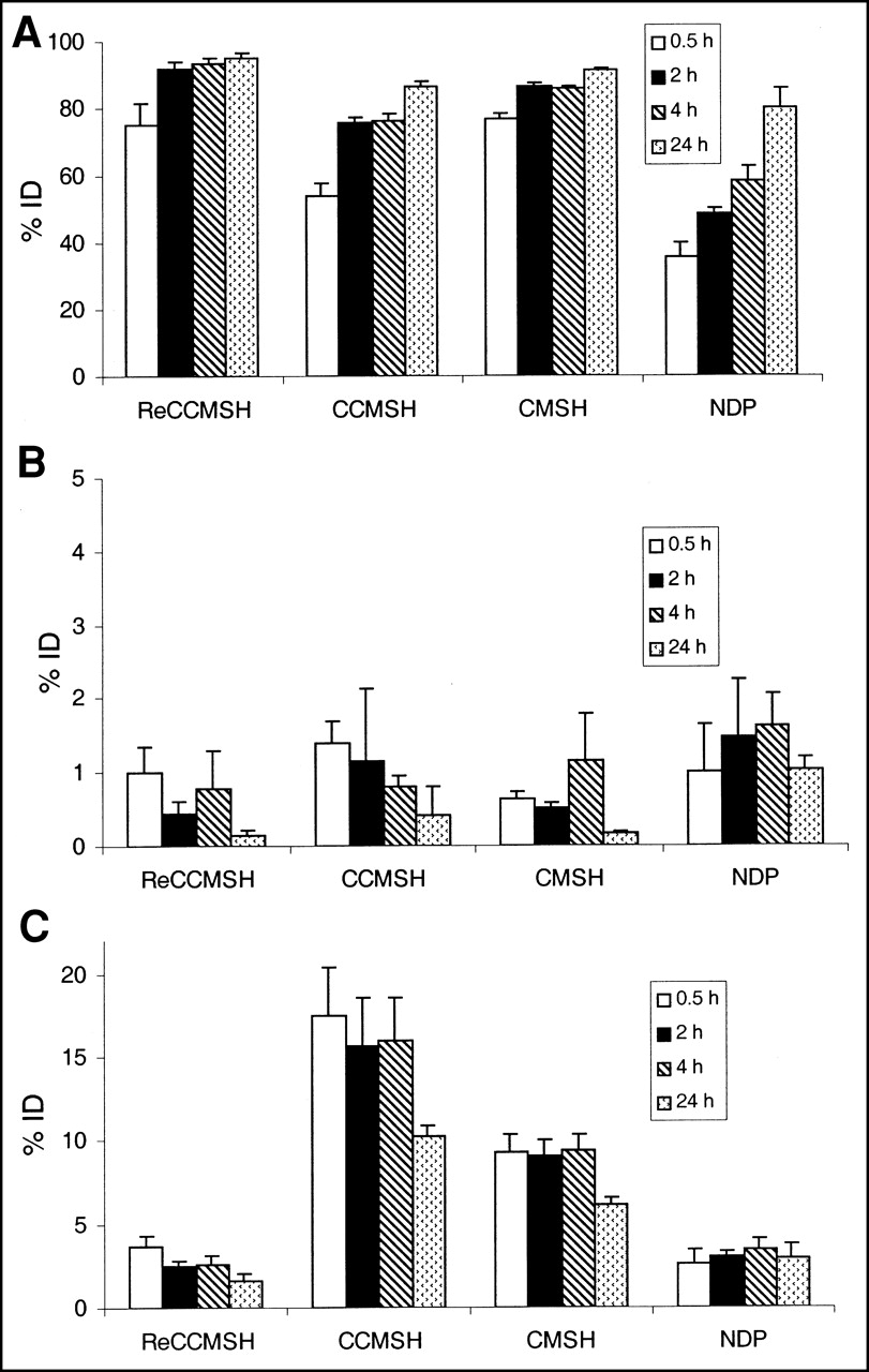
All 111In-DOTA–α-MSH complexes were primarily cleared through the kidney into the urine, with low radioactivity accumulation in the gastrointestinal tract (Fig. 2). Approximately 92% of the total administrated 111In-DOTA–ReCCMSH radioactivity was eliminated into the urine at 2 h after injection, which was significantly faster than the other 3 111In-DOTA–peptides. Because of the muscle accumulation, the radioactivity clearance of 111In-DOTA–NDP was much slower, and approximately 52 %ID of the radioactivity still remained in the whole body at 2 h after radioactivity administration (Fig. 2).

Radioactivity uptake (%ID) in urine (A), intestines (B), and kidneys (C) for 111In-DOTA–ReCCMSH (ReCCMSH), DOTA-CCMSH (CCMSH), DOTA-CMSH (CMSH), and DOTA-NDP (NDP) in B16/F1 murine melanoma–bearing C57 mice at 0.5, 2, 4, and 24 h after injection (n = 5).
The nonspecific kidney radioactivity uptake was much higher for the free sulfhydryl and disulfide bond–containing peptides (e.g., 111In-DOTA–CCMSH and 111In-DOTA–CMSH) compared with that of 111In-DOTA–ReCCMSH or 111In-DOTA–NDP. The rhenium-mediated cyclization enhanced the renal radioactivity clearance, and the kidney radioactivity uptake of 111In-DOTA–ReCCMSH was 3–8 times lower than that of 111In-DOTA–CCMSH and 111In-DOTA–CMSH and was even significantly lower than that of 111In-DOTA–NDP at 24 h after injection (Table 2; Fig. 2).
Figure 3 illustrates the radioactivity concentration (%ID/g) of 111In-DOTA–ReCCMSH in the tumor and normal tissues of interest over time. Because a high tumor uptake (8.6 %ID/g at as early as 5 min after injection) and high tumor retention (2.1 and 1.4 %ID/g at 48 and 72 h, respectively, after injection) were observed, the area under the curve (AUC) of tumor uptake for 111In-DOTA– ReCCMSH was 311.1. On the other hand, with fast radioactivity clearance from the blood and blood-rich organs, the half-life for whole-body radioactivity clearance of 111In-DOTA–ReCCMSH was 19.1 min. Figure 3 also lists the AUC and the ratio of tumor AUC to tissue AUC for blood, lung, liver, and carcass. The high tumor uptake and whole-body clearance of 111In-DOTA–ReCCMSH highlight the therapeutic potential of DOTA-ReCCMSH radiolabeled with β-emitting radiolanthanides or α-emitting radiobismuth for melanoma therapy.
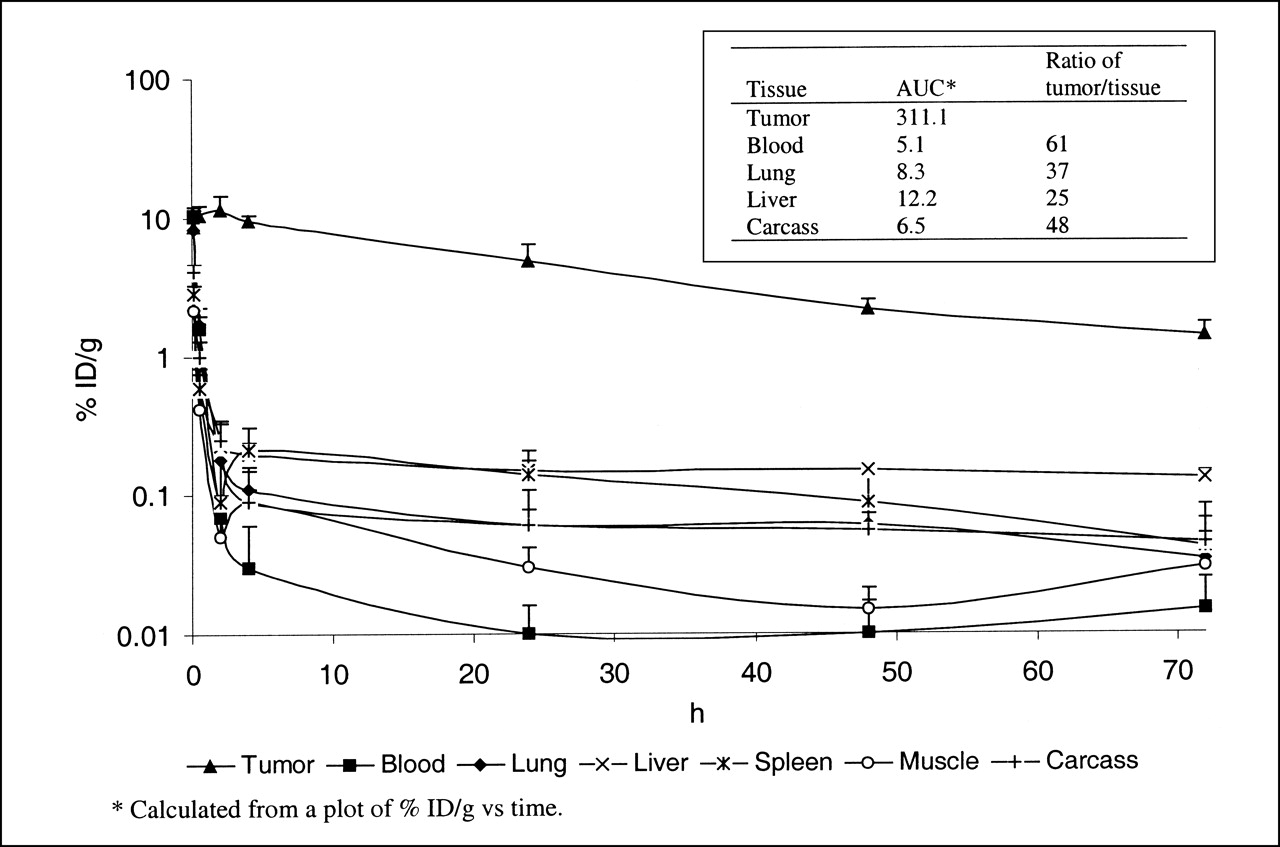
Tumor retention of radioactivity and clearance of radioactivity from normal tissues for 111In-DOTA–ReCCMSH over time (%ID/g) (n = 5). Area under curve (AUC) and ratio of tumor AUC to tissue AUC for blood, lung, liver, and carcass are also listed.
DISCUSSION
The development of radiolabeled α-MSH receptor-targeting peptides for melanoma diagnostic imaging and radiotherapy is attractive because receptor agonists are rapidly internalized on binding (14,17,18). Unfortunately, both directly labeled and chelator-labeled α-MSH peptide analogs are degraded intracellularly, causing rapid release of the radioactivity from the target tissue (12,14). We have reported high melanoma tumor uptake and retention values for a 99mTc- or Recyclized α-MSH analog, CCMSH (11,13,14). We hypothesized that the compact structure of 99mTc/ReCCMSH and the unique ReO-cysteine coordination chemistry resisted intracellular degradation while enhancing intracellular retention (14). To better understand the molecular basis for the favorable tumor-targeting and clearance properties of 99mTc/ReCCMSH, we compared 111In-DOTA-labeled ReCCMSH with 3 other closely related linear and cyclic 111In-DOTA-labeled α-MSH peptide analogs. The linear, nonmetalated homolog of 111In-DOTA–ReCCMSH, 111In-DOTA–CCMSH, exhibited poor tumor uptake and retention, clearly showing the benefit of ReO-mediated cyclization or peptide cyclization itself. A comparison of 111In-DOTA–ReCCMSH with the disulfide bond-cyclized analog 111In-DOTA–CMSH showed that different methods of peptide cyclization yielded molecules with very different in vivo biodistribution properties. Together, these results provided strong evidence that ReO cyclization, not just peptide cyclization itself, was responsible for the high tumor uptake, retention, and whole-body clearance properties of ReCCMSH in vivo. The tumor uptake value of the linear 111In-DOTA–NDP was similar to that of DOTA-ReCCMSH at early times but was lower, with significance, at 24 h after injection. The similar tumor uptake values at earlier times likely result from the extremely high affinity NDP possesses for the α-MSH receptor. Concurrently, high muscle uptake was observed for 111In-DOTA–NDP at 0.5–4 h after injection. It is not likely that the high muscle uptake values were caused by NDP-specific interactions because high muscle uptake was not observed for 99mTc[Cys-Gly-Cys-Gly]-NDP, 99mTc-MAG2–NDP, or 125I-(Try2)-NDP (12). The accompanying high muscle uptake of 111In-DOTA–NDP makes its therapeutic application unlikely.
Nonspecific radioactivity accumulation in the kidneys is often associated with the in vivo application of radiolabeled peptides and antibody fragments (19–21). We postulated that the positive charge of the lysine residue was the main cause for the nonspecific radioactivity retention in the kidneys. In our previous studies, substitution of Lys11 with Nle11 or Gly11 in the 99mTc-CCMSH sequence yielded analogs with significantly reduced kidney uptake but sacrificed the high tumor-targeting property of the 99mTc-CCMSH (14). In this investigation, we found that high renal radioactivity uptake could also be caused by free sulfhydryls or the disulfide bond moiety. The kidney uptake of 111In-DOTA–ReCCMSH was 3–8 times lower than that of 111In-DOTA–CMSH and 111In-DOTA–CCMSH. Rhenium oxocoordination of the Cys-thiols appeared to shield them from interacting with the kidney, facilitating excretion of 111In-DOTA–ReCCMSH.
A comparison of clearance properties between 111In-DOTA–ReCCMSH and its related 99mTc-CCMSH analog revealed that 111In-DOTA–ReCCMSH exhibited superior clearance kinetics. Approximately 92 %ID of 111In-DOTA–ReCCMSH radioactivity was eliminated through the urine at 2 h after injection, compared with only 73 %ID for 99mTc-CCMSH at the same time point. Likewise, the blood clearance rate for 111In-DOTA–ReCCMSH was twice that for 99mTc-CCMSH (14). In addition, 111In-DOTA–ReCCMSH exhibited superior tumor retention values compared with 99mTc-CCMSH at 24 h after injection, although tumor uptake values for both complexes were not statistically different at early time points. The increased whole-body clearance kinetics and increased tumor retention properties of 111In-DOTA–ReCCMSH were likely to have resulted from greater hydrophilicity because of an increase in the number of charges. At a neutral pH, both 111In-DOTA–ReCCMSH and 99mTc-CCMSH have a net zero charge. However, 111In-DOTA–ReCCMSH has a larger number of ionizable groups because of the presence of the DOTA chelator. It is likely that a complex with a greater number of charges would have more difficulty diffusing across a cell membrane, thus resulting in an increase in tumor retention for 111In-DOTA–ReCCMSH. Moreover, an increase in hydrophilicity should promote rapid blood clearance and renal excretion, which are consistent with the biodistribution data.
Several benefits are associated with DOTA conjugation to ReCCMSH. The DOTA chelator is able to strongly chelate a variety of β- or α-particle–emitting radiometals, such as 111In, 90Y, 149Pm, 177Lu, 212Pb, and 212/213Bi, under physiologic conditions (22–26). The metal chelation flexibility of DOTA would allow DOTA–ReCCMSH to be labeled with radionuclides that deposit their energies over a range of approximately 10 μm to 1 cm. DOTA conjugation also appears to improve tumor cell retention. Redistribution of radioactive catabolites derived from internalized peptide complexes will dramatically influence tumor radioactivity uptake and retention (27). The radiometals used to complex with DOTA exhibit multiple charges. Free radiolabeled DOTA species produced on degradation of the radiolabeled peptide conjugates will remain largely intracellular because of the difficulty of moving a charged complex across cellular membranes (28). Hence, a reduction in the diffusion of charged radiolabeled DOTA species across the cell membrane will further reduce washout of tumor-targeted radioactivity, resulting in enhanced retention times. Compared with our previous results reported for 99mTc/ReCCMSH (11,13,14), the biodistribution data reported here for 111In-DOTA–ReCCMSH clearly show that the DOTA-conjugated complex has equally good tumor uptake kinetics, with increased tumor retention >24 h after injection and enhanced clearance kinetics. Because of both its higher tumor-targeting capacity and its low background, 111In-DOTA–ReCCMSH exhibited the highest uptake ratio of tumor to normal tissues of any reported radiolabeled α-MSH complex, highlighting its ability to selectively deposit radionuclides in melanoma tumor cells.
CONCLUSION
A novel method of peptide cyclization through ReO coordination has enhanced the melanoma tumor-targeting capacity and clearance of 111In-DOTA–ReCCMSH, resulting in an extremely high uptake ratio of tumor to normal tissues. Rhenium-mediated cyclization also significantly reduced nonspecific renal radioactivity accumulation, compared with its free sulfhydryl and disulfide bond–containing counterparts. High tumor uptake and retention, coupled with rapid clearance kinetics, make β- or α-emitter–radiolabeled DOTA–ReCCMSH an attractive potential therapeutic agent for melanoma.
Acknowledgments
The authors thank Drs. Wynn Volkert, Susan L. Deutscher, and Vladislav V. Glinsky for helpful discussions and assistance. This study was supported by grant ER61661 from the Department of Energy and grant NCI R41 CA85106 from the National Institutes of Health.
Footnotes
Received Mar. 9, 2001; revision accepted Aug. 20, 2001.
For correspondence or reprints contact: Thomas P. Quinn, PhD, Department of Biochemistry, 117 Schweitzer Hall, University of Missouri–Columbia, Columbia, MO 65211.
REFERENCES
In this issue


Jump to section
Related Articles
Cited By...
- Small Peptide-Based Nanodelivery Systems for Cancer Therapy and Diagnosis
- A tumor-targeted immune checkpoint blocker
- Effects of the Amino Acid Linkers on the Melanoma-Targeting and Pharmacokinetic Properties of 111In-Labeled Lactam Bridge-Cyclized {alpha}-MSH Peptides
- Reduction of the Ring Size of Radiolabeled Lactam Bridge-Cyclized {alpha}-MSH Peptide, Resulting in Enhanced Melanoma Uptake
- PET of Malignant Melanoma Using 18F-Labeled Metallopeptides
- 111In-Labeled Galectin-3-Targeting Peptide as a SPECT Agent for Imaging Breast Tumors
- Evaluation of an 111In-Radiolabeled Peptide as a Targeting and Imaging Agent for ErbB-2 Receptor Expressing Breast Carcinomas
- Small-Animal PET of Melanocortin 1 Receptor Expression Using a 18F-Labeled {alpha}-Melanocyte-Stimulating Hormone Analog
- Synthesis and Biologic Evaluation of 64Cu-Labeled Rhenium-Cyclized {alpha}-MSH Peptide Analog Using a Cross-Bridged Cyclam Chelator
- 99mTc- and 111In-Labeled {alpha}-Melanocyte-Stimulating Hormone Peptides as Imaging Probes for Primary and Pulmonary Metastatic Melanoma Detection
- Melanoma Therapy via Peptide-Targeted {alpha}-Radiation
- Melanoma Targeting with DOTA-{alpha}-Melanocyte-Stimulating Hormone Analogs: Structural Parameters Affecting Tumor Uptake and Kidney Uptake
- Radioiodination of Rhenium Cyclized {alpha}-Melanocyte-Stimulating Hormone Resulting in Enhanced Radioactivity Localization and Retention in Melanoma
- A Gallium-Labeled DOTA-{alpha}-Melanocyte- Stimulating Hormone Analog for PET Imaging of Melanoma Metastases
- A Novel DOTA-{alpha}-Melanocyte-Stimulating Hormone Analog for Metastatic Melanoma Diagnosis