
Main menu

  • Home
  • Content
    • Current
    • Ahead of print
    • Past Issues
    • JNM Supplement
    • SNMMI Annual Meeting Abstracts
    • Continuing Education
    • JNM Podcasts
  • Subscriptions
    • Subscribers
    • Institutional and Non-member
    • Rates
    • Journal Claims
    • Corporate & Special Sales
  • Authors
    • Submit to JNM
    • Information for Authors
    • Assignment of Copyright
    • AQARA requirements
  • Info
    • Reviewers
    • Permissions
    • Advertisers
  • About
    • About Us
    • Editorial Board
    • Contact Information
  • More
    • Alerts
    • Feedback
    • Help
    • SNMMI Journals
  • SNMMI
    • JNM
    • JNMT
    • SNMMI Journals
    • SNMMI

User menu

  • Subscribe
  • My alerts
  • Log in
  • My Cart

Search

  • Advanced search
Journal of Nuclear Medicine
  • SNMMI
    • JNM
    • JNMT
    • SNMMI Journals
    • SNMMI
  • Subscribe
  • My alerts
  • Log in
  • My Cart
Journal of Nuclear Medicine

Advanced Search

  • Home
  • Content
    • Current
    • Ahead of print
    • Past Issues
    • JNM Supplement
    • SNMMI Annual Meeting Abstracts
    • Continuing Education
    • JNM Podcasts
  • Subscriptions
    • Subscribers
    • Institutional and Non-member
    • Rates
    • Journal Claims
    • Corporate & Special Sales
  • Authors
    • Submit to JNM
    • Information for Authors
    • Assignment of Copyright
    • AQARA requirements
  • Info
    • Reviewers
    • Permissions
    • Advertisers
  • About
    • About Us
    • Editorial Board
    • Contact Information
  • More
    • Alerts
    • Feedback
    • Help
    • SNMMI Journals
  • View or Listen to JNM Podcast
  • Visit JNM on Facebook
  • Join JNM on LinkedIn
  • Follow JNM on Twitter
  • Subscribe to our RSS feeds
Meeting ReportPhysics, Instrumentation & Data Sciences - Data Analysis & Management

High photon emission rate effects on PET/CT using Lu-177, a phantom study

David Sennung and Magnus Dahlbom
Journal of Nuclear Medicine June 2024, 65 (supplement 2) 242514;
David Sennung
1UCLA Health
  • Find this author on Google Scholar
  • Find this author on PubMed
  • Search for this author on this site
Magnus Dahlbom
2University of California, Los Angeles
  • Find this author on Google Scholar
  • Find this author on PubMed
  • Search for this author on this site
  • Article
  • Figures & Data
  • Info & Metrics
Loading

Abstract

242514

Introduction: Patients undergoing radionuclide therapy (RNT) may also be scheduled for diagnostic PET imaging, post-infusion, for evaluation of the treatment as well as other clinical findings of significance. Lu-177 is currently the most used isotope for RNT emits a significant amount of low (6.6% 113 keV) medium (11.1% 208 keV) energy photons. Although only 511 keV photons are of concern in PET, detected lower energy emissions from Lu-177 still must be processed by the detector electronics. The presence of high activity concentrations of Lu-177 could therefore interfere with PET imaging performed post-therapy. This interference could produce dead-time, event pileup, random coincidences which could affect image quality and quantification. The purpose of this work is to investigate the effect on image quality in the presence of Lu-177 during PET imaging.

Methods: A phantom study was done using a flangeless ACR PET phantom. All data were acquired on a Siemens mCT PET/CT system with LSO scintillation crystals. A theranostic infused patient during a PET scan were simulated by adding a phantom background with an initial activity of 88.2 µCi/ml of Lu-177. The phantom was the filled with F-18 according to ACR accreditation protocol with a whole-body patient dose of 14 mCi. No additional activity was added outside the scanner field-of-view (FOV).

Scans were repeated at multiple Lu-177 activity concentrations until a final phantom background activity reached 0.05 µCi/ml. Quality as well as quantification analysis methods were performed on reconstructed 10 mm slices. Quantification analysis consisted of measuring average SUV in the phantom background. The contrast, resolution and image quality of the reconstructed images were visually evaluated.

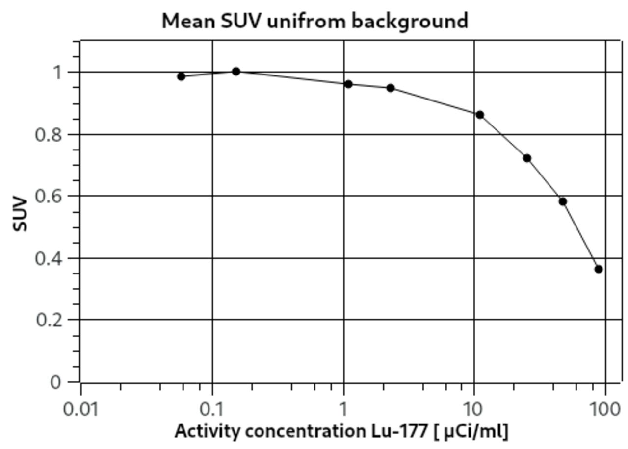
Results: Results from the quantification analysis showed a rapid decrease in SUV accuracy at activity concentrations exceeding 11 µCi/ml. Calculations showed a 5, 10 and 20% reduction in SUV in the uniform background section at activities 2.0, 6.8 and 17 µCi/mlCi respectively.

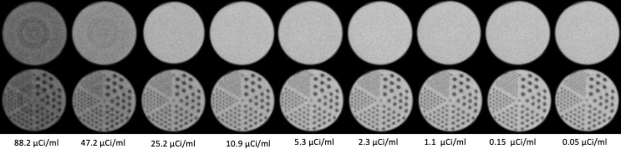
High photon emission rates lead to event pile-up events and detector mis-positioning and results in visible ring artifacts. The ring artifacts can be seen in throughout the phantom at Lu-177 activity concentrations exceeding 11 µCi/ml.

All rod sectors in the resolution portion of the phantom are visible up to 11 µCi/ml of Lu-177. Exceeding this concentration, a noticeable degradation in resolution and contrast for the smallest 4.8 mm rod pattern was observed. A similar degradation was observed for the 6.5 mm rods at activity concentrations exceeding 25 µCi/ml.

The results of this study may change in the presence of background activity outside the scanner FOV. Furthermore, results may be also different for PET scanners using slower scintillators, such as BGO.

Conclusions: Photons from the Lu-177 background clearly affected both image quality and quantification. Although the photon emissions from Lu-177 have energies outside the energy window used for 511 keV photons, these low energy photons are detected and need to be processed by the detector electronics. At high activity concentrations the Lu-177 background affected both image quality (ring artifacts, loss in contrast and spatial resolution). Quantification was also observed to be affected at background concentrations exceeding 2.0 µCi/ml, which is comparable to the average activity concentrations expected in a patient during the first few hours following an infusion (2.8 µCi/ml immediately after a standard Lu-177 infusion, 200 mCi infusion in a 70 kg patient). Since activity concentration rapidly decreases through excretion and isotope decay, delaying a PET scan at least 24 h post-infusion should minimize the effects of the Lu-177 background.

Figure
  • Download figure
  • Open in new tab
  • Download powerpoint
Figure
  • Download figure
  • Open in new tab
  • Download powerpoint
Previous
Back to top

In this issue

Journal of Nuclear Medicine
Vol. 65, Issue supplement 2
June 1, 2024
  • Table of Contents
  • Index by author
Article Alerts
Sign In to Email Alerts with your Email Address
Email Article

Thank you for your interest in spreading the word on Journal of Nuclear Medicine.

NOTE: We only request your email address so that the person you are recommending the page to knows that you wanted them to see it, and that it is not junk mail. We do not capture any email address.

Enter multiple addresses on separate lines or separate them with commas.
High photon emission rate effects on PET/CT using Lu-177, a phantom study
(Your Name) has sent you a message from Journal of Nuclear Medicine
(Your Name) thought you would like to see the Journal of Nuclear Medicine web site.
Citation Tools
High photon emission rate effects on PET/CT using Lu-177, a phantom study
David Sennung, Magnus Dahlbom
Journal of Nuclear Medicine Jun 2024, 65 (supplement 2) 242514;

Citation Manager Formats

  • BibTeX
  • Bookends
  • EasyBib
  • EndNote (tagged)
  • EndNote 8 (xml)
  • Medlars
  • Mendeley
  • Papers
  • RefWorks Tagged
  • Ref Manager
  • RIS
  • Zotero
Share
High photon emission rate effects on PET/CT using Lu-177, a phantom study
David Sennung, Magnus Dahlbom
Journal of Nuclear Medicine Jun 2024, 65 (supplement 2) 242514;
Twitter logo Facebook logo LinkedIn logo Mendeley logo
  • Tweet Widget
  • Facebook Like
  • Google Plus One
Bookmark this article

Jump to section

  • Article
  • Figures & Data
  • Info & Metrics

Related Articles

  • No related articles found.
  • Google Scholar

Cited By...

  • No citing articles found.
  • Google Scholar

Similar Articles

SNMMI

© 2025 SNMMI

Powered by HighWire