


Abstract
242362
Introduction: Although pain is the most common chief complaint for a medical visit, there remain limited diagnostic tools for the localization of nociceptive pain [1]. Voltage gated calcium channels (VGCC) are responsible for the detection and transmission of cellular signaling related to pain within the nervous system. Mn2+ is a surrogate for Ca2+ and is also readily transported through VGCCs, and positron-emitting 5xMn is proposed as a molecular probe for the detection and localization of nociceptive activity [2]–[4]. Hence the purpose of this study was to determine the uptake kinetics of the 52gMn as a PET radiotracer (Study A) and to investigate VGCC upregulation in the spinal cord correlated with pain-associated cellular signaling (Study B).
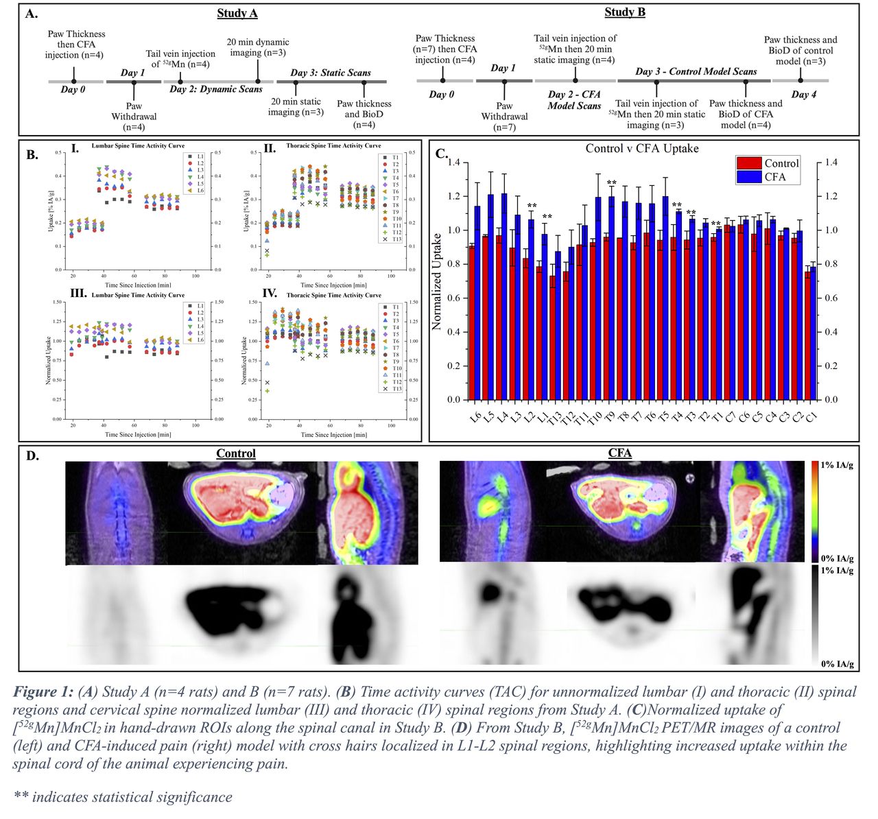
Methods: 52gMn (t1/2 =5.6 d) was produced using a 16 MeV GE PETtrace, chemically separated from natCr target material as [52gMn]MnCl2and administered via tail vein injections. Inflammatory pain was achieved via Complete Freund’s Adjuvant (CFA) injection in the footpad of the right hind paw. The presence (or lack) of pain behaviors was confirmed using Von Frey paw withdrawal analysis and inflammation was assessed with measurements of paw thickness. Previous µPET/CT imaging of CFA rat models measured increased uptake in the lumbar spinal canal but could not eliminate inflammatory changes in the spinal column or bone uptake as confounds due to inadequate soft tissue contrast and bone uptake [5]. Here, we report an improved study design using time-of-flight (TOF) PET/3.0T MR imaging (Signa PET/MRI, GE) analyzed by hand-drawn ROIs (Encore, MIM Software Inc.) of the spinal cord from C1-L6 using MR images acquired using a sagittal SPGR sequence. Student T-tests determined significance between spinal cord and non-target tissue (95% CI). At the completion of each study, all animals were sacrificed, tissue samples were collected, and biodistribution of the tracer was determined (data not shown). The timelines of both studies are shown in Figure 1.A.
Results: Paw withdrawal and paw thickness measurements showed increased sensitivity and swelling, a sign of inflammation, in CFA-injected animals. Study A determined peak uptake occurred between 25-50 minutes post injection (p.i.), however changes in uptake were not only dependent on time p.i., but also on animal. This dependence is best shown between 20-60 min, where data collected at 19, 24, 29, 34, and 39 min P.I. are from the first animal scanned and data collected at 37, 42, 47, 52, and 57 min P.I. are from the second animal scanned. (Figure 1.B.I-II). Animals were scanned in 20-minute intervals to avoid >30 min periods under anesthesia. Several tissues (e.g., liver, subcutaneous fat, and cervical spine) were studied to normalize uptake in lumbar and thoracic segments across animals. Cervical spine was ultimately used because it was hypothesized that tissue uptake within the nervous system would have similar dependence on subject-specific characteristics (mass, unknown underlying conditions, etc.) (Figure 1.B.III-IV). Using the optimized time window from Study A, Study B determined that uptake at the L2, L1, T9, T4, T3, and T1 level in the experimental (CFA) group was significantly greater than in the control group (Figure 1.C).
Conclusions: Rats exhibiting inflammatory pain post-CFA injection showed significantly higher [52gMn]MnCl2 uptake in several regions of the lumbar and thoracic spinal canal vs. control using optimized imaging time windows and reference regions. Future 5xMn- PET/MR studies will test a spared nerve injury (SNI) surgical pain model, which is considered the gold standard of animal pain models.
References
[1] K.W. Tung et al., Semin. Musculoskelet.Radiol., no. April, 2015
[2] R. Hernandez et al., Diabetes, vol. 66, 2017
[3] M.E. Daube et al., Int. J. Nucl. Med. Biol., vol. 12, 1985
[4] S.A. Graves et al., Sci. Rep., vol. 7, 2017
[5] K.E. Barrett et al., J. Nucl. Med., vol. 63, no. supplement 2, 2022

In this issue


Jump to section
Related Articles
Cited By...
- No citing articles found.