


Abstract
242113
Introduction: Background: The endocannabinoid system plays an important role in many basic brain functions and is implicated in human pathological conditions including neurodegeneration, mood disturbances, schizophrenia, and drug dependence. The cannabinoid receptor CB1, presumably the most abundant G-protein coupled neurotransmitter receptor in the brain, is central to endocannabinoid function and is the primary target of Δ9-tetrahydrocannabinol (THC), the major psychoactive component of cannabis. It is thus of great interest to image CB1 in human brains and in brains of preclinical animal models, including mice, using specific radiotracers such as [18F]FMPEP-d2 [1]. However, the small size of mice and their limited availability of blood have made image quantification of murine PET challenging. The goal of this work is to develop a quantitative approach to PET imaging of CB1 binding using [18F]FMPEP-d2 in wild type (WT) and dopamine transporter-knockout (DATKO) mice [2].
Methods: Methods: [18F]FMPEP-d2 was synthesized using our new radiochemical method [3]. Dynamic [18F]FMPEP-d2 PET images were acquired on a Mediso nanoScan PET-CT for a total of 23 mice [n=11 (6 females) for DATKO and n=12 (6 females) for WT]. Another 16 C57BL6J mice (6 females) were used for HPLC plasma radiometabolite analysis of cardiac blood samples collected at 2, 5, 10, 30 and 60 min after [18F]FMPEP-d2 injection through the tail vein. Time-activity curves (TACs) of 20 brain regions and heart blood pool were extracted from the dynamically reconstructed images (39 frames) and the whole blood activity of the mice was used to derive population-based metabolites-corrected plasma input function (PBIF). Kinetic analysis was performed using PMOD4.203.
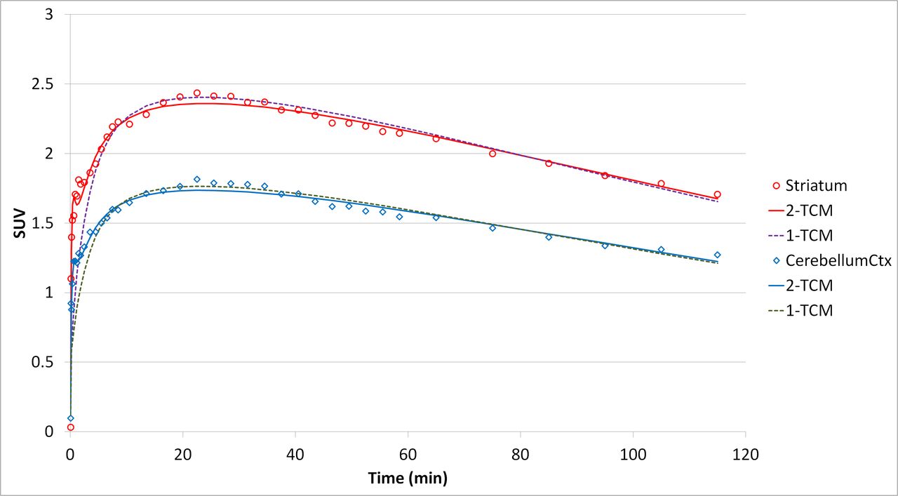
Results: Results: [18F]FMPEP-d2 had a plasma-blood concentration ratio of 1.27 ± 0.11, which was constant in the time frame (2-60 min), and a blood half-life of 3.9 min in mice. There was no significant difference in cardiac blood TACs between WT and DATKO (Fig. 1A), thus a unified PBIF was constructed by combining both groups. A two-tissue compartment model was found to fit brain TACs significantly better than a one-tissue compartment model (whole brain model selection criterion: 3.9 ± 0.3 vs. 1.8 ± 0.5, respectively, p<0.05; see Fig. 1B) and 60 min of acquisition was sufficient to derive a stable binding parameter of total distribution volume (VT) for [18F]FMPEP-d2. Overall, the transgenic mice had significantly lower whole brain VT than their WT littermates (11.7 ± 1.4 vs. 13.6 ± 2.2 mL/cm3, respectively, p<0.05), in particular in female mice, which was consistent with TAC analysis of the images and preliminary qPCR measurement of CB1 expression in the brain.
Conclusions: Conclusion: A population-based approach was effectively implemented for the quantitative analysis of dynamic PET images using [18F]FMPEP-d2 in mice and can be considered for application in human PET imaging studies.


In this issue


Jump to section
Related Articles
Cited By...
- No citing articles found.