


Abstract
242075
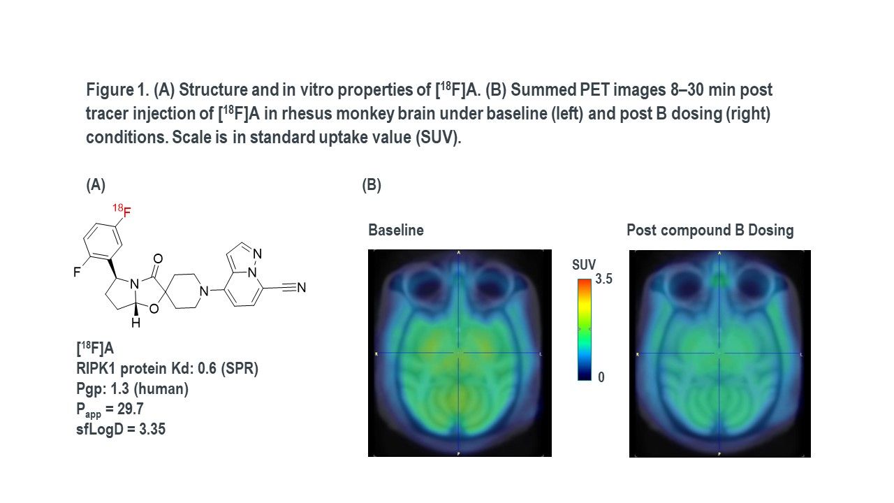
Introduction: Receptor-interacting serine/threonine-protein kinase 1 (RIPK1) regulates inflammation, cytokine release, and necroptotic cell death and is implicated in pathogenic cellular pathways in amyotrophic lateral sclerosis (ALS), Alzheimer’s disease (AD), multiple sclerosis and autoinflammatory disease. RIPK1 kinase has emerged as a promising therapeutic target for the treatment of a wide range of neurodegenerative diseases such as AD or ALS. The development of a RIPK1-selective PET tracer would greatly facilitate therapeutic efforts by confirming in vivo target engagement of potential clinical candidates. This abstract describes the evaluations of a RIPK1 PET tracer candidate, [18F]A (Figure 1A), for use in the determination of central RIPK1 enzyme occupancy with therapeutic candidates in vitro and in vivo PET imaging studies.
Methods: was synthesized from the pinacol boronate (BPin) precursor using copper mediated radio-fluorination. In vitro autoradiography and homogenate binding studies with [3H] or [18F]A were conducted in RIPK1 protein and in monkey and human brain tissues (including non AD and AD) to assess the RIPK1 PET tracer feasibility Bmax/Kd ratio (target density Bmax, radioligand binding affinity Kd). Baseline PET scans and blocking studies were performed to determine the regional brain distribution, RIPK1 specific binding, and examine the utility of [18F]A to measure target engagement of RIPK1 therapeutic candidates in rhesus monkey.
Results: In vitro autoradiography studies with [3H]A or [18F]A in monkey, and human brain slices showed that this radioligand has low displaceable binding. Binding density in AD cortex slices was slightly higher than in non-AD cortex slices, but statistically insignificant. Minimal displaceable binding of [3H] or [18F]A was observed in ALS spinal cord slices or monkey cortex slices. [3H]A or [18F]A bind with high affinity to single, saturable sites in RIPK1 protein (Kd ~ 1 nM), however [3H]A or [18F]A display weak binding affinity in rhesus monkey and human brain tissue homogenates (Table 1), resulting in smalle Bmax/Kd ratios, indicating a lack of specific binding to RIPK1 target which was consistent with minimal displaceable binding of [3H]A or [18F]A in tissue slices. PET baseline studies in rhesus monkeys showed good brain penetration with a homogeneous tracer distribution throughout the brain and rapid washout. RIPK1 specificity in vivo was evaluated by co-administering an distinct RIPK1 inhibitor, compound B (uM level plasma concentration), which resulted in a reduction of volume of distribution (VT) in the thalamus and striatum regions. However the specific signal was too small to accurately measure enzyme occupancy of compound B (Figure 1B).
Table 1. Bmax and Kd Values for [18F]A binding in RIPK1 protein, monkey and human frontal cortex homogenates.
Conclusions: Both the in vitro binding and in vivo PET imaging data observed with [3H]A or [18F]A suggest extremely low Bmax/Kd ratio in brain tissues indicating a low probability of success for a CNS-targeted RIPK1 PET tracer within the chemical matters we investigated.


In this issue


Jump to section
Related Articles
Cited By...
- No citing articles found.