


Abstract
241908
Introduction: Patients with esophageal cancer usually undergo double-contrast barium esophagography as the initial assessment. However, high contrast concentrations lead to high computed tomography (CT) numbers and streaking artifacts on CT images because of photon absorption. This increase in CT numbers also contributes to high PET attenuation coefficients, resulting in an overestimation of tracer uptake and thereby producing false-positive PET results. In addition, the CT part of the combined PET/CT protocol is commonly applied as a whole-body low dose scan. In comparison to standard-radiation-dose CT, lowering the tube potential in CT scans may result in reduced ray penetration, more metal artifacts on CT scans and could potentially affect PET images. This study aims to evaluate the status of gastrointestinal (GI) tract retention of contrast medium after barium esophagography conducted within a one-month period, and its temporal influence on CT and PET images.
Methods: A retrospective review was performed on 179 clinical PET/CT studies from February 2010 to June 2022, involving patients who had undergone barium esophagography within the preceding 30 days. The entire GI system was segmented into six regions: stomach, small intestine, ascending colon (A-colon), transverse colon (T-colon), descending colon (D-colon) and rectosigmoid colon. Three events were documented for each segment: contrast medium (barium) retention, CT streaking artifact and PET images overcorrection. Barium retention and CT streaking artifact were evaluated by visual assessment. Overcorrection of PET image was defined as increased 18F-FDG uptake altering the pattern of hot areas on PET images with computed tomography-based attenuation correction (CTAC) compared to non-attenuation corrected (NAC) images. The time intervals between barium esophagography and PET/CT images were categorized and grouped as follow: 0, 1, 2, 3, 4 to 7, 8 to 14, 15 to 21 and 21 to 29 days.
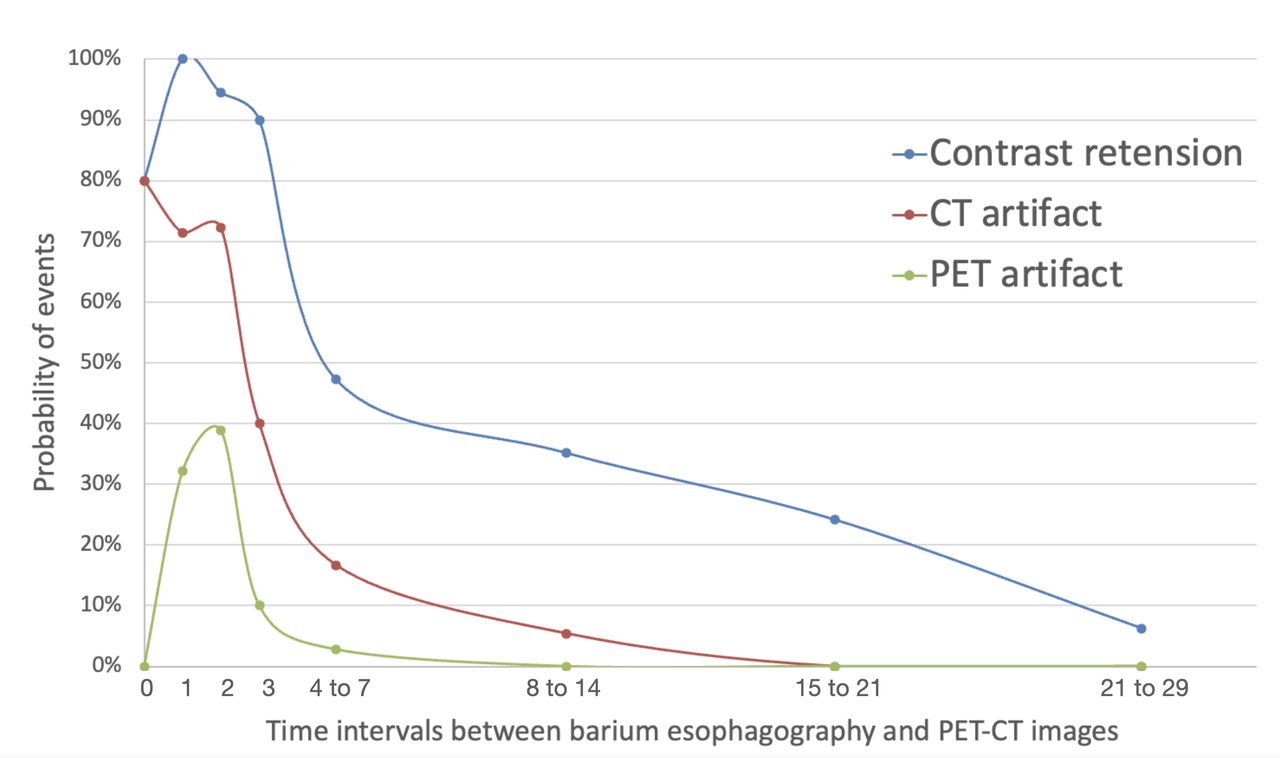
Results: In all 179 PET examinations, 18 (10.0%) of them exhibited contrast media PET artifact. Grouping the examinations based on time interval between barium esophagography and PET revealed probabilities of 90% for contrast medum retention, 40% for CT streaking artifact, and 10% for PET overcorrection after three days. Subsequently, after 7 days, the probability dropped to 35.1%, 5.4% and 0%, respectively. When categorized by GI tracts segments, 22 (2%) out of 1074 segments displayed contrast media PET artifacts, predominantly concentrated in the T-colon and rectosigmoid colon. Categorizing these studies into two groups based on the time intervals of 0 to 3 and 4 to 29 days, the chi-square test revealed a statistically significant difference (p < 0.001) in the probability of all three events: contrast medium retention, CT streaking artifact, and PET overcorrection artifact.
Conclusions: The likelihood of barium retention, CT streaking artifact, and overcorrected PET data diminished over time, notably plunging after the initial 3 days. A suggested strategy to mitigate CT streaking and PET overcorrection artifacts is to conduct PET/CT imaging three days after oral contrast medium ingested.


In this issue


Jump to section
Related Articles
Cited By...
- No citing articles found.