


Abstract
241597
Introduction: Immune-targeted therapies, such as immune checkpoint inhibitors and chimeric antigen receptor (CAR)-T cells, have shown promising outcomes in improving survival for patients with advanced malignant tumors, including lymphoma. However, these therapies exhibit mechanisms of action distinct from traditional cytotoxic chemotherapies, leading to new response patterns that challenge the established imaging-based assessment criteria. Yet, limited understanding exists regarding novel tumor response patterns to immunotherapy on hybrid functional and anatomic images. This study aims to investigate the patterns of non-Hodgkin lymphoma (NHL) response to CAR-T cell therapy using F-18 FDG PET/CT imaging.
Methods: Forty-seven patients with Stages III or IV NHL underwent CAR-T cell therapy with F-18 FDG PET/CT imaging for tumor response assessment from 2020 to 2023. Inclusion and exclusion criteria were established for patient selection. Patients with pre-therapy and first post-therapy FDG PET/CT imaging within 3 months of CAR-T cell infusion were included. SUVmax and size of target lesions were measured using MIM software for image analysis. Treatment responses of FDG avid lesions were assessed per the PERCIST Criteria and compared with clinical outcomes. Statistical analyses were performed using R 4.2.2 software.
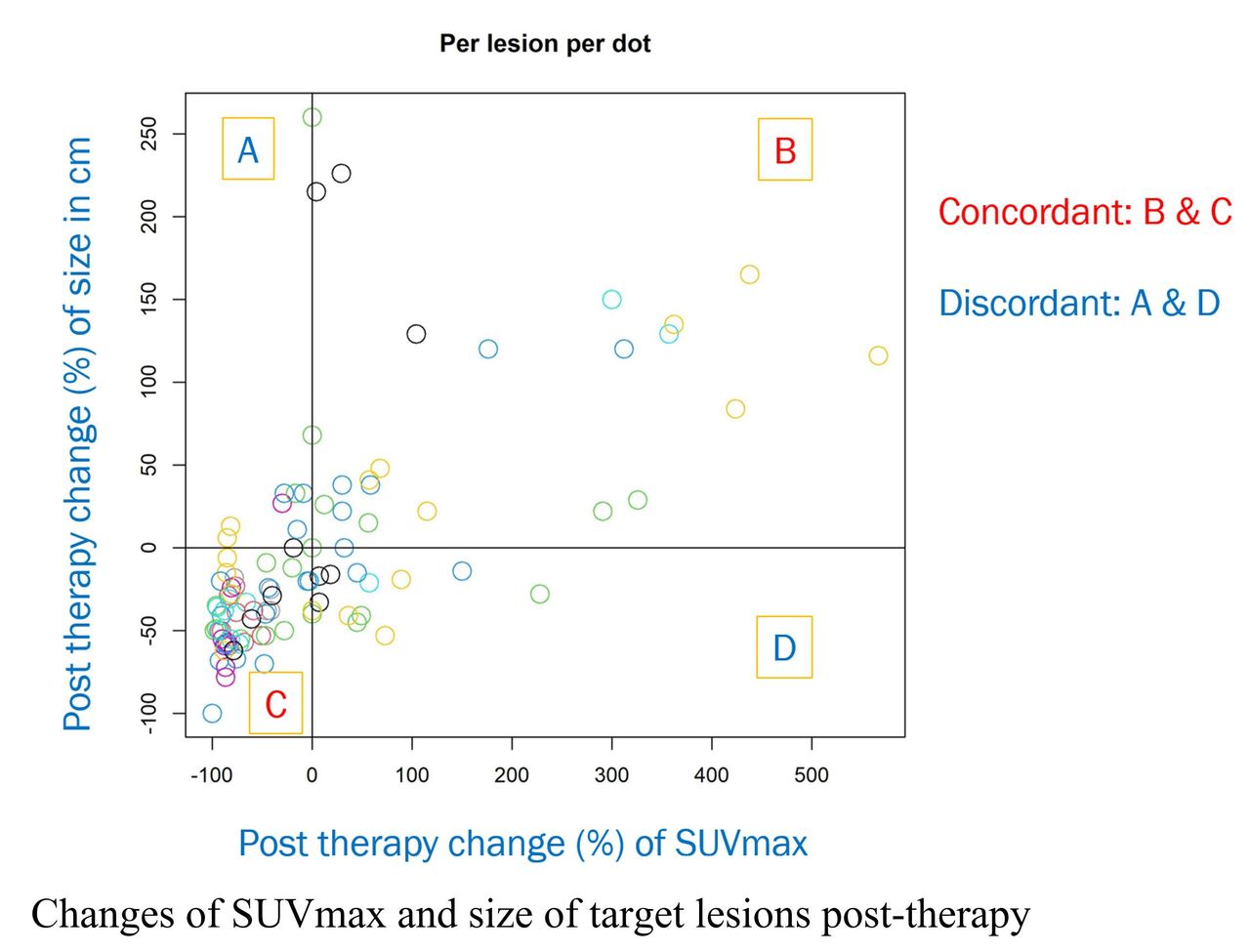
Results: Twenty-six out of 47 cases meeting the inclusion criteria were investigated (6 female, 20 male; age range 22-74 years, mean age 60 ± 13 years). Conventional response patterns according to the PERCIST Criteria were observed, including 9 cases (35%) of complete metabolic response (CMR), 4 cases (15%) of partial metabolic response (PMR), 1 case (4%) of stable metabolic disease (SMD), and 12 cases (46%) of progressive metabolic disease (PMD). Novel response patterns were also observed, such as mixed responses (n=10), pseudoprogression (n=2), and concordant/discordant responses of metabolic activity versus lesion size. Furthermore, survival demonstrated a significant correlation with tumor metabolic response at about 3 months post-therapy.
Conclusions: F-18 FDG PET/CT imaging revealed conventional response patterns (CMR, PMR, SMD, PMD) in NHL cases undergoing CAR-T cell therapy. Moreover, novel response patterns, such as concordant and discordant responses of metabolic activity versus size, mixed responses, and pseudoprogression, were observed. Our data suggests that early post-therapy tumor metabolic response can serve as a valuable indicator for assessing immunotherapy efficacy and predicting prognosis in NHL patients. However, further confirmation with larger data sets is necessary for validation of our results.


In this issue


Jump to section
Related Articles
Cited By...
- No citing articles found.