


Abstract
241586
Introduction: Prostate-specific membrane antigen (PSMA) PET/CT imaging has provided significant advances in diagnosing and treating patients with prostate cancer. PSMA PET/CT has been used in AI models to evaluate lesions; however, few studies have assessed the transferability of AI models across the commonly used tracer types. In this analysis, we deployed a CNN-based AI model designed to identify lesions on both 68Ga-PSMA-11 PET/CT and 18F-DCFPyL PET/CT images and evaluated the generalizability and performance on 18F-PSMA-1007 PET/CT images, which has a different biodistribution.
Methods: A set of N=169 images, comprising 68Ga-PSMA-11 PET/CT images from 89 patients (126 images, 1-2 imaging time-points/patient) and 18F-DCFPyL PET/CT images from 43 patients (43 images, 1 imaging time-point/patient), all diagnosed with metastatic prostate cancer, were used for training a lesion detection CNN employing a retina U-net architecture. The CNN’s performance was assessed separately in external validation datasets with 68Ga-PSMA-11 PET/CT images (200 patients, 337 images), 18F-DCFPyL PET/CT images (27 patients, 27 images) and an additional dataset of 18F-PSMA-1007 PET/CT images (13 patients, 18 images). Lesions were manually delineated for comparison and reviewed by a clinician with extensive training in reading PSMA PET/CT. Detection performance was evaluated using the sensitivity and the number of false positives per image (FPs/image). Outcomes were consolidated for all lesions, for lesions with SUVmax>2.5 g/ml, and for lesions with SUVmax>4 g/ml.
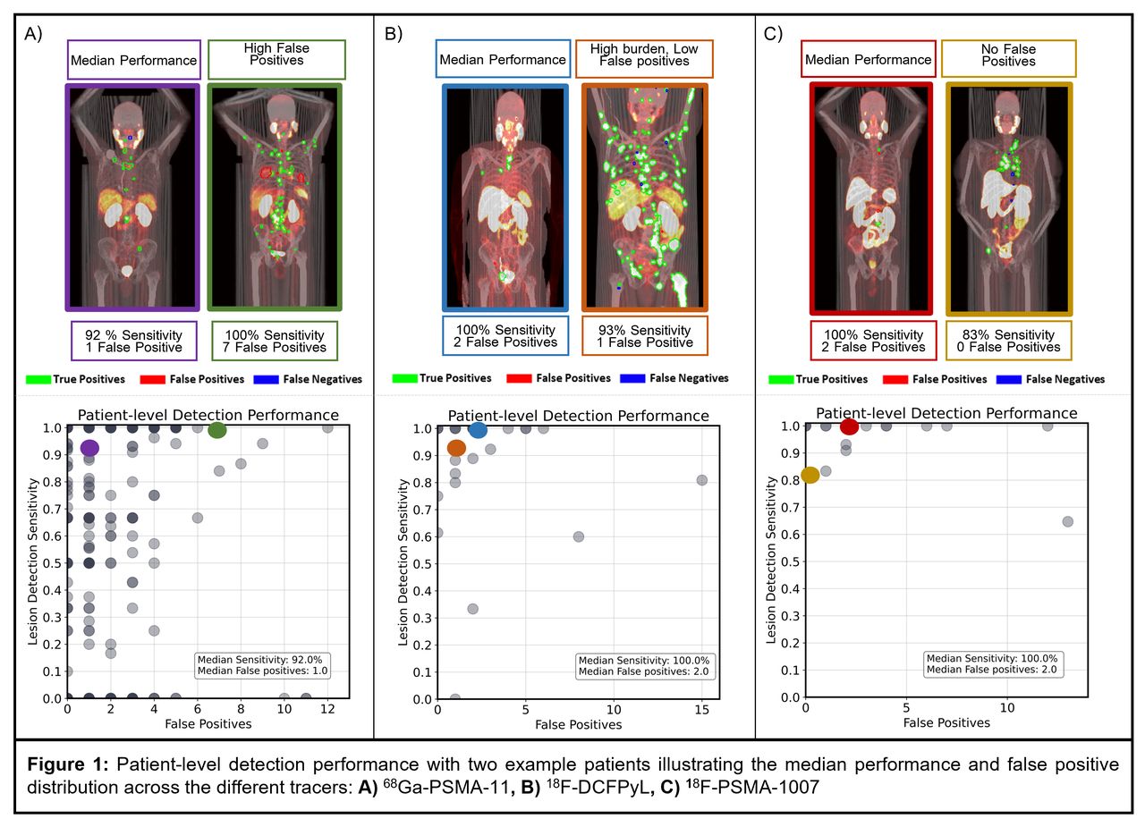
Results: In the 68Ga-PSMA-11 test images (N= 337), a total of 1499 (1-100 lesions/image) lesions were delineated. The median detection sensitivity for all lesions was 92% (IQR: 43%-100%) with 1 (0-2) FPs/image. For lesions SUVmax>2.5 g/ml (1299 lesions) the median detection sensitivity was 100% (60%-100%) with 1 (0-2) FPs/image, and for lesions SUVmax>4 g/ml (932 lesions) the median detection sensitivity was 100% (100%-100%) with 1 (0-2) FPs/image. For the 18F-DCFPyL test images (N= 27), a total of 546 (1-157 lesions /image) lesions were delineated. The median detection sensitivity for all lesions was 100% (82%-100%) with 2 (1-2.5) FPs/image, for lesions SUVmax>2.5 g/ml (514 lesions) the median detection sensitivity was 100% (85%-100%) with 2 (1-2.5) FPs/image, and for lesions SUVmax>4 g/ml (370 lesions) the median detection sensitivity was 100% (90%-100%) with 1 (0.5-2) FPs/image. Similarly, for the 18F-PSMA-1007 test images (N=18), a total of 130 (1-29 lesions /image) lesions were delineated. The median detection sensitivity for all lesions was 100% (95%-100%) with 2 (1-4) FPs/image, for lesions SUVmax>2.5 g/ml (120 lesions) the median detection sensitivity was 100% (95%-100%) with 1.5 (0-3) FPs/image, and for lesions SUVmax>4 g/ml (91 lesions) the median detection sensitivity was 100% (100%-100%) with 0 (0-1) FPs/image.
Conclusions: A CNN detection model, trained on 68Ga-PSMA-11 and 18F-DCFPyL PET/CT images, demonstrated high median sensitivity and minimal false positive rates in the 68Ga-PSMA-11, and 18F-DCFPyL external holdout datasets, and on the 18F-PSMA-1007 images. Although the 18F-PSMA-1007 dataset was small, this preliminary data shows the potential for generalizability across different PSMA tracers, including those with different biodistributions such as 18F-PSMA-1007. Investigating generalizability in larger, diverse cohorts is vital to fully understand the implications for model stability and clinical reliability. Analysis is ongoing to include more 18F-PSMA-1007 images in this study.


In this issue


Jump to section
Related Articles
Cited By...
- No citing articles found.