


Abstract
241493
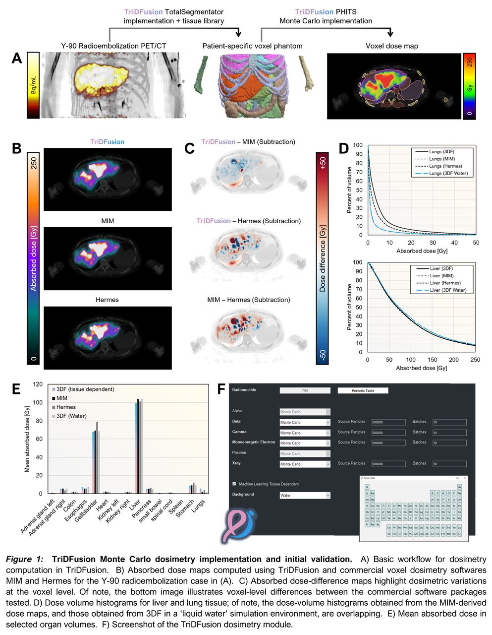
Introduction: The TriDFusion (3DF) image viewer software is evolving as a robust, open-source alternative for supporting the nuclear medicine community research infrastructure, offering a comprehensive suite of tools for image analysis. (1) In the realm of nuclear medicine and theranostics, precise patient-specific absorbed dose calculations are crucial. Tailoring interventions based on accurate dosimetry is foundational to establishing reliable dose-response relationships, minimizing the probability of over- or undertreatment, and optimizing treatment efficacy while minimizing collateral organ damage. In recognizing the need for accessible solutions, we integrated a Monte Carlo-based voxel dosimetry workflow into TriDFusion. This enhancement provides an open-source solution for dosimetry, ensuring that advanced dosimetry capabilities are accessible by the research community.
Methods: The Monte Carlo dosimetry module implemented in TriDFusion integrates machine learning-based segmentation (2), a tissue composition library, and the PHITS Monte Carlo particle transport engine (3) to calculate absorbed dose deposition at the voxel level. Transport of electrons, positrons, photons, and alpha particles is supported. Contributions from each of these source types are summed to yield the absorbed dose map. A comprehensive database of radiation energies and yields for 1252 radionuclides (4) is utilized to support dosimetry in medicine and radiation protection.
The software supports various segmentation approaches, including the AI-based TotalSegmentator TriDFusion implementation, which assigns reference material density and composition to its defined segments. Existing DICOM RT structure objects or manual contouring can also be used for segment definition. This adaptability ensures that the operator can choose the segmentation method that best aligns with their preferences and existing workflows, facilitating a user-friendly and efficient dosimetric analysis.
The initial validation of the dosimetry module was conducted by comparing absorbed dose maps and mean absorbed dose within organ contours obtained with TriDFusion; the dose maps were generated from PET scans after trans-arterial radioembolization (TARE) with both FDA-approved resin and glass 90Y microspheres for the treatment of colorectal liver metastases. With TriDFusion, dose maps were generated using two methods: 1) assigning reference tissue composition and density to regions contoured with TotalSegmentator (i.e., the default), or, 2) a ‘dose to water’ approach as a control which replicate certain vendor assumptions.
Results: TriDFusion demonstrated agreement with different vendor dosimetry software in the initial validation tests. The 90Y mean absorbed dose to liver tissue calculated with MIM or Hermes software was generally within 5% of the TriDFusion results. Graphical comparisons are provided in Figure 1.
Conclusions: The TriDFusion implementation of accurate automatic segmentation and PHITS Monte Carlo particle transport engines provides a comprehensive solution for image analysis and dosimetry support. Utilizing AI-based segmentation may improve accuracy, reduce inter-operator variability and enhance throughput. As an open-source alternative to commercial software, TriDFusion offers accessibility and increased flexibility, empowering researchers with advanced tools that can be readily available in clinical practice toward improving patient care.

In this issue


Jump to section
Related Articles
Cited By...
- No citing articles found.