


Abstract
241441
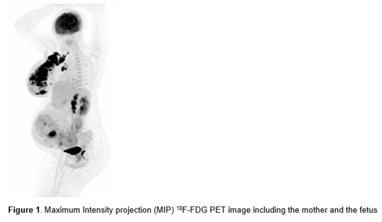
Introduction: 18F-FDG PET/CT imaging is widely used in oncology for diagnoses, staging and treatment response evaluation. However, in pregnant patients, the use of 18F-FDG PET/CT imaging can cause a dilemma because of the radiation exposure to the fetus. Fortunately, the new long-axial field-of-view (LAFOV) PET/CT systems exhibit improved sensitivity compared with conventional short-axial field-of-view (SAFOV) PET/CT systems. Therefore, the radiation burden on the fetus can be reduced by reducing the amount of injected 18F-FDG dose. With this study, we aimed to examine the fetal radiation dose when performing an 18F-FDG PET/CT scan on an LAFOV PET/CT system with reduced injected dose.
Methods: Two pregnant patients in the second and third trimester were retrospectively included. Both patients received an 18F-FDG PET/CT scan on the LAFOV PET/CT system with an intravenous bolus injection of 0.30 MBq/kg. In addition, both patients underwent a low-dose CT (LDCT) scan for attenuation correction. The fetus was included in the field-of-view (FOV) of one patient. Fetal radiation dose from radioactive 18F-FDG administration was estimated using dose conversion factors (mGy/MBq) taken from three published papers including second and third trimester pregnancies (Stabin, JNM, 2018; Takalkar et al., JNM, 2011; Zanottti-Fregonara et al., JNM, 2015). Radiation exposure to the fetus from the LDCT scans was calculated based on the computed tomography dose index (CTDI) measured in milligray (mGy) and estimated using CT-Expo (version 2.7).
Results: The fetal radiation dose conversion factors from the PET from the three studies ranged between 0.004 mGy/MBq to 0.014 mGy/MBq with a mean fetal radiation exposure of 0.008 ± 0.003 mGy/MBq. The first patient received 27.58 MBq radioactive 18F-FDG, resulting in a fetal radiation dose from the PET ranging between 0.11 - 0.39 mGy, with a mean fetal radiation dose of 0.21 ± 0.07 mGy. The fetus was not included in the FOV, resulting in a fetal radiation dose from the LDCT scan of <0.10 mGy due to scatter radiation. The total fetal radiation dose ranged between 0.11 – 0.49 mGy. The second patient received 31.08 MBq radioactive 18F-FDG, resulting in a fetal radiation dose from the PET scan ranging between 0.13 – 0.44 mGy, with a mean fetal radiation dose of 0.24 ± 0.08 mGy. The fetus was included in the FOV, resulting in an estimated fetal radiation dose from the LDCT of 0.90 mGy. Therefore, the total fetal radiation dose ranged from 1.03 mGy to 1.34 mGy.
Conclusions: We managed to scan patients with only 10% of the normal injected dose when conducting the PET/CT scan on an LAFOV PET/CT system while maintaining acceptable image quality. The ultra-low administrated radioactivity led to maximum fetal radiation exposure from the PET of 0.44 mGy. The fetal radiation dose from the LDCT was estimated at 0.90 mGy. In the future, it might be possible to reduce fetal radiation exposure even further since Mostafapour et al. (2023) found that CT radiation can be reduced by 90% when applying a tin filter, resulting in a fetal radiation exposure ranging between 0.09 – 0.11 mGy. The increased sensitivity of LAFOV PET/CT systems reduced the radiation dose to the fetus significantly and, in the future, the radiation exposure from an 18F-FDG PET/CT might be negligible for the fetus.


In this issue


Jump to section
Related Articles
Cited By...
- No citing articles found.