


Abstract
241310
Introduction: Mammography has extended in recent years to include a dual-energy approach, which is based on K-edge imaging. This technique utilizes contrast agents to increase tumor detection in women with dense breasts through weighted subtraction between the low- and high-energy images. However, dual-energy mammography (DEM) requires two sequential exposures at two different energies, leading to higher doses as well as misregistration artifacts from patient movement. On the other hand, spectral mammography, an emerging technology similar to DEM, but making use of photon-counting detectors, provides energy information via a single x-ray exposure. This system uses less radiation than DEM, without impacting image quality, and eliminates motion artifacts associated with DEM. This technique has also been shown to improve contrast-to-noise ratios (CNR) and specificity without contrast injection, resulting in better breast lesion characterization. Currently, contrast-enhanced mammography is performed using iodinated small molecules, but these agents produce sub-optimal contrast due to iodine’s relatively high K-edge. Thus, there exists a need to explore novel imaging agents that are explicitly designed for contrast-enhanced photon-counting mammography. Recently, our group identified silver sulfide nanoparticles (Ag2S-NP) to generate stronger contrast than iodine in DEM, as silver’s lower K-edge is well matched to the energies used. We therefore decided to investigate the feasibility of using Ag2S-NP as a spectral mammography specific contrast agent.
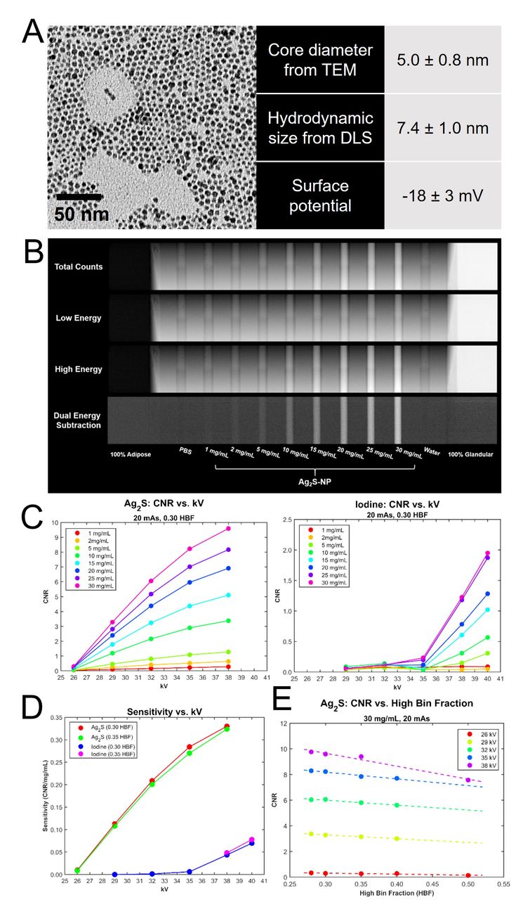
Methods: In this study, monodisperse, ultrasmall Ag2S-NP were synthesized by heating a mixture of silver nitrate and glutathione in ethylene glycol (Fig. S1) and were purified via repeated centrifugation. These nanoparticles were characterized using various analytical tools such as TEM, DLS, and zeta potential (Fig. A). The biocompatibility of Ag2S-NP was evaluated using the LIVE/DEAD assay in five cell lines after 24 hours of treatment. To evaluate the potential of Ag2S-NP as a photon-counting mammography contrast agent, a Philips MicroDose SI prototype mammography system was used to image a customized ramp phantom composed of adipose and glandular tissue-equivalent materials. The following acquisition parameters were used: 26-40 kV, 12-24 mAs, and 0.28-0.40 high bin fractions (HBF). Ag2S-NP with concentrations ranging from 1 to 30 mg Ag/ml along with PBS and water were inserted in the phantom (Fig. B).
Results: These sub-5 nm nanoparticles remain well suspended in PBS and serum, proving their robust stability under physiological conditions. Nearly 100% relative cell viability was observed even at a dose of 1 mg Ag/ml, indicating good biocompatibility (Fig. S2). Analysis of the phantom images shows a linear correlation between Ag2S-NP concentration and DE signal at each scanning condition (Fig. S3). Ag2S-NP produce significantly higher CNR than iodine over the concentrations (Fig. S4) and energies used, while iodine signal is not detectable below 35 kV (Fig. C). The imaging sensitivity of Ag2S-NP is significantly greater than that of iodine, even at energies above iodine’s K-edge (Fig. D). The optimal energy for detection of iodine is beyond the clinical mammographic energy range. Lastly, HBF calibration, which determines the threshold energy for count binning, could be further reduced to enhance the contrast of Ag2S-NP (Fig. E). Therefore, silver can be imaged at lower mammographic energies (< 35 kV) without compromising image quality, which results in radiation dose savings (Fig. S5).
Conclusions: We have synthesized Ag2S-NP that have good stability and excellent biocompatibility. When compared to iodine, these nanoparticles provide improved contrast production, dose efficiency and sensitivity in photon-counting mammography at clinically relevant beam energies, demonstrating their utility as a contrast agent for this new technique for breast cancer screening.


In this issue


Jump to section
Related Articles
Cited By...
- No citing articles found.