
Main menu

  • Home
  • Content
    • Current
    • Ahead of print
    • Past Issues
    • JNM Supplement
    • SNMMI Annual Meeting Abstracts
    • Continuing Education
    • JNM Podcasts
  • Subscriptions
    • Subscribers
    • Institutional and Non-member
    • Rates
    • Journal Claims
    • Corporate & Special Sales
  • Authors
    • Submit to JNM
    • Information for Authors
    • Assignment of Copyright
    • AQARA requirements
  • Info
    • Reviewers
    • Permissions
    • Advertisers
  • About
    • About Us
    • Editorial Board
    • Contact Information
  • More
    • Alerts
    • Feedback
    • Help
    • SNMMI Journals
  • SNMMI
    • JNM
    • JNMT
    • SNMMI Journals
    • SNMMI

User menu

  • Subscribe
  • My alerts
  • Log in
  • My Cart

Search

  • Advanced search
Journal of Nuclear Medicine
  • SNMMI
    • JNM
    • JNMT
    • SNMMI Journals
    • SNMMI
  • Subscribe
  • My alerts
  • Log in
  • My Cart
Journal of Nuclear Medicine

Advanced Search

  • Home
  • Content
    • Current
    • Ahead of print
    • Past Issues
    • JNM Supplement
    • SNMMI Annual Meeting Abstracts
    • Continuing Education
    • JNM Podcasts
  • Subscriptions
    • Subscribers
    • Institutional and Non-member
    • Rates
    • Journal Claims
    • Corporate & Special Sales
  • Authors
    • Submit to JNM
    • Information for Authors
    • Assignment of Copyright
    • AQARA requirements
  • Info
    • Reviewers
    • Permissions
    • Advertisers
  • About
    • About Us
    • Editorial Board
    • Contact Information
  • More
    • Alerts
    • Feedback
    • Help
    • SNMMI Journals
  • View or Listen to JNM Podcast
  • Visit JNM on Facebook
  • Join JNM on LinkedIn
  • Follow JNM on Twitter
  • Subscribe to our RSS feeds
Research ArticleSupplement

Advantages and Challenges of Total-Body PET/CT at a Tertiary Cancer Center: Insights from Sun Yat-sen University Cancer Center

Wanqi Chen, Yinghe Li, Zhijian Li, Yongluo Jiang, Yingpu Cui, Jiling Zeng, Yiwen Mo, Si Tang, Shatong Li, Lei Liu, Yumo Zhao, Yingying Hu and Wei Fan
Journal of Nuclear Medicine May 2024, 65 (Supplement 1) 54S-63S; DOI: https://doi.org/10.2967/jnumed.123.266948
Wanqi Chen
1State Key Laboratory of Oncology in South China, Guangdong Provincial Clinical Research Center for Cancer, Guangzhou, China;
2Department of Nuclear Medicine, Sun Yat-sen University Cancer Center, Guangzhou, China; and
  • Find this author on Google Scholar
  • Find this author on PubMed
  • Search for this author on this site
Yinghe Li
1State Key Laboratory of Oncology in South China, Guangdong Provincial Clinical Research Center for Cancer, Guangzhou, China;
2Department of Nuclear Medicine, Sun Yat-sen University Cancer Center, Guangzhou, China; and
  • Find this author on Google Scholar
  • Find this author on PubMed
  • Search for this author on this site
Zhijian Li
1State Key Laboratory of Oncology in South China, Guangdong Provincial Clinical Research Center for Cancer, Guangzhou, China;
2Department of Nuclear Medicine, Sun Yat-sen University Cancer Center, Guangzhou, China; and
  • Find this author on Google Scholar
  • Find this author on PubMed
  • Search for this author on this site
Yongluo Jiang
1State Key Laboratory of Oncology in South China, Guangdong Provincial Clinical Research Center for Cancer, Guangzhou, China;
2Department of Nuclear Medicine, Sun Yat-sen University Cancer Center, Guangzhou, China; and
  • Find this author on Google Scholar
  • Find this author on PubMed
  • Search for this author on this site
Yingpu Cui
1State Key Laboratory of Oncology in South China, Guangdong Provincial Clinical Research Center for Cancer, Guangzhou, China;
2Department of Nuclear Medicine, Sun Yat-sen University Cancer Center, Guangzhou, China; and
  • Find this author on Google Scholar
  • Find this author on PubMed
  • Search for this author on this site
Jiling Zeng
1State Key Laboratory of Oncology in South China, Guangdong Provincial Clinical Research Center for Cancer, Guangzhou, China;
2Department of Nuclear Medicine, Sun Yat-sen University Cancer Center, Guangzhou, China; and
  • Find this author on Google Scholar
  • Find this author on PubMed
  • Search for this author on this site
Yiwen Mo
1State Key Laboratory of Oncology in South China, Guangdong Provincial Clinical Research Center for Cancer, Guangzhou, China;
2Department of Nuclear Medicine, Sun Yat-sen University Cancer Center, Guangzhou, China; and
  • Find this author on Google Scholar
  • Find this author on PubMed
  • Search for this author on this site
Si Tang
1State Key Laboratory of Oncology in South China, Guangdong Provincial Clinical Research Center for Cancer, Guangzhou, China;
2Department of Nuclear Medicine, Sun Yat-sen University Cancer Center, Guangzhou, China; and
  • Find this author on Google Scholar
  • Find this author on PubMed
  • Search for this author on this site
Shatong Li
1State Key Laboratory of Oncology in South China, Guangdong Provincial Clinical Research Center for Cancer, Guangzhou, China;
2Department of Nuclear Medicine, Sun Yat-sen University Cancer Center, Guangzhou, China; and
  • Find this author on Google Scholar
  • Find this author on PubMed
  • Search for this author on this site
Lei Liu
1State Key Laboratory of Oncology in South China, Guangdong Provincial Clinical Research Center for Cancer, Guangzhou, China;
2Department of Nuclear Medicine, Sun Yat-sen University Cancer Center, Guangzhou, China; and
  • Find this author on Google Scholar
  • Find this author on PubMed
  • Search for this author on this site
Yumo Zhao
3United Imaging Healthcare Co. Ltd., Shanghai, China
  • Find this author on Google Scholar
  • Find this author on PubMed
  • Search for this author on this site
Yingying Hu
1State Key Laboratory of Oncology in South China, Guangdong Provincial Clinical Research Center for Cancer, Guangzhou, China;
2Department of Nuclear Medicine, Sun Yat-sen University Cancer Center, Guangzhou, China; and
  • Find this author on Google Scholar
  • Find this author on PubMed
  • Search for this author on this site
Wei Fan
1State Key Laboratory of Oncology in South China, Guangdong Provincial Clinical Research Center for Cancer, Guangzhou, China;
2Department of Nuclear Medicine, Sun Yat-sen University Cancer Center, Guangzhou, China; and
  • Find this author on Google Scholar
  • Find this author on PubMed
  • Search for this author on this site
  • Article
  • Figures & Data
  • Supplemental
  • Info & Metrics
  • PDF
Loading

Article Figures & Data

Figures

  • Tables
  • Additional Files
  • Figure
    • Download figure
    • Open in new tab
    • Download powerpoint
  • FIGURE 1.
    • Download figure
    • Open in new tab
    • Download powerpoint
    FIGURE 1.

    Application of total-body PET/CT at SYSUCC. (A) Disease distribution. (B) Age distribution. (C) Disease stage distribution. (D) Disease distribution in pediatric patients younger than 15 y. Others = nononcologic patients or patients who have not yet been followed up.

  • FIGURE 2.
    • Download figure
    • Open in new tab
    • Download powerpoint
    FIGURE 2.

    Selection of PET/CT devices according to patients’ medical conditions at SYSUCC.
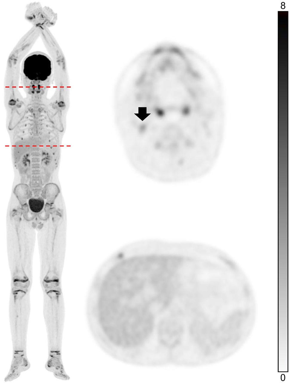
  • FIGURE 3.
    • Download figure
    • Open in new tab
    • Download powerpoint
    FIGURE 3.

    Example of total-body PET scan with one-fifth dose of 18F-FDG (0.79 MBq/kg) activity. Thirteen-year-old boy diagnosed with relapsed anaplastic lymphoma kinase-positive anaplastic large cell lymphoma underwent ninth PET/CT examination for follow-up after chemotherapy. Slight radiotracer uptake of inflammatory lymph node in right neck (arrow) is shown. Axial PET image of liver displays good background quality.
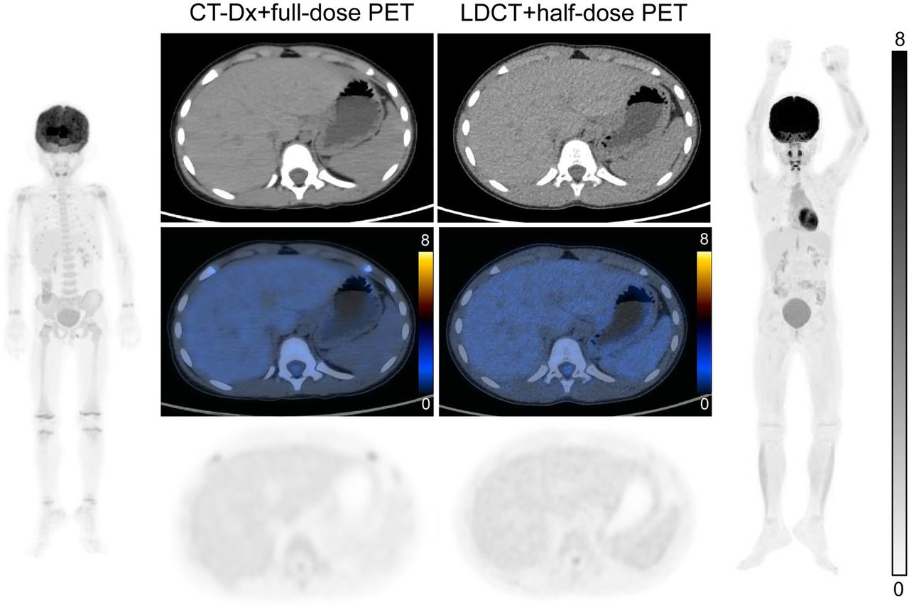
  • FIGURE 4.
    • Download figure
    • Open in new tab
    • Download powerpoint
    FIGURE 4.

    Comparison of image quality between CT-Dx and LDCT combined with different 18F-FDG injection doses. Five-year-old boy diagnosed with B-cell lymphoblastic leukemia underwent complete remission after systemic chemotherapy. Total-body PET/CT examinations were performed during 2-y follow-up. One-half dose of 18F-FDG (1.85 MBq/kg) combined with reduced CT dose was used for latter examination (right) to minimize radiation exposure. Representative CT images, axial PET images, and fused PET/CT images of liver show diagnostic and staging efficacy is not compromised. CT-Dx = diagnostic CT scan with higher x-ray dose; LDCT = low-dose CT.

  • FIGURE 5.
    • Download figure
    • Open in new tab
    • Download powerpoint
    FIGURE 5.

    Advantages of full-body coverage using 18F-FDG (3.7 MBq/kg) total-body PET/CT. (A) PET images of 32-y-old man diagnosed with right pelvic melanoma and lymph nodal metastases. Arrowheads indicate venous thrombosis within right leg veins. (B) 65-y-old man with malignant melanoma. Diagnostic accuracy of total-body PET was confirmed by extensive detection of cutaneous primary lesion and lymph node metastases throughout entire body. (C) 9-y-old girl with lymphoma revealed by biopsy of left orbital mass. Total-body PET image shows lymph, cutaneous, and bone involvement. (D) 3-y-old child diagnosed with B lymphoblastic lymphoma. Multiple masses in liver, spleen, and kidney with multisystem invasion, including lymph nodes and focal distal bones, were detected.

  • FIGURE 6.
    • Download figure
    • Open in new tab
    • Download powerpoint
    FIGURE 6.

    Example of intraindividual comparison of 18F-FDG (3.7 MBq/kg) total-body PET (left) and conventional PET (right) images in lung cancer patient with liver and bone metastases. Axial PET images of total-body PET scans revealed 4 lesions in liver (arrows) that were barely recognizable in conventional PET/CT scans, because radiotracer uptake of liver lesions was similar to physiologic liver background.

  • FIGURE 7.
    • Download figure
    • Open in new tab
    • Download powerpoint
    FIGURE 7.

    Example of retroperitoneal small lymph nodes posing challenges for diagnosis. Fifty-five-year-old woman with surgically removed cervical cancer was referred for 18F-FDG (3.7 MBq/kg) total-body PET/CT scan. Axial PET images and fused PET/CT images display paraaortic small lymph nodes (arrowheads) with higher 18F-FDG uptake (SUVmax, 6.5) than physiologic background.

Tables

  • Figures
  • Additional Files
    • View popup
    TABLE 1.

    Key Parameters of Total-Body PET/CT Acquisition Protocols at SYSUCC

    CT-AC parametersCT-Dx parametersReconstruction parameters of PET
    AgeProtocolDose (MBq/kg)Time (min)Tube voltage (kV)Fixed tube (mAs)Tube voltage (kV)Ref.mAs of TCM (mAs)Rotation time (s)IterationsSubsetsMatrixFOV (mm)
    >20 y, torso or TB routineTorso routine3.70512010120800.5320256 × 256600
    Torso NPC3.7031205120800.5220256 × 256600
    TB routine1.858NANA1201200.5320256 × 256600
    TB NPC1.856NANA1201200.5320256 × 256600
    ≤20 y, TB routine only0- to 4-y nonlymphoma1.8510NANA100750.3320512 × 512600
    0- to 4-y lymphoma1.8510NANA100700.3320512 × 512600
    0- to 4-y lymphoma follow-up1.8510NANA100550.3320512 × 512600
    4- to 7-y nonlymphoma1.8510NANA100800.3320512 × 512600
    4- to 7-y lymphoma1.8510NANA100700.3320512 × 512600
    4- to 7-y lymphoma follow-up1.8510NANA100600.3320512 × 512600
    7- to 15-y nonlymphoma1.8510NANA120600.3320512 × 512600
    7- to 15-y lymphoma1.8510NANA120550.3320512 × 512600
    7- to 15-y lymphoma follow-up1.8510NANA120500.3320512 × 512600
    15- to 20-y nonlymphoma3.706NANA120650.5320360 × 360600
    15- to 20-y lymphoma3.706NANA120600.5320360 × 360600
    15- to 20-y lymphoma follow-up3.706NANA120550.5320360 × 360600
    >45-kg nonlymphoma3.70/1.856/10NANA120600.5320256 × 256600
    >45-kg lymphoma3.70/1.856/10NANA120500.5320256 × 256600
    >45-kg lymphoma follow-up3.70/1.856/10NANA120400.5320256 × 256600
    • CT-AC = low-dose CT scan for attenuation correction; CT-Dx = diagnostic CT scan with higher x-ray dose; Ref.mAs = reference milliampere·seconds; TCM = tube current modulation; FOV = field of view; TB = total body; NA = not applicable.

    • For patients >45 kg, injection dose is based on age (1.85 MBq/kg applied for pediatric patients younger than 15 y and 3.70 MBq/kg for patients from 15- to 20-y old).

    • View popup
    TABLE 2.

    Clinical Studies Exploring Low-Dose Injection Activity of 18F-FDG Using Total-Body PET/CT

    YearAuthorPatients (n)PopulationDiseaseInjection doseConclusion
    2022Chen et al. (12)100Pediatric patientsOncologyHalf dose (1.85 MBq/kg)Sufficient image quality and lesion conspicuity could be maintained at fast acquisition time of 1 min with half-dose activity of 18F-FDG
    2022He et al. (17)46AdultsOncologyHalf dose (1.85 MBq/kg)Half dose of 18F-FDG with acquisition times ≥ 5 min could be applied in clinical practice
    2021Tan et al. (13)56AdultsLung cancerHalf dose (1.85 MBq/kg)Total-body PET/CT with half dose of 18F-FDG in 2 and 4 min achieved comparable image quality to conventional PET/CT
    2022Hu et al. (18)30AdultsOncologyUltralow dose (0.37 MBq/kg)Ultralow 18F-FDG activity with 8-min acquisition in total-body PET/CT can achieve acceptable image quality equivalent to that in full-activity group after 2-min acquisition
    2022Abdelhafez et al. (54)30AdultsAIAUltralow dose (78.1 ± 4.7 MBq)Systemic joint evaluation in AIA (and non-AIA) is feasible with total-body PET/CT and ultra-low-dose 18F-FDG protocol
    2022Tan et al. (55)62AdultsCRCUltralow dose (0.37 MBq/kg)Total-body PET/CT with ultralow dose of 18F-FDG can maintain satisfactory image quality and lesion detectability in CRC
    2023Tan et al. (56)30AdultsOncologyUltralow dose (0.37 MBq/kg) vs. half dose (1.85 MBq/kg)Total-body PET/CT with ultra-low-dose activity of 18F-FDG, corresponding acquisition time of 8 min, provides acceptable image quality and lesion detection
    • AIA = autoimmune inflammatory arthritis; CRC = colorectal cancer.

Additional Files

  • Figures
  • Tables
  • Supplemental Data

    Files in this Data Supplement:

    • Supplemental Table 1
PreviousNext
Back to top

In this issue

Journal of Nuclear Medicine: 65 (Supplement 1)
Journal of Nuclear Medicine
Vol. 65, Issue Supplement 1
May 1, 2024
  • Table of Contents
  • Table of Contents (PDF)
  • About the Cover
  • Index by author
  • Complete Issue (PDF)
Print
Download PDF
Article Alerts
Sign In to Email Alerts with your Email Address
Email Article

Thank you for your interest in spreading the word on Journal of Nuclear Medicine.

NOTE: We only request your email address so that the person you are recommending the page to knows that you wanted them to see it, and that it is not junk mail. We do not capture any email address.

Enter multiple addresses on separate lines or separate them with commas.
Advantages and Challenges of Total-Body PET/CT at a Tertiary Cancer Center: Insights from Sun Yat-sen University Cancer Center
(Your Name) has sent you a message from Journal of Nuclear Medicine
(Your Name) thought you would like to see the Journal of Nuclear Medicine web site.
Citation Tools
Advantages and Challenges of Total-Body PET/CT at a Tertiary Cancer Center: Insights from Sun Yat-sen University Cancer Center
Wanqi Chen, Yinghe Li, Zhijian Li, Yongluo Jiang, Yingpu Cui, Jiling Zeng, Yiwen Mo, Si Tang, Shatong Li, Lei Liu, Yumo Zhao, Yingying Hu, Wei Fan
Journal of Nuclear Medicine May 2024, 65 (Supplement 1) 54S-63S; DOI: 10.2967/jnumed.123.266948

Citation Manager Formats

  • BibTeX
  • Bookends
  • EasyBib
  • EndNote (tagged)
  • EndNote 8 (xml)
  • Medlars
  • Mendeley
  • Papers
  • RefWorks Tagged
  • Ref Manager
  • RIS
  • Zotero
Share
Advantages and Challenges of Total-Body PET/CT at a Tertiary Cancer Center: Insights from Sun Yat-sen University Cancer Center
Wanqi Chen, Yinghe Li, Zhijian Li, Yongluo Jiang, Yingpu Cui, Jiling Zeng, Yiwen Mo, Si Tang, Shatong Li, Lei Liu, Yumo Zhao, Yingying Hu, Wei Fan
Journal of Nuclear Medicine May 2024, 65 (Supplement 1) 54S-63S; DOI: 10.2967/jnumed.123.266948
Twitter logo Facebook logo LinkedIn logo Mendeley logo
  • Tweet Widget
  • Facebook Like
  • Google Plus One
Bookmark this article

Jump to section

  • Article
    • Visual Abstract
    • Abstract
    • APPLICATION OF TOTAL-BODY PET/CT IN SYSUCC
    • SCANNING PROTOCOLS OF TOTAL-BODY PET/CT IN SYSUCC
    • SELECTION OF PET/CT SCANNERS ACCORDING TO PATIENTS’ MEDICAL CONDITIONS
    • ADVANTAGES AND APPLICATIONS OF TOTAL-BODY PET SCANNING
    • CHALLENGES OF TOTAL-BODY PET/CT
    • SUMMARY
    • DISCLOSURE
    • ACKNOWLEDGMENTS
    • REFERENCES
  • Figures & Data
  • Supplemental
  • Info & Metrics
  • PDF

Related Articles

  • PubMed
  • Google Scholar

Cited By...

  • Advancing Global Nuclear Medicine: The Role and Future Contributions of China
  • Google Scholar

More in this TOC Section

  • Is the Amyloid Imaging Tsunami Really Happening?
  • Imaging and Fluid Biomarkers of Alzheimer Disease: Complementation Rather Than Competition
  • A New Dawn
Show more Supplement

Similar Articles

Keywords

  • total-body PET/CT
  • oncology
  • application
  • imaging protocol
  • pediatric
SNMMI

© 2025 SNMMI

Powered by HighWire