
Main menu

  • Home
  • Content
    • Current
    • Ahead of print
    • Past Issues
    • JNM Supplement
    • SNMMI Annual Meeting Abstracts
    • Continuing Education
    • JNM Podcasts
  • Subscriptions
    • Subscribers
    • Institutional and Non-member
    • Rates
    • Journal Claims
    • Corporate & Special Sales
  • Authors
    • Submit to JNM
    • Information for Authors
    • Assignment of Copyright
    • AQARA requirements
  • Info
    • Reviewers
    • Permissions
    • Advertisers
  • About
    • About Us
    • Editorial Board
    • Contact Information
  • More
    • Alerts
    • Feedback
    • Help
    • SNMMI Journals
  • SNMMI
    • JNM
    • JNMT
    • SNMMI Journals
    • SNMMI

User menu

  • Subscribe
  • My alerts
  • Log in
  • My Cart

Search

  • Advanced search
Journal of Nuclear Medicine
  • SNMMI
    • JNM
    • JNMT
    • SNMMI Journals
    • SNMMI
  • Subscribe
  • My alerts
  • Log in
  • My Cart
Journal of Nuclear Medicine

Advanced Search

  • Home
  • Content
    • Current
    • Ahead of print
    • Past Issues
    • JNM Supplement
    • SNMMI Annual Meeting Abstracts
    • Continuing Education
    • JNM Podcasts
  • Subscriptions
    • Subscribers
    • Institutional and Non-member
    • Rates
    • Journal Claims
    • Corporate & Special Sales
  • Authors
    • Submit to JNM
    • Information for Authors
    • Assignment of Copyright
    • AQARA requirements
  • Info
    • Reviewers
    • Permissions
    • Advertisers
  • About
    • About Us
    • Editorial Board
    • Contact Information
  • More
    • Alerts
    • Feedback
    • Help
    • SNMMI Journals
  • View or Listen to JNM Podcast
  • Visit JNM on Facebook
  • Join JNM on LinkedIn
  • Follow JNM on Twitter
  • Subscribe to our RSS feeds
Research ArticleClinical Investigation

Theranostic Intratumoral Convection-Enhanced Delivery of 124I-Omburtamab in Patients with Diffuse Intrinsic Pontine Glioma: Pharmacokinetics and Lesion Dosimetry

Neeta Pandit-Taskar, Pat B. Zanzonico, Milan Grkovski, Maria Donzelli, Scott M. Vietri, Christopher Horan, Brian Serencsits, Kavya Prasad, Serge Lyashchenko, Kim Kramer, Ira J. Dunkel and Mark M. Souweidane
Journal of Nuclear Medicine September 2024, 65 (9) 1364-1370; DOI: https://doi.org/10.2967/jnumed.123.266365
Neeta Pandit-Taskar
1Department of Radiology, Memorial Sloan Kettering Cancer Center, New York, New York;
2Department of Radiology, Weill Cornell Medical College, New York, New York;
  • Find this author on Google Scholar
  • Find this author on PubMed
  • Search for this author on this site
Pat B. Zanzonico
3Department of Medical Physics, Memorial Sloan Kettering Cancer Center, New York, New York;
  • Find this author on Google Scholar
  • Find this author on PubMed
  • Search for this author on this site
Milan Grkovski
3Department of Medical Physics, Memorial Sloan Kettering Cancer Center, New York, New York;
  • Find this author on Google Scholar
  • Find this author on PubMed
  • Search for this author on this site
Maria Donzelli
4Department of Pediatrics, Memorial Sloan Kettering Cancer Center, New York, New York;
  • Find this author on Google Scholar
  • Find this author on PubMed
  • Search for this author on this site
Scott M. Vietri
1Department of Radiology, Memorial Sloan Kettering Cancer Center, New York, New York;
5Radiochemistry & Molecular Imaging Probe Core Facility, Memorial Sloan Kettering Cancer Center, New York, New York;
  • Find this author on Google Scholar
  • Find this author on PubMed
  • Search for this author on this site
Christopher Horan
3Department of Medical Physics, Memorial Sloan Kettering Cancer Center, New York, New York;
  • Find this author on Google Scholar
  • Find this author on PubMed
  • Search for this author on this site
Brian Serencsits
3Department of Medical Physics, Memorial Sloan Kettering Cancer Center, New York, New York;
  • Find this author on Google Scholar
  • Find this author on PubMed
  • Search for this author on this site
Kavya Prasad
3Department of Medical Physics, Memorial Sloan Kettering Cancer Center, New York, New York;
  • Find this author on Google Scholar
  • Find this author on PubMed
  • Search for this author on this site
Serge Lyashchenko
5Radiochemistry & Molecular Imaging Probe Core Facility, Memorial Sloan Kettering Cancer Center, New York, New York;
6Molecular Pharmacology and Chemistry Program, Memorial Sloan Kettering Cancer Center, New York, New York;
  • Find this author on Google Scholar
  • Find this author on PubMed
  • Search for this author on this site
Kim Kramer
4Department of Pediatrics, Memorial Sloan Kettering Cancer Center, New York, New York;
7Department of Pediatrics, Weill Cornell Medical College, New York, New York;
  • Find this author on Google Scholar
  • Find this author on PubMed
  • Search for this author on this site
Ira J. Dunkel
4Department of Pediatrics, Memorial Sloan Kettering Cancer Center, New York, New York;
7Department of Pediatrics, Weill Cornell Medical College, New York, New York;
  • Find this author on Google Scholar
  • Find this author on PubMed
  • Search for this author on this site
Mark M. Souweidane
8Department of Neurosurgery, Memorial Sloan Kettering Cancer Center, New York, New York; and
9Department of Neurological Surgery, Weill Cornell Medical College, New York, New York
  • Find this author on Google Scholar
  • Find this author on PubMed
  • Search for this author on this site
  • Article
  • Figures & Data
  • Supplemental
  • Info & Metrics
  • PDF
Loading

Article Figures & Data

Figures

  • Tables
  • Additional Files
  • Figure
    • Download figure
    • Open in new tab
    • Download powerpoint
  • FIGURE 1.
    • Download figure
    • Open in new tab
    • Download powerpoint
    FIGURE 1.

    (A) Whole-body clearance curve for all cohorts. (B) Whole-body clearance curves by cohort. (C) Blood clearance curve of 124I-omburtamab displayed in aggregate decay-corrected mean values for all patients. (D) Blood clearance curves for patients by cohort. %ID/L = percentage injected dose per liter.

  • FIGURE 2.
    • Download figure
    • Open in new tab
    • Download powerpoint
    FIGURE 2.

    124I-omburtamab lesion and whole-body distribution in 8-y-old female patient. Baseline MRI showing 1.3 × 1.2 cm eccentric mass centered within left lateral pons extending to left cerebral peduncle. 124I-omburtamab infusion activity was 25.9 MBq. Imaging was performed on day of infusion (D0) and on days 2 (D2), 3 (D3), 6 (D6), and 8 (D8) with whole-body (A) and head imaging (B). High activity is noted at tumor site until last imaging time point (D8). Low activity systemic distribution is noted on D2 as excreted activity in kidneys and bladder, thyroid and stomach, and liver that decreased from D3 to D8.

  • FIGURE 3.
    • Download figure
    • Open in new tab
    • Download powerpoint
    FIGURE 3.

    (A) Lesion uptake and radiation-absorbed dose of 124I-omburtamab show high concentration within tumor. (B) Radiation-absorbed dose in lesion increases with increased activity over cohorts. (C) Lesion–to–whole-body ratio of radiation-absorbed dose is high, highlighting safe margin and high therapeutic index with local administration.
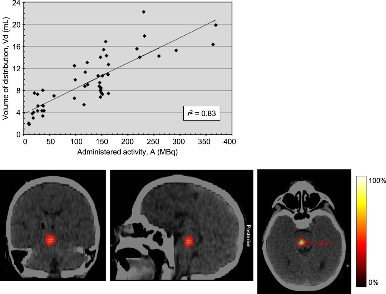
  • FIGURE 4.
    • Download figure
    • Open in new tab
    • Download powerpoint
    FIGURE 4.

    Distribution volume of administered activity. Linear regression between Vd (in mL) and administered activity (in MBq) of 124I-omburtamab, with slope of 0.046 mL/MBq, ordinate intercept of 3.8 mL, and correlation coefficient of 0.83. Regression is highly statistically significant (P < 0.0005), as determined by ANOVA using F statistic.

  • FIGURE 5.
    • Download figure
    • Open in new tab
    • Download powerpoint
    FIGURE 5.

    Example of imaging in 8-y-old patient. Baseline MRI showing 3.5 × 2.8 cm mass centered in pons extending to right cerebral peduncle and brachium pontis. 124I-omburtamab infusion activity was 148 MBq. Imaging was performed on day of infusion (D0), day 4 (D4), and day 6 (D6), with whole-body and head imaging. High activity is noted at site of tumor until last imaging time point (D6). Infusion time was 10 h. First imaging time point was 16 h after initiation. D1 scan shows activity in spinal canal and systemic activity in liver, stomach, and bladder. Further systemic distribution is noted on D2 as excreted activity in kidneys and bladder, thyroid and stomach, and liver that decreased from D4 to D6.

Tables

  • Figures
  • Additional Files
    • View popup
    TABLE 1.

    Patient Demographics and Administered-Activity Cohorts

    CohortAdministered activity level (MBq)*Number of patientsAge range (y)Administered activity range (MBq)*Maximum lesion size (cm)Volume infused (mL)
    19.25 (0.25)33–88.88–9.99 (0.24–0.27)3.1–3.30.24–0.27
    218.5 (0.5)35–6; 1715.9–20.0 (0.43–0.54)3.1–5.50.47–0.63
    327.75 (0.75)33–725.9–27.0 (0.70–0.73)1.3–4.20.69–0.77
    437 (1.0)43–7; 16, 1734.8–38.9 (0.94–1.05)2.3–3.20.99–1.05
    592.5 (2.5)35–6; 1796.9–98.4 (2.62–2.66)4.1–4.22.56–2.61
    6120.25 (3.25)34–7117.7–122.1 (3.18–3.3)3.0–4.13.42–3.57
    7148 (4.0)143–11142.5–158.4 (3.85–4.28)2.1–5.03.84–4.54
    8222 (6)63–8; 12, 16217.9–243.46 (5.87–6.58)2.1–4.64.18–8.56
    9296 (8)33, 11, 18281.2–288.6 (7.6–7.8)2.2–3.66.17–8.11
    10370 (10)25, 10362.6–370.7 (9.83–10.02)3.6,3.99.71–11.95
    • ↵* Activity in mCi given in parentheses.

    • View popup
    TABLE 2.

    Normal-Organ Radiation-Absorbed Dose

    Organ or tissueAbsorbed dose (mGy/MBq)
    MeanSDMinimumMaximum
    Adrenal glands0.380.220.060.98
    Brain4.112.091.049.38
    Breasts0.250.140.030.66
    Gallbladder wall0.430.250.071.01
    Lower large intestine wall0.370.230.030.97
    Small intestine0.380.230.040.97
    Stomach wall1.140.930.083.72
    Upper large intestine wall0.380.230.040.96
    Heart wall0.450.240.101.02
    Kidneys0.370.220.061.11
    Liver0.920.770.093.17
    Lungs0.280.140.100.76
    Muscle0.340.180.060.84
    Ovaries0.390.240.030.98
    Pancreas0.450.250.061.09
    Red marrow0.390.170.110.90
    Osteogenic cells0.600.260.191.36
    Skin0.290.140.070.71
    Spleen0.370.240.041.28
    Testes0.320.200.020.89
    Thymus0.330.180.060.85
    Thyroid1.151.120.137.14
    Urinary bladder wall2.012.420.0812.10
    Uterus0.470.300.041.49
    Total body0.580.250.201.22
    Effective dose (mSv/MBq)0.690.280.261.59

Additional Files

  • Figures
  • Tables
  • Supplemental Data

    Files in this Data Supplement:

    • Supplemental Data
PreviousNext
Back to top

In this issue

Journal of Nuclear Medicine: 65 (9)
Journal of Nuclear Medicine
Vol. 65, Issue 9
September 1, 2024
  • Table of Contents
  • Table of Contents (PDF)
  • About the Cover
  • Index by author
  • Complete Issue (PDF)
Print
Download PDF
Article Alerts
Sign In to Email Alerts with your Email Address
Email Article

Thank you for your interest in spreading the word on Journal of Nuclear Medicine.

NOTE: We only request your email address so that the person you are recommending the page to knows that you wanted them to see it, and that it is not junk mail. We do not capture any email address.

Enter multiple addresses on separate lines or separate them with commas.
Theranostic Intratumoral Convection-Enhanced Delivery of 124I-Omburtamab in Patients with Diffuse Intrinsic Pontine Glioma: Pharmacokinetics and Lesion Dosimetry
(Your Name) has sent you a message from Journal of Nuclear Medicine
(Your Name) thought you would like to see the Journal of Nuclear Medicine web site.
Citation Tools
Theranostic Intratumoral Convection-Enhanced Delivery of 124I-Omburtamab in Patients with Diffuse Intrinsic Pontine Glioma: Pharmacokinetics and Lesion Dosimetry
Neeta Pandit-Taskar, Pat B. Zanzonico, Milan Grkovski, Maria Donzelli, Scott M. Vietri, Christopher Horan, Brian Serencsits, Kavya Prasad, Serge Lyashchenko, Kim Kramer, Ira J. Dunkel, Mark M. Souweidane
Journal of Nuclear Medicine Sep 2024, 65 (9) 1364-1370; DOI: 10.2967/jnumed.123.266365

Citation Manager Formats

  • BibTeX
  • Bookends
  • EasyBib
  • EndNote (tagged)
  • EndNote 8 (xml)
  • Medlars
  • Mendeley
  • Papers
  • RefWorks Tagged
  • Ref Manager
  • RIS
  • Zotero
Share
Theranostic Intratumoral Convection-Enhanced Delivery of 124I-Omburtamab in Patients with Diffuse Intrinsic Pontine Glioma: Pharmacokinetics and Lesion Dosimetry
Neeta Pandit-Taskar, Pat B. Zanzonico, Milan Grkovski, Maria Donzelli, Scott M. Vietri, Christopher Horan, Brian Serencsits, Kavya Prasad, Serge Lyashchenko, Kim Kramer, Ira J. Dunkel, Mark M. Souweidane
Journal of Nuclear Medicine Sep 2024, 65 (9) 1364-1370; DOI: 10.2967/jnumed.123.266365
Twitter logo Facebook logo LinkedIn logo Mendeley logo
  • Tweet Widget
  • Facebook Like
  • Google Plus One
Bookmark this article

Jump to section

  • Article
    • Visual Abstract
    • Abstract
    • MATERIALS AND METHODS
    • RESULTS
    • DISCUSSION
    • CONCLUSION
    • DISCLOSURE
    • Footnotes
    • REFERENCES
  • Figures & Data
  • Supplemental
  • Info & Metrics
  • PDF

Related Articles

  • PubMed
  • Google Scholar

Cited By...

  • No citing articles found.
  • Google Scholar

More in this TOC Section

  • SNMMI Procedure Standard/EANM Practice Guideline for Brain [18F]FDG PET Imaging, Version 2.0
  • Meeting Upcoming Clinical and Diagnostic Needs in Oncologic Imaging: A Structured Reporting System for Fibroblast-Activation-Protein–Targeted Imaging—FAP-RADS Version 1.0
  • Imaging Efficacy of [18F]CTT1057 PET for the Detection of PSMA-Positive Tumors Using Histopathology as Standard of Truth: Results from the GuideView Phase 2/3 Prospective Multicenter Study
Show more Clinical Investigation

Similar Articles

Keywords

  • 124I-omburtamab
  • DIPG
  • diffuse intrinsic pontine glioma
  • convection-enhanced delivery
  • dosimetry
  • organ-absorbed doses
SNMMI

© 2025 SNMMI

Powered by HighWire