
Main menu

  • Home
  • Content
    • Current
    • Ahead of print
    • Past Issues
    • JNM Supplement
    • SNMMI Annual Meeting Abstracts
    • Continuing Education
    • JNM Podcasts
  • Subscriptions
    • Subscribers
    • Institutional and Non-member
    • Rates
    • Journal Claims
    • Corporate & Special Sales
  • Authors
    • Submit to JNM
    • Information for Authors
    • Assignment of Copyright
    • AQARA requirements
  • Info
    • Reviewers
    • Permissions
    • Advertisers
  • About
    • About Us
    • Editorial Board
    • Contact Information
  • More
    • Alerts
    • Feedback
    • Help
    • SNMMI Journals
  • SNMMI
    • JNM
    • JNMT
    • SNMMI Journals
    • SNMMI

User menu

  • Subscribe
  • My alerts
  • Log in
  • Log out
  • My Cart

Search

  • Advanced search
Journal of Nuclear Medicine
  • SNMMI
    • JNM
    • JNMT
    • SNMMI Journals
    • SNMMI
  • Subscribe
  • My alerts
  • Log in
  • Log out
  • My Cart
Journal of Nuclear Medicine

Advanced Search

  • Home
  • Content
    • Current
    • Ahead of print
    • Past Issues
    • JNM Supplement
    • SNMMI Annual Meeting Abstracts
    • Continuing Education
    • JNM Podcasts
  • Subscriptions
    • Subscribers
    • Institutional and Non-member
    • Rates
    • Journal Claims
    • Corporate & Special Sales
  • Authors
    • Submit to JNM
    • Information for Authors
    • Assignment of Copyright
    • AQARA requirements
  • Info
    • Reviewers
    • Permissions
    • Advertisers
  • About
    • About Us
    • Editorial Board
    • Contact Information
  • More
    • Alerts
    • Feedback
    • Help
    • SNMMI Journals
  • View or Listen to JNM Podcast
  • Visit JNM on Facebook
  • Join JNM on LinkedIn
  • Follow JNM on Twitter
  • Subscribe to our RSS feeds
Research ArticleClinical Investigation

Diagnostic Potential of Supplemental Static and Dynamic 68Ga-FAPI-46 PET for Primary 18F-FDG–Negative Pulmonary Lesions

Manuel Röhrich, Johanna Daum, Ewgenija Gutjahr, Anna-Maria Spektor, Frederik M. Glatting, Yasemin Aylin Sahin, Hans Georg Buchholz, Jorge Hoppner, Cathrin Schroeter, Eleni Mavriopoulou, Kai Schlamp, Matthias Grott, Florian Eichhorn, Claus Peter Heußel, Hans Ulrich Kauczor, Michael Kreuter, Frederik Giesel, Mathias Schreckenberger, Hauke Winter and Uwe Haberkorn
Journal of Nuclear Medicine June 2024, 65 (6) 872-879; DOI: https://doi.org/10.2967/jnumed.123.267103
Manuel Röhrich
1Department of Nuclear Medicine, University Hospital Heidelberg, Heidelberg, Germany;
2Department of Nuclear Medicine, University Hospital Mainz, Mainz, Germany;
3German Center of Lung Research, Heidelberg, Germany;
  • Find this author on Google Scholar
  • Find this author on PubMed
  • Search for this author on this site
Johanna Daum
1Department of Nuclear Medicine, University Hospital Heidelberg, Heidelberg, Germany;
3German Center of Lung Research, Heidelberg, Germany;
  • Find this author on Google Scholar
  • Find this author on PubMed
  • Search for this author on this site
Ewgenija Gutjahr
4Institute of Pathology, University Hospital Heidelberg, Heidelberg, Germany;
  • Find this author on Google Scholar
  • Find this author on PubMed
  • Search for this author on this site
Anna-Maria Spektor
1Department of Nuclear Medicine, University Hospital Heidelberg, Heidelberg, Germany;
3German Center of Lung Research, Heidelberg, Germany;
  • Find this author on Google Scholar
  • Find this author on PubMed
  • Search for this author on this site
Frederik M. Glatting
1Department of Nuclear Medicine, University Hospital Heidelberg, Heidelberg, Germany;
5Clinical Cooperation Unit Molecular and Radiation Oncology, German Cancer Research Center, Heidelberg, Germany;
6Department of Radiation Oncology, University Hospital Heidelberg, Heidelberg, Germany;
  • Find this author on Google Scholar
  • Find this author on PubMed
  • Search for this author on this site
Yasemin Aylin Sahin
2Department of Nuclear Medicine, University Hospital Mainz, Mainz, Germany;
  • Find this author on Google Scholar
  • Find this author on PubMed
  • Search for this author on this site
Hans Georg Buchholz
2Department of Nuclear Medicine, University Hospital Mainz, Mainz, Germany;
  • Find this author on Google Scholar
  • Find this author on PubMed
  • Search for this author on this site
Jorge Hoppner
1Department of Nuclear Medicine, University Hospital Heidelberg, Heidelberg, Germany;
3German Center of Lung Research, Heidelberg, Germany;
  • Find this author on Google Scholar
  • Find this author on PubMed
  • Search for this author on this site
Cathrin Schroeter
1Department of Nuclear Medicine, University Hospital Heidelberg, Heidelberg, Germany;
3German Center of Lung Research, Heidelberg, Germany;
  • Find this author on Google Scholar
  • Find this author on PubMed
  • Search for this author on this site
Eleni Mavriopoulou
1Department of Nuclear Medicine, University Hospital Heidelberg, Heidelberg, Germany;
3German Center of Lung Research, Heidelberg, Germany;
  • Find this author on Google Scholar
  • Find this author on PubMed
  • Search for this author on this site
Kai Schlamp
3German Center of Lung Research, Heidelberg, Germany;
7Department of Radiology, Thoraxklinik, University Hospital Heidelberg, Heidelberg, Germany;
  • Find this author on Google Scholar
  • Find this author on PubMed
  • Search for this author on this site
Matthias Grott
3German Center of Lung Research, Heidelberg, Germany;
8Department of Thoracic Surgery, Thoraxklinik, University Hospital Heidelberg, Heidelberg, Germany;
  • Find this author on Google Scholar
  • Find this author on PubMed
  • Search for this author on this site
Florian Eichhorn
3German Center of Lung Research, Heidelberg, Germany;
8Department of Thoracic Surgery, Thoraxklinik, University Hospital Heidelberg, Heidelberg, Germany;
  • Find this author on Google Scholar
  • Find this author on PubMed
  • Search for this author on this site
Claus Peter Heußel
3German Center of Lung Research, Heidelberg, Germany;
7Department of Radiology, Thoraxklinik, University Hospital Heidelberg, Heidelberg, Germany;
  • Find this author on Google Scholar
  • Find this author on PubMed
  • Search for this author on this site
Hans Ulrich Kauczor
3German Center of Lung Research, Heidelberg, Germany;
9Department of Diagnostic and Interventional Radiology, University Hospital Heidelberg, Heidelberg, Germany;
10Center for Interstitial and Rare Lung Diseases, Pneumology, and Respiratory Critical Care Medicine, Thoraxklinik, University of Heidelberg, Heidelberg, Germany;
  • Find this author on Google Scholar
  • Find this author on PubMed
  • Search for this author on this site
Michael Kreuter
11Department of Pneumology, Mainz Center for Pulmonary Medicine, Mainz University, Mainz, Germany;
12Medical Center and Department of Pulmonary, Critical Care, and Sleep Medicine, Marienhaus Clinic Mainz, Mainz, Germany;
  • Find this author on Google Scholar
  • Find this author on PubMed
  • Search for this author on this site
Frederik Giesel
1Department of Nuclear Medicine, University Hospital Heidelberg, Heidelberg, Germany;
3German Center of Lung Research, Heidelberg, Germany;
13Department of Nuclear Medicine, Medical Faculty, University Hospital Düsseldorf, Heinrich Heine University, Düsseldorf, Germany;
14Institute for Radiation Sciences, Osaka University, Osaka, Japan;
15German Cancer Consortium, Heidelberg, Germany; and
  • Find this author on Google Scholar
  • Find this author on PubMed
  • Search for this author on this site
Mathias Schreckenberger
2Department of Nuclear Medicine, University Hospital Mainz, Mainz, Germany;
  • Find this author on Google Scholar
  • Find this author on PubMed
  • Search for this author on this site
Hauke Winter
3German Center of Lung Research, Heidelberg, Germany;
7Department of Radiology, Thoraxklinik, University Hospital Heidelberg, Heidelberg, Germany;
  • Find this author on Google Scholar
  • Find this author on PubMed
  • Search for this author on this site
Uwe Haberkorn
1Department of Nuclear Medicine, University Hospital Heidelberg, Heidelberg, Germany;
3German Center of Lung Research, Heidelberg, Germany;
9Department of Diagnostic and Interventional Radiology, University Hospital Heidelberg, Heidelberg, Germany;
16Clinical Cooperation Unit Nuclear Medicine, German Cancer Research Center, Heidelberg, Germany
  • Find this author on Google Scholar
  • Find this author on PubMed
  • Search for this author on this site
  • Article
  • Figures & Data
  • Supplemental
  • Info & Metrics
  • PDF
Loading

Article Figures & Data

Figures

  • Tables
  • Additional Files
  • Figure
    • Download figure
    • Open in new tab
    • Download powerpoint
  • FIGURE 1.
    • Download figure
    • Open in new tab
    • Download powerpoint
    FIGURE 1.

    FAP expression in lepidic LC. (A and B) Representative hematoxylin and eosin staining (left) and immunohistochemical staining against FAP (right) of central part of lepidic LC biopsy, which shows strong stromal FAP positivity (A), and tumor front of lepidic LC, showing transition from FAP-positive LC tumor rim into FAP-negative physiologic lung tissue (B) (magnification: upper rows, ×10; lower rows, ×40) (scale bars: upper rows, 100 μm; lower rows, 20 μm).
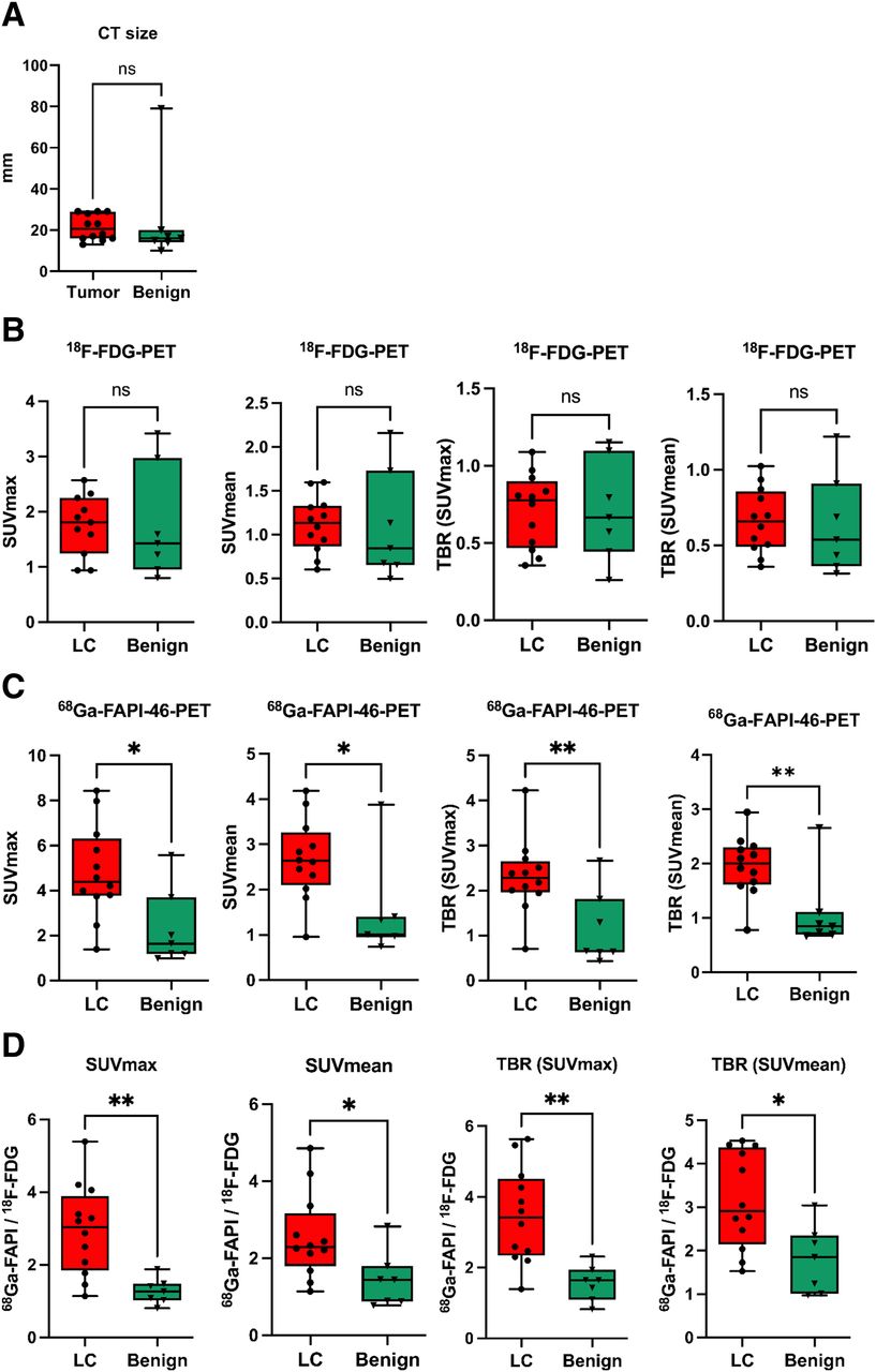
  • FIGURE 2.
    • Download figure
    • Open in new tab
    • Download powerpoint
    FIGURE 2.

    Quantitative analysis of 18F-FDG and 68Ga-FAPI-46 uptake in LC and benign pulmonary lesions of 19 patients. (A–C) Box plots of SUVmax, SUVmean, and their corresponding TBRs against mediastinal blood pool for LC and benign pulmonary lesions calculated for 18F-FDG (A) and 68Ga-FAPI-46 (B) and fold changes of all parameters calculated for ratio of 68Ga-FAPI-46 to18F-FDG (C). Boxes represent interquartile range, whiskers represent interquartile range of 1.5, and horizontal line within box indicates median. Data outliers are shown separately within graph. *P < 0.05. **P < 0.01. ns = not significant.
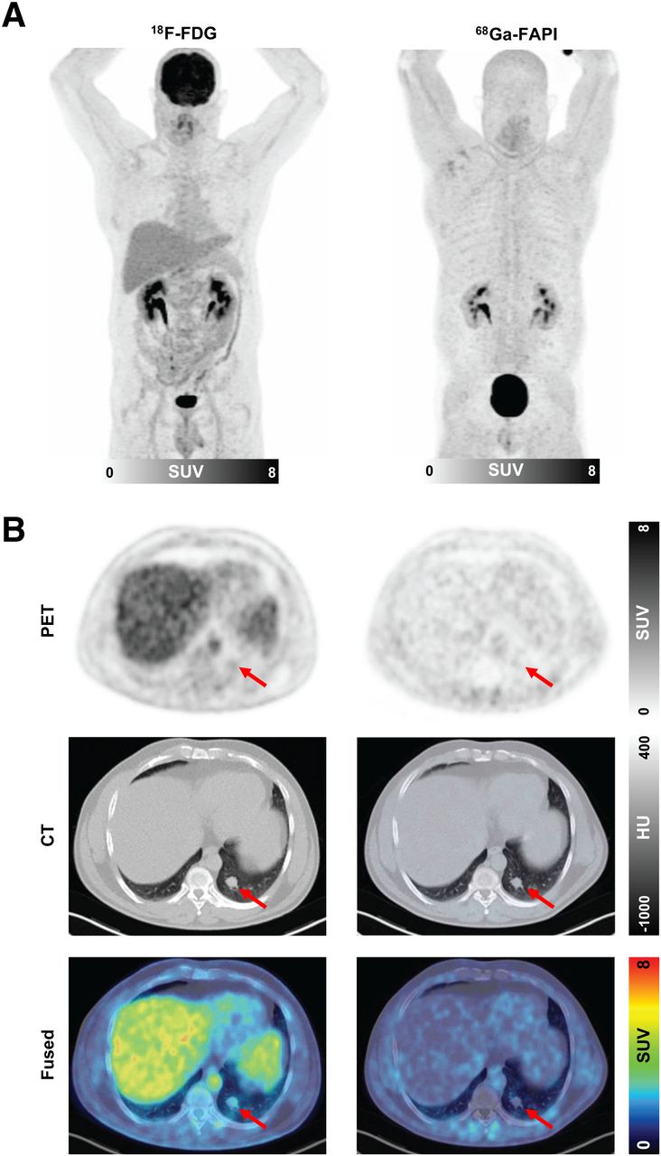
  • FIGURE 3.
    • Download figure
    • Open in new tab
    • Download powerpoint
    FIGURE 3.

    Example 18F-FDG and 68Ga-FAPI-46 images of 51-y-old woman with adenocarcinoma with lepidic growth pattern in right upper lobe. (A) Maximum-intensity-projection PET images. (B) Axial images of suggestive lesion (red arrows) with low CT density in right lower lobe. Green arrows show blood pool in aortic arch. Lesion had 18F-FDG uptake below blood pool niveau but was strongly 68Ga-FAPI-46–positive. CT-guided biopsy led to pathologic diagnosis of adenocarcinoma, and patient was treated by stereotactic body radiation therapy because of functional inoperability. HU = Hounsfield units.

  • FIGURE 4.
    • Download figure
    • Open in new tab
    • Download powerpoint
    FIGURE 4.

    Example 18F-FDG and 68Ga-FAPI-46 images of 41-y-old man with hamartoma in left lower lobe. (A) Maximum-intensity-projection PET images. (B) Axial images of suggestive lesion (arrows) in left lower lobe. After wedge resection, hamartoma was diagnosed by pathology. HU = Hounsfield units.

  • FIGURE 5.
    • Download figure
    • Open in new tab
    • Download powerpoint
    FIGURE 5.

    Dynamic 68Ga-FAPI-46 PET imaging properties of LC and benign pulmonary lesions. (A) Averaged time–activity curves (relative to peak) of LC and benign pulmonary lesions. (B) Box plot of time to peak and slope of LC and benign pulmonary lesions. Boxes represent interquartile range, whiskers represent interquartile range of 1.5, and horizontal line within box indicates median. (C) Representative cases: 63-y-old man with adenocarcinoma (arrow) of left upper lobe and 62-y-old man with focally 68Ga-FAPI-46–avid sarcoid mass (encircled) in right lower lobe. Images are from static PET, and time–activity curves are from dynamic PET. Although both lesions show intermediate 68Ga-FAPI-46 uptake, time–activity curves clearly differ, with delayed peak of LC and markedly pronounced slope of sarcoidosis. *P < 0.05.

  • FIGURE 6.
    • Download figure
    • Open in new tab
    • Download powerpoint
    FIGURE 6.

    Receiver-operating-characteristic (ROC) curves of 4 quantitative PET parameters with highest discriminatory power: 68Ga-FAPI-46/18F-FDG SUVmax (A), 68Ga-FAPI-46/18F-FDG TBR SUVmax (B), SUVmean TBR (C), and SUVmax TBR (D).

Tables

  • Figures
  • Additional Files
    • View popup
    TABLE 1.

    Clinical Parameters and Diagnoses of 19 Patients with 18F-FDG–Negative Pulmonary Lesions

    Patient no.Age (y)SexDynamic PET imagingLocalizationLargest diameter (mm)Histologic confirmationDiagnosisGrowth patternTNM stage
    164MNoRight upper lobe20Wedge resectionTuberculosisNot applicableNot applicable
    273MYesLeft hilus14Bronchoscopy with biopsyCalcified lymph nodeNot applicableNot applicable
    368FYesRight lower lobe29LobectomyAdenocarcinomaLepidicT1cN0M0
    458MYesRight upper lobe10Wedge resectionGranulomaNot applicableNot applicable
    575MYesLeft lower lobe16LobectomyAdenocarcinomaAcinar/lepidicT1bN0M0
    670FYesLeft lower lobe23LobectomyAdenocarcinomaAcinarT1cN0M0
    762MYesRight lower lobe79LobectomySarcoidosisNot applicableNot applicable
    850MYesLeft upper lobe13CT-guided biopsyAdenocarcinomaAcinarT1bN0M0
    957FYesRight lower lobe17LobectomyAdenocarcinomaAcinarT1cN0M0
    1063MYesRight upper lobe15CT-guided biopsyAdenocarcinomaAcinarT1bN1M0
    1141MNoLeft lower lobe17Wedge resectionHamartomaNot applicableNot applicable
    1245MYesRight lower lobe16EnucleationHamartomaNot applicableNot applicable
    1370MYesRight upper lobe16LobectomyAdenocarcinomaAcinarT2aN0M0
    1472MYesLeft upper lobe23LobectomyAdenocarcinomaAcinarT1bN0M0
    1565MYesRight upper lobe29LobectomyAdenocarcinomaLepidicT1cN0M0
    1650MNoRight lower lobe18CT-guided biopsyTypical carcinoidNot applicableT1bN0M0
    1751FYesRight upper lobe29CT-guided biopsyAdenocarcinomaLepidic/acinarT2aN0M0
    1864FYesLeft upper lobe28Segment resectionAdenocarcinomaLepidicT1aN0M0
    1977MYesLeft lower lobe15CT-guided biopsyLung tissueNot applicableNot applicable

Additional Files

  • Figures
  • Tables
  • Supplemental Data

    Files in this Data Supplement:

    • Supplemental Data
PreviousNext
Back to top

In this issue

Journal of Nuclear Medicine: 65 (6)
Journal of Nuclear Medicine
Vol. 65, Issue 6
June 1, 2024
  • Table of Contents
  • Table of Contents (PDF)
  • About the Cover
  • Index by author
  • Complete Issue (PDF)
Print
Download PDF
Article Alerts
Sign In to Email Alerts with your Email Address
Email Article

Thank you for your interest in spreading the word on Journal of Nuclear Medicine.

NOTE: We only request your email address so that the person you are recommending the page to knows that you wanted them to see it, and that it is not junk mail. We do not capture any email address.

Enter multiple addresses on separate lines or separate them with commas.
Diagnostic Potential of Supplemental Static and Dynamic 68Ga-FAPI-46 PET for Primary 18F-FDG–Negative Pulmonary Lesions
(Your Name) has sent you a message from Journal of Nuclear Medicine
(Your Name) thought you would like to see the Journal of Nuclear Medicine web site.
Citation Tools
Diagnostic Potential of Supplemental Static and Dynamic 68Ga-FAPI-46 PET for Primary 18F-FDG–Negative Pulmonary Lesions
Manuel Röhrich, Johanna Daum, Ewgenija Gutjahr, Anna-Maria Spektor, Frederik M. Glatting, Yasemin Aylin Sahin, Hans Georg Buchholz, Jorge Hoppner, Cathrin Schroeter, Eleni Mavriopoulou, Kai Schlamp, Matthias Grott, Florian Eichhorn, Claus Peter Heußel, Hans Ulrich Kauczor, Michael Kreuter, Frederik Giesel, Mathias Schreckenberger, Hauke Winter, Uwe Haberkorn
Journal of Nuclear Medicine Jun 2024, 65 (6) 872-879; DOI: 10.2967/jnumed.123.267103

Citation Manager Formats

  • BibTeX
  • Bookends
  • EasyBib
  • EndNote (tagged)
  • EndNote 8 (xml)
  • Medlars
  • Mendeley
  • Papers
  • RefWorks Tagged
  • Ref Manager
  • RIS
  • Zotero
Share
Diagnostic Potential of Supplemental Static and Dynamic 68Ga-FAPI-46 PET for Primary 18F-FDG–Negative Pulmonary Lesions
Manuel Röhrich, Johanna Daum, Ewgenija Gutjahr, Anna-Maria Spektor, Frederik M. Glatting, Yasemin Aylin Sahin, Hans Georg Buchholz, Jorge Hoppner, Cathrin Schroeter, Eleni Mavriopoulou, Kai Schlamp, Matthias Grott, Florian Eichhorn, Claus Peter Heußel, Hans Ulrich Kauczor, Michael Kreuter, Frederik Giesel, Mathias Schreckenberger, Hauke Winter, Uwe Haberkorn
Journal of Nuclear Medicine Jun 2024, 65 (6) 872-879; DOI: 10.2967/jnumed.123.267103
Twitter logo Facebook logo LinkedIn logo Mendeley logo
  • Tweet Widget
  • Facebook Like
  • Google Plus One
Bookmark this article

Jump to section

  • Article
    • Visual Abstract
    • Abstract
    • MATERIALS AND METHODS
    • RESULTS
    • DISCUSSION
    • CONCLUSION
    • DISCLOSURE
    • Footnotes
    • REFERENCES
  • Figures & Data
  • Supplemental
  • Info & Metrics
  • PDF

Related Articles

  • PubMed
  • Google Scholar

Cited By...

  • SNMMI Procedure Standard/EANM Practice Guideline for Fibroblast Activation Protein (FAP) PET
  • Google Scholar

More in this TOC Section

  • Detection of HER2-Low Lesions Using HER2-Targeted PET Imaging in Patients with Metastatic Breast Cancer: A Paired HER2 PET and Tumor Biopsy Analysis
  • First-in-Human Study of 18F-Labeled PET Tracer for Glutamate AMPA Receptor [18F]K-40: A Derivative of [11C]K-2
  • [11C]Carfentanil PET Whole-Body Imaging of μ-Opioid Receptors: A First in-Human Study
Show more Clinical Investigation

Similar Articles

Keywords

  • fibroblast activation protein
  • FAPI
  • PET
  • lung cancer
  • pulmonary lesions
SNMMI

© 2025 SNMMI

Powered by HighWire