


Article Figures & Data
Figures
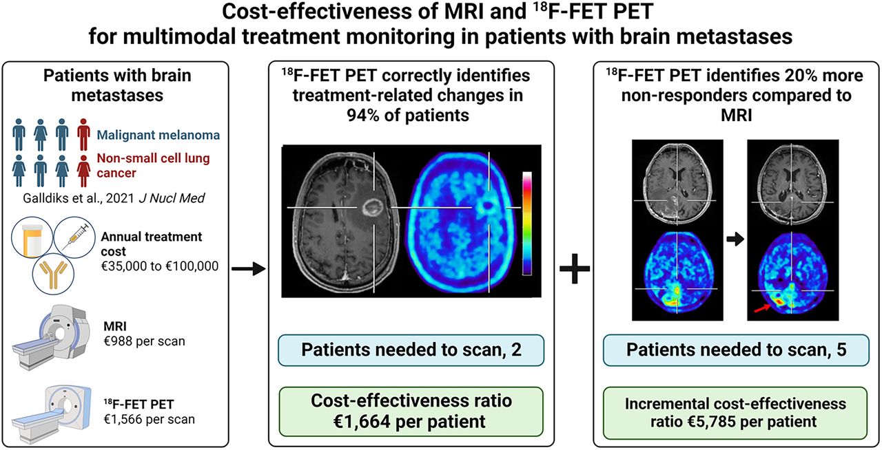
- FIGURE 1.
Model 1 (upper panel): Decision tree for assessing effectiveness of additional 18F-FET PET for differentiating treatment-related changes (TRC) from brain metastasis (BM) relapse after multimodal therapy. Twenty-seven patients underwent 18F-FET PET. N1 divides patients into those diagnosed with brain metastasis relapse or treatment-related changes according to 18F-FET PET criteria (i.e., mean tumor-to-brain ratio of more or less than 1.95, respectively). N2 and N3 assign both groups to patients’ outcomes based on both clinical course during subsequent follow-up and either neuropathologic diagnosis or Response Assessment in Neuro-Oncology criteria for immunotherapy. Model 2 (lower panel): Decision tree model for assessing effectiveness of 18F-FET PET and MRI to identify nonresponder to multimodal therapy based on stable clinical course for <6 mo. Eleven patients underwent serial 18F-FET PET and MRI. N1 and N2 represent chance nodes to be responder or nonresponder according to 18F-FET PET and MRI criteria (i.e., relative reduction or increase in mean tumor-to-background ratio of 10% for 18F-FET PET, respectively; Response Assessment in Neuro-Oncology criteria for immunotherapy for MRI). N3–N6 divide each of 4 groups of 18F-FET PET and MRI responders (and nonresponders) into true and false responders (and nonresponders), respectively.
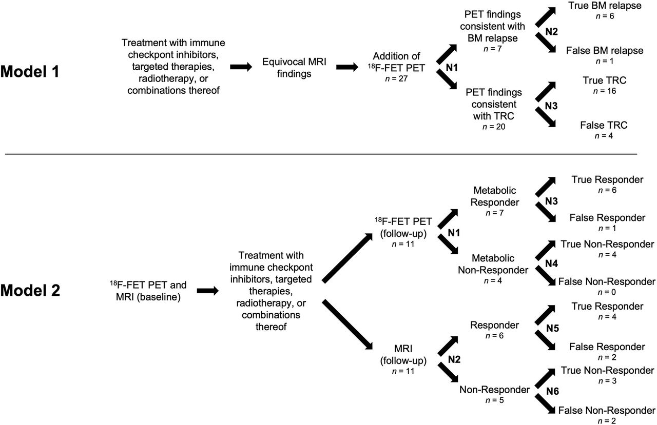
- FIGURE 2.
Tornado diagrams of cost-effectiveness ratio of additional 18F-FET PET scans for identification of treatment-related changes (upper panel) and ICER of 18F-FET PET for identification of nonresponder (lower panel) after multimodal therapy. Cost-effectiveness ratios and ICERs were calculated by applying upper and lower interval values, as shown in Table 1, onto N1–N3 and N1–N6, respectively.
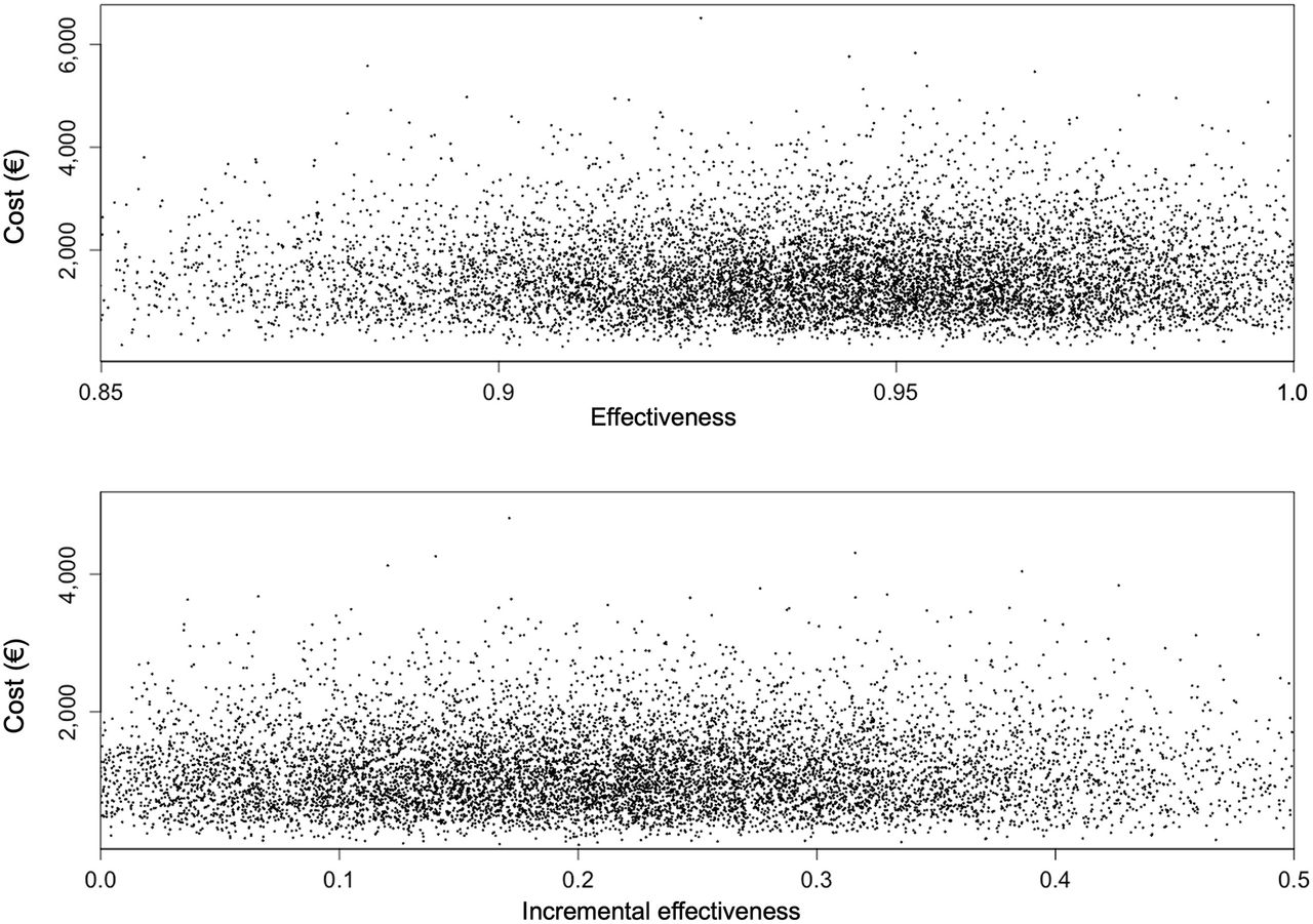
- FIGURE 3.
Distribution of results from Monte Carlo analysis (dots) about effectiveness of additional 18F-FET PET for identification of treatment-related changes (upper panel) and IE of 18F-FET PET for identification of nonresponder (lower panel) after multimodal therapy. Note different scaling of axes. Margin values for effectiveness (upper panel, values < 0.85 and > 1.0, 5.0% of values; lower panel, values < 0 and > 0.5, 7.4% of values) are not shown.


Tables
- TABLE 1.
Chance Node Intervals and Corresponding Effectiveness and CER in 1-Way Deterministic Sensitivity Analysis for Decision Tree Models 1 and 2
Decision tree model 1 (identification of treatment-related changes) Decision tree model 2 (identification of nonresponder) Chance node Parameter Lower interval Upper interval Lower interval Upper interval N1 Value (%) 20.4 31.4 58.1 69.1 Effectiveness (%) 95.6 92.4 23.4 15.8 CER (€) 1,638.13 1,694.51 4,935.10 7,343.11 N2 Value (%) 76.2 95.2 49.0 60.0 Effectiveness (%) 90.6 97.9 14.8 25.5 CER (€) 1,729.33 1,599.13 7,795.63 4,537.22 N3 Value (%) 71.5 88.5 76.2 95.2 Effectiveness (%) 93.5 94.7 10.6 32.3 CER (€) 1,675.86 1,654.82 10,906.40 3,585.31 N4 Value (%) 91.5 108.5* Effectiveness (%) NA NA 18.5 21.3* CER (€) 6,240.62 5,439.00* N5 Value (%) 64.7 68.7 Effectiveness (%) NA NA 21.4 18.5 CER (€) 5,405.24 6,246.08 N6 Value (%) 58.0 62.0 Effectiveness (%) NA NA 20.8 19.2 CER (€) 5,558.43 6,021.43 ↵* Theoretic result, because value for N4 is >100%.
NA = not applicable.
Effectiveness and CER were based on indicated chance node values. Decision tree model 1 comprises only N1–N3. Values for effectiveness and CER of decision tree model 2 correspond to incremental values (difference in 18F-FET PET and MRI).
Decision tree model 1 (identification of treatment-related changes) Decision tree model 2 (identification of nonresponder) Chance node Calculated value (%) SD (%) Calculated value (%) SD (%) N1 25.9 5 63.6 5 N2 85.7 8 54.5 5 N3 80.0 8 85.7 8 N4 NA NA 100.0 8 N5 NA NA 66.7 8 N6 NA NA 60.0 8 NA = not applicable.
Calculated values for chance nodes were taken from decision tree models, and SDs were set similar to earlier study (5). Decision tree model 1 comprises only N1–N3.
- TABLE 3.
Statistics Resulting from Monte Carlo Analysis (10,000 Samples) for Effectiveness
Decision tree model 1 (identification of treatment-related changes) Decision tree model 2 (identification of nonresponder) Value or percentile 18F-FET PET (%) Cost for 18F-FET PET (€) MRI (%) 18F-FET PET (%) IE (%) Difference in cost 18F-FET PET − MRI (€) Mean 94.1 1,551.39 60.1 80.7 20.6 1,146.03 SD 3.6 777.79 8.2 10.3 574.56 Minimum 76.7 89.00 28.5 45.3 16.9 65.74 2.5th 86.3 440.92 44.2 61.6 17.4 325.71 10th 89.5 685.46 49.5 68.3 18.8 506.36 Median 94.4 1,420.00 60.0 80.2 20.2 1,048.97 90th 98.4 2,585.29 70.5 93.5 23.0 1,909.78 97.5th 100.5* 3,441.53 76.4 102.7* 26.3* 2,542.28 Maximum 109.2* 6,515.79 98.5 150.6* 52.1* 4,813.26 ↵* Theoretic result, because calculated value for effectiveness of 18F-FET PET is >100%.
Columns for decision tree model 1 indicate, first, probability of correctly detecting treatment-related changes after treatment with immune checkpoint inhibition, targeted therapy, radiotherapy, or combinations thereof by 18F-FET PET alone and, second, γ-distributed cost for 1 18F-FET PET scan. Columns for decision tree model 2 indicate probability of correctly detecting nonresponder to immune checkpoint inhibition, targeted therapy, radiotherapy, or combinations thereof by MRI or 18F-FET PET, respectively. Column IE indicates their difference and thus IE in using 18F-FET PET. Rightmost column indicates γ-distributed difference in cost between 2 serial 18F-FET PET and MRI scans.
In this issue


Jump to section
Related Articles
Cited By...
- No citing articles found.