


Abstract
T26
Introduction: Whole-body dynamic PET images are acquired using multiple continuous bed motion (CBM) methods. Previous studies have reported the clinical usefulness of whole-body dynamic PET imaging, but the optimization of image quality of this PET acquisition method has not been evaluated yet. In this study, we evaluated the image quality of whole-body dynamic PET imaging using a phantom and clinical data to optimize image reconstruction parameters.
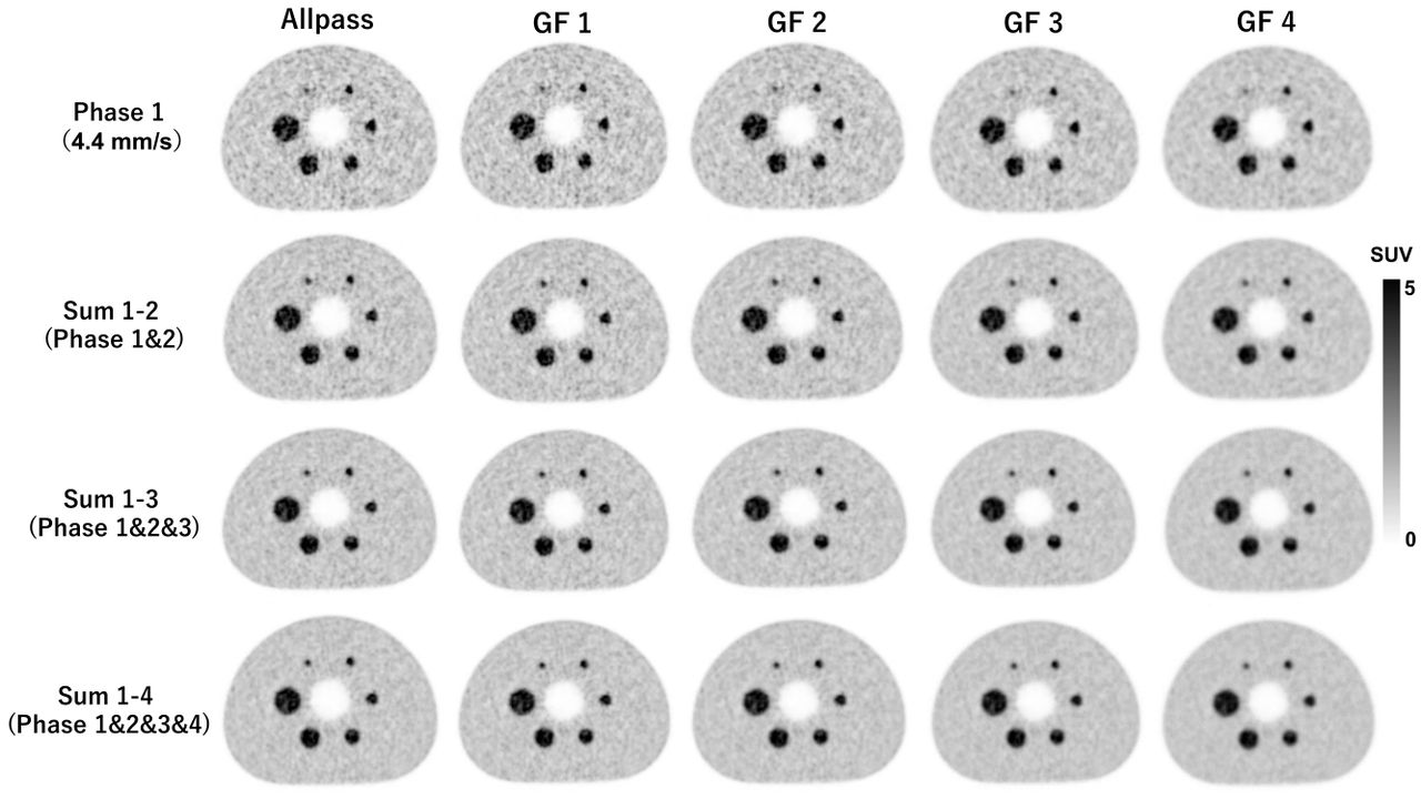
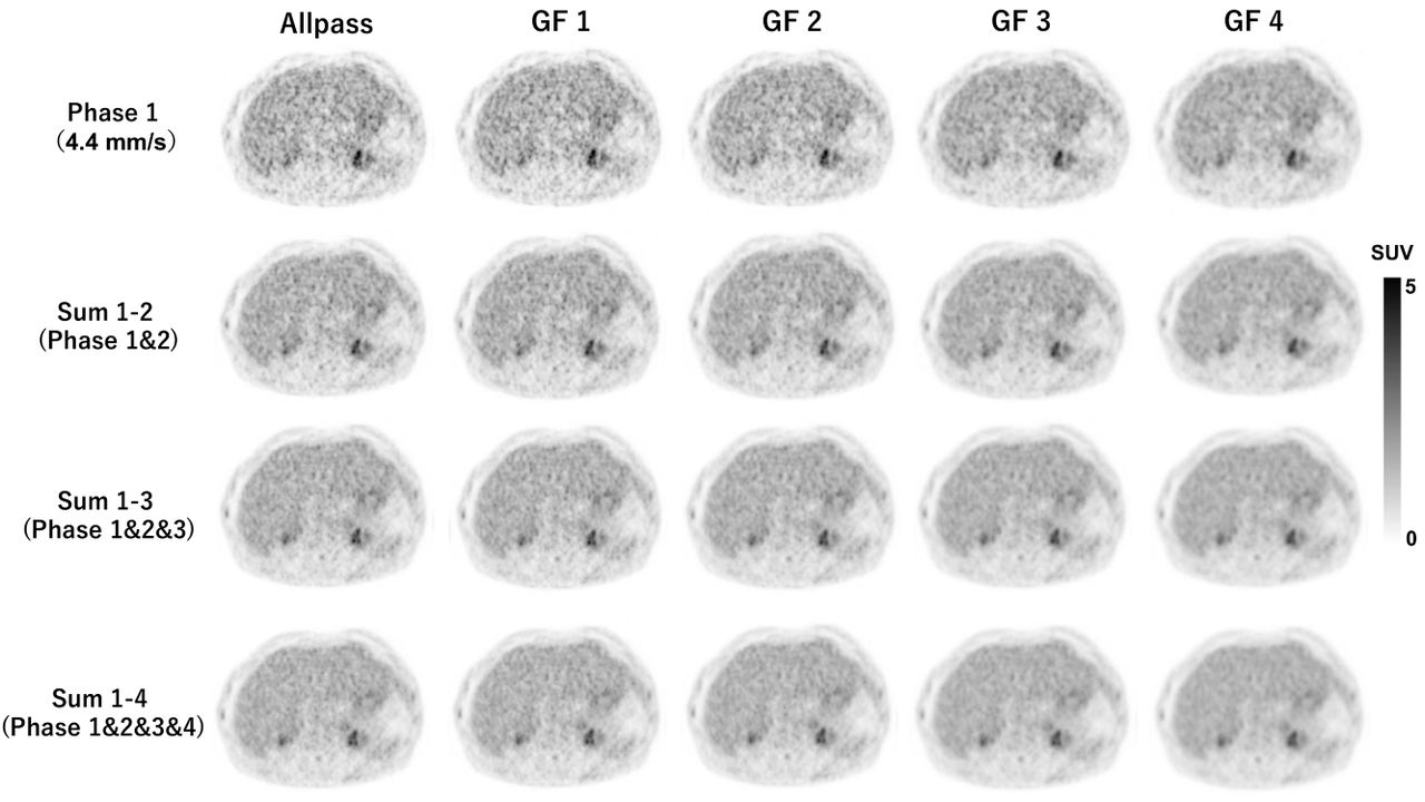
Methods: PET images were acquired with a bed speed of 4.4 mm/s using multiple CBM methods. In phantom tests, acquired PET images were reconstructed using the ordered-subsets expectation maximization (OSEM, iteration 4 and subset 5) + point-spread function (PSF) + time of flight (TOF) algorithms and successively superimposed. As a result, the following PET images were obtained according to the number of piled up images: Sum 1-2 (Phases 1 and 2), Sum 1-3 (Phases 1 to 3), and Sum 1-4 (Phases 1 to 4). The Gaussian filter (GF) was varied from 1 to 4 mm in full width at half maximum (FWHM). Image quality of phantom images were evaluated using image noise level (CVBG) and contrast for 10 mm hot sphere. In clinical study, the PET image data of 12 consecutive patients with no hepatic lesions were retrospectively analyzed. The image quality was evaluated using hepatic signal-to-noise ratio (liver SNR). This clinical study was approved by the institutional review board.
Results: In phantom studies, CVBG was smaller than 10.0% by using 4 mm of GF for Sum 1-3 and 3 mm of GF for Sum 1-4. Four mm of GF reduced contrast no more than 14% for all superimposed images. In the clinical studies, liver SNR exceeded 10.0 by using 4 mm of GF for Sum 1-2 and 3 mm of GF for Sum 1-3 and Sum 1-4.
Conclusions: The optimization of GF according to the number of superimposed images provided PET images suitable for clinical diagnosis in whole-body dynamic PET tests.


In this issue


Jump to section
Related Articles
Cited By...
- No citing articles found.