
Main menu

  • Home
  • Content
    • Current
    • Ahead of print
    • Past Issues
    • JNM Supplement
    • SNMMI Annual Meeting Abstracts
    • Continuing Education
    • JNM Podcasts
  • Subscriptions
    • Subscribers
    • Institutional and Non-member
    • Rates
    • Journal Claims
    • Corporate & Special Sales
  • Authors
    • Submit to JNM
    • Information for Authors
    • Assignment of Copyright
    • AQARA requirements
  • Info
    • Reviewers
    • Permissions
    • Advertisers
  • About
    • About Us
    • Editorial Board
    • Contact Information
  • More
    • Alerts
    • Feedback
    • Help
    • SNMMI Journals
  • SNMMI
    • JNM
    • JNMT
    • SNMMI Journals
    • SNMMI

User menu

  • Subscribe
  • My alerts
  • Log in
  • Log out
  • My Cart

Search

  • Advanced search
Journal of Nuclear Medicine
  • SNMMI
    • JNM
    • JNMT
    • SNMMI Journals
    • SNMMI
  • Subscribe
  • My alerts
  • Log in
  • Log out
  • My Cart
Journal of Nuclear Medicine

Advanced Search

  • Home
  • Content
    • Current
    • Ahead of print
    • Past Issues
    • JNM Supplement
    • SNMMI Annual Meeting Abstracts
    • Continuing Education
    • JNM Podcasts
  • Subscriptions
    • Subscribers
    • Institutional and Non-member
    • Rates
    • Journal Claims
    • Corporate & Special Sales
  • Authors
    • Submit to JNM
    • Information for Authors
    • Assignment of Copyright
    • AQARA requirements
  • Info
    • Reviewers
    • Permissions
    • Advertisers
  • About
    • About Us
    • Editorial Board
    • Contact Information
  • More
    • Alerts
    • Feedback
    • Help
    • SNMMI Journals
  • View or Listen to JNM Podcast
  • Visit JNM on Facebook
  • Join JNM on LinkedIn
  • Follow JNM on Twitter
  • Subscribe to our RSS feeds
Meeting ReportOncology: Clinical Therapy & Diagnosis (includes Phase 2, Phase 3, post approval studies) - GU

Sestamibi quantitative evaluation of renal masses

Taylor Standiford, Spencer Bell, Robert Wang, Alexander Kutikov, Jared Schober, Mohan Doss, Yi Li and Jian (Michael) Yu
Journal of Nuclear Medicine June 2023, 64 (supplement 1) P984;
Taylor Standiford
1Temple University Hospital
  • Find this author on Google Scholar
  • Find this author on PubMed
  • Search for this author on this site
Spencer Bell
2Fox Chase Cancer Center - Temple Health
  • Find this author on Google Scholar
  • Find this author on PubMed
  • Search for this author on this site
Robert Wang
3Fox Chase Cancer Center
  • Find this author on Google Scholar
  • Find this author on PubMed
  • Search for this author on this site
Alexander Kutikov
3Fox Chase Cancer Center
  • Find this author on Google Scholar
  • Find this author on PubMed
  • Search for this author on this site
Jared Schober
3Fox Chase Cancer Center
  • Find this author on Google Scholar
  • Find this author on PubMed
  • Search for this author on this site
Mohan Doss
3Fox Chase Cancer Center
  • Find this author on Google Scholar
  • Find this author on PubMed
  • Search for this author on this site
Yi Li
3Fox Chase Cancer Center
  • Find this author on Google Scholar
  • Find this author on PubMed
  • Search for this author on this site
Jian (Michael) Yu
3Fox Chase Cancer Center
  • Find this author on Google Scholar
  • Find this author on PubMed
  • Search for this author on this site
  • Article
  • Figures & Data
  • Info & Metrics
Loading

Abstract

P984

Introduction: The differentiation of renal cell carcinoma (RCC) from oncocytoma is challenging with the current imaging techniques. By result, resection has traditionally been the treatment of choice for patients presenting with solid renal masses concerning for malignancy. Recently, 99m Tc-sestamibi SPECT/CT imaging has been employed in the work-up of solid renal masses with the hope of reducing unnecessary procedures. The ideal target to background ratio used to differentiate oncocytomas from renal cell carcinomas remains unknown. We report our institutional quantitative analysis of 99m Sestamibi SPECT/CT Imaging in conjunction with disease confirmation through tissue diagnosis.

Methods: 99m Tc-sestamibi SPECT/CT scans were performed on patients with one or more renal masses detected on prior CT or MR imaging. Using MIM software (a nuclear medicine image display, processing, and fusion software), the counts in the area of the masses were compared to the background counts in the normal area of the kidney using manually generated regions of interest. The target to kidney background ratio (TBR) of the renal mass was calculated. Of the 75 patients imaged, 52 patients had histologic diagnosis via either percutaneous biopsy or surgical resection. Histology was not available for the remaining 23 patients at the time of this study. In total, histology was available for 56 masses as several patients had multiple masses. The TBRs of these masses were then compared to the histologic diagnoses.

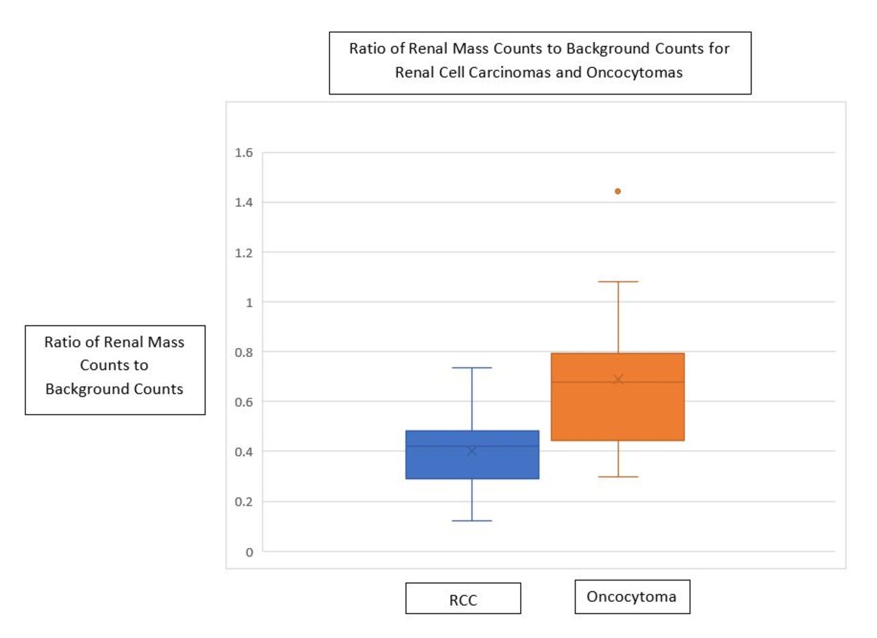
Results: Of the 56 masses sampled or resected, 35 were diagnosed as RCC and 14 were diagnosed as oncocytomas. The remaining masses were diagnosed as other benign tumors (N=2) or metastatic disease from a non-renal primary (N=1). The average target to background ratio for the RCC’s was 0.40 (90%CI, 0.36-0.44), (Range: 0.12-0.73) and the average target to background ratio for the oncocytomas was 0.72 (90%CI, 0.58-0.86), (Range: 0.30-1.44).

Conclusions: The quantitative analysis of renal masses evaluated with 99m Sestamibi SPECT/CT Imaging revealed a difference in the radiotracer uptake of oncocytoma and renal cell carcinoma. Further research is needed to validate the results and determine the utility of 99m Sestamibi SPECT/CT Imaging in clinical practice.

Figure
  • Download figure
  • Open in new tab
  • Download powerpoint
Previous
Back to top

In this issue

Journal of Nuclear Medicine
Vol. 64, Issue supplement 1
June 1, 2023
  • Table of Contents
  • Index by author
Article Alerts
Sign In to Email Alerts with your Email Address
Email Article

Thank you for your interest in spreading the word on Journal of Nuclear Medicine.

NOTE: We only request your email address so that the person you are recommending the page to knows that you wanted them to see it, and that it is not junk mail. We do not capture any email address.

Enter multiple addresses on separate lines or separate them with commas.
Sestamibi quantitative evaluation of renal masses
(Your Name) has sent you a message from Journal of Nuclear Medicine
(Your Name) thought you would like to see the Journal of Nuclear Medicine web site.
Citation Tools
Sestamibi quantitative evaluation of renal masses
Taylor Standiford, Spencer Bell, Robert Wang, Alexander Kutikov, Jared Schober, Mohan Doss, Yi Li, Jian (Michael) Yu
Journal of Nuclear Medicine Jun 2023, 64 (supplement 1) P984;

Citation Manager Formats

  • BibTeX
  • Bookends
  • EasyBib
  • EndNote (tagged)
  • EndNote 8 (xml)
  • Medlars
  • Mendeley
  • Papers
  • RefWorks Tagged
  • Ref Manager
  • RIS
  • Zotero
Share
Sestamibi quantitative evaluation of renal masses
Taylor Standiford, Spencer Bell, Robert Wang, Alexander Kutikov, Jared Schober, Mohan Doss, Yi Li, Jian (Michael) Yu
Journal of Nuclear Medicine Jun 2023, 64 (supplement 1) P984;
Twitter logo Facebook logo LinkedIn logo Mendeley logo
  • Tweet Widget
  • Facebook Like
  • Google Plus One
Bookmark this article

Jump to section

  • Article
  • Figures & Data
  • Info & Metrics

Related Articles

  • No related articles found.
  • Google Scholar

Cited By...

  • No citing articles found.
  • Google Scholar

More in this TOC Section

  • Head-to-head Comparison of 68Ga-FAPI PET/CT and 18F-FDG PET/CT in Fumarate Hydratase-Deficient Renal Cell Carcinoma
  • Comparison of day of treatment vs 1 day post-treatment Lu177 SPECT/CT images in patients treated with lutetium Lu 177 vipivotide tetraxetan.
  • Impact of theranostics tumor board on clinical management of patients referred for radiopharmaceutical therapy
Show more Oncology: Clinical Therapy & Diagnosis (includes Phase 2, Phase 3, post approval studies) - GU

Similar Articles

SNMMI

© 2025 SNMMI

Powered by HighWire