


Abstract
P926
Introduction: Autoimmune encephalitis (AIE) encompasses a group of disorders in which the host immune system targets self-antigens expressed in the central nervous system. It is a rapidly progressive inflammatory pathology with severe and sometimes irreversible neurological damage. Even though it is relatively rare, it is associated with poor prognosis and death in up to 7–12% of cases. Thus, an early diagnosis and initiation of specific treatment are extremely important in its management. In children, there is rapid onset of neuropsychiatric symptoms. Diagnosis of AIE is difficult, as children are less likely to show well-defined neurologic symptoms, complexity of normal behavioral changes during childhood and limited capacity to describe their symptoms. A long list of adjunct diagnostic tools including EEG, MRI, standard biochemistry test are available for initial evaluation but are nonspecific. Despite many antibodies being described against the central nervous system, a significant proportion of childhood auto-immune encephalitis (AIE) do not exhibit detectable known antibodies, which is a diagnostic challenge for the clinicians. 18F-FDG-PET has also been shown to be superior to morphological imaging for early diagnosis of AIE. But limited data is available in literature, describing 99mTc ECD brain perfusion SPECT pattern in children with AIE. Authors here describe brain perfusion pattern in a series of 5 cases, presented with features of acute AIE
Methods: 5 Children (3 males & 2 females) age range 1-17 yrs, satisfying the diagnostic criteria of probable AIE based on thorough clinical and lab evaluation, including (EEG, MRI, ANA, TPO, CSF for autoimmune antibodies against NMDA, AMPA1,2, CASPR, LGI-1, GABA-B,) referred from Paediatric Neurology unit of our Institute from Aug’2022 to Nov’2022, for 99mTc-ECD Brain perfusion SPECT, were retrospectively analysed. Radiotracer was injected intravenously at rest, during the seizure-free period (>24 hrs) and brain SPECT was acquired 45 mins after injection. Reconstructed SPECT images were analysed by an experienced Nuclear Medicine Physician.
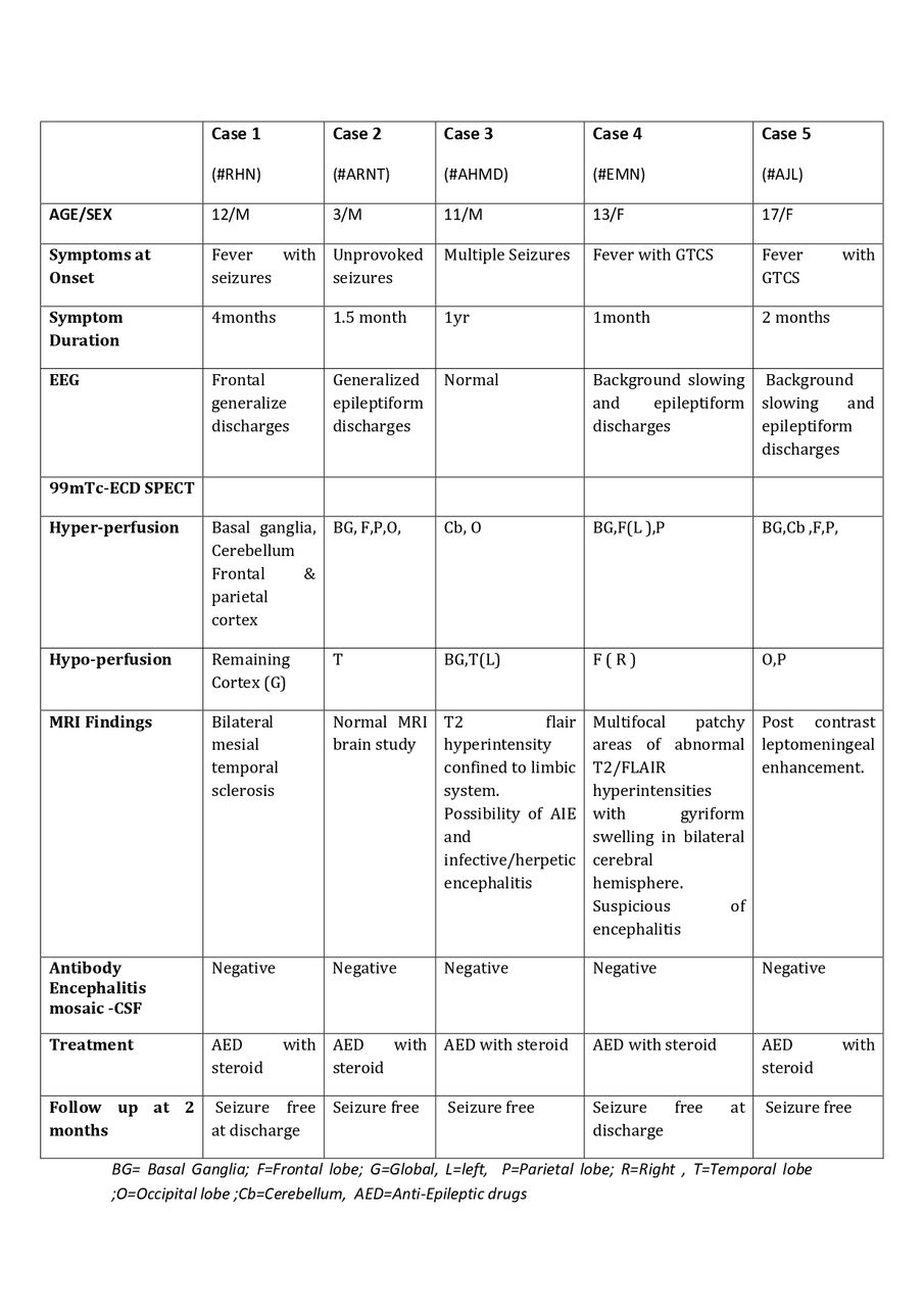
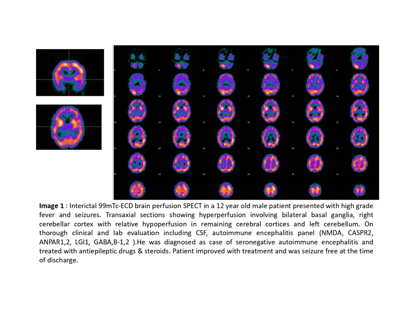
Results: 99mTc-ECD SPECT scan revealed a mixed pattern of perfusion defects in this entire patient group. Predominantly focal areas of hyperperfusion were noted in the basal ganglia, cerebral & cerebellar cortices(Image-1). Additional foci of moderate hypoperfusion and relative diffuse hypoperfusion was also noted in remaining cerebral cortices. All 5 patients were started on Antiepileptic drugs(AED) & steroids. Most of the patients improved with treatment and were seizure free at the time of discharge from the hospital(Table-1)
Conclusions: 99mTc-ECD brain perfusion SPECT was abnormal in all five seronegative patients with probable AIE. The prognosis in this group of patients depends on an early diagnosis and initiation of specific treatment. Interictal 99mTc-ECD SPECT scan offered additional functional information to the treating physician and was complimentary in diagnosing the disease. MRI was able to aid the diagnostic dilemma in few patients, whereas 99mTc-ECD SPECT was able to provide additional corroborative evidence in almost all the patients. Further, larger prospective studies might help in assessing the pattern of involvement and an early use of functional imaging in children suspected with AIE, for a timely diagnosis, disease-specific treatment and improvement in outcome.


In this issue


Jump to section
Related Articles
Cited By...
- No citing articles found.