


Abstract
P810
Introduction: Whole body positron emission tomography (PET) plays a crucial role in identifying malignancies and assessing functional disorders. Recent research has shown deep learning-based PET enhancement technology can reduce scan duration while maintaining image quality. Although reduced injected dose and reduced scan time are physically related, drug metabolism at different levels can result in various image distortion features. In this study, we apply a deep learning model trained by using fast scan data to low injected dose images as well as compare the image quality of enhanced images against that of the standard acquisitions.
Methods: In this prospective study, 30 patients were injected with 0.08-0.1 mCi/kg 19F-FDG and scanned (20 PET-MR, GE SIGNA PET/MR, 3min/bed; 10 PET-CT, GE Discovery MI, 1min/bed) to acquire the standard PET acquisitions (STD). 13 patients (10 PET-CT; 3 PET-MR) were injected half the standard activity and underwent PET examination twice the standard scan time. Half-dose images (LD) were reconstructed using the first half list mode data and simulated standard images (simu-STD) were reconstructed using all list mode data. LD images were then enhanced (LD-enhanced) using a pretrained deep learning model (fast-DL), which is trained using fast scanned images as the input and the standard acquisitions as the output. 5 regions of interest (ROIs) were drawn in relatively homogeneous liver area to calculate signal-to-noise ratio (SNR). ROIs in LD and LD-enhanced images were copied from corresponding simu-STD images. Peak SNR (PSNR) and Structure Similarity Index Measure (SSIM) of LD and LD-enhanced images were evaluated with simu-STD as the standard acquisition. SNR between STD and simu-STD were compared using Mann-Whitney u test. SNR, PSNR and SSIM between simu-STD, LD and LD-enhanced images are compared using paired Wilcoxon test.
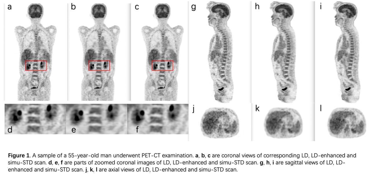
Results: As Figure 1 shows, LD images could be enhanced by fast-DL model without generating extra artifacts. The LD-enhanced images preserved the physiological and pathological structures while noise level was suppressed. Statistically, STD and simu-STD shared the same SNR distribution (P=0.478). SNR level of LD-enhanced images were significantly higher than LD (P=0.028) and the same as simu-STD (P=0.917). LD-enhanced had significant better PSNR and SSIM than LD (P<0.0001) and an average of 1.75 dB PSNR improvement was found.
Conclusions: Experimental results suggested a true low injected activity (1/2 dose) acquisition of whole-body FDG PET images could be enhanced to a standard of care quality by deep learning method.

In this issue


Jump to section
Related Articles
Cited By...
- No citing articles found.