


Abstract
P625
Introduction: Pancreatic ductal adenocarcinoma (PDAC) is an invasive and rapidly progressive malignancy. One of the major challenges in the management of these patients is the lack of a reliable imaging tool to monitor tumor response to treatment. A high degree of fibrosis, mainly collagen type I, has been recognized as the hallmark of PDAC, which further increases in response to neoadjuvant chemoradiotherapy (CRT). This study aims to develop an image-guided paradigm for monitoring treatment response using the collagen type I specific PET imaging probe, 68Ga-CBP8 in mouse models and patients with PDAC.
Methods: Mouse models of human PDAC were generated by subcutaneous implantation of FOLFIRNOX-sensitive (PANC1 and PDAC6) and FOLFIRINOX-resistant (SU8686) patient-derived PDAC cells in nude mice (male, nu:nu, n=88). Mice were randomized into 2 groups of treatment with FOLFIRINOX (67 mg/kg leucovorin, 33 mg/kg fluorouracil, 33 mg/kg irinotecan, 3 mg/kg oxaliplatin, 2X a week, i.v.) or vehicle. Animals in each group underwent PET/MRI with 68Ga-CBP8 (3.7-11.1 MBq, i.v.) prior to-, and at 7- and 15-days post-treatment (n=4-8/group). To assess the specificity of the probe and changes in tumor permeability, additional FOLFIRINOX-treated PANC1 tumor-bearing mice (n=4) were imaged with 68Ga-CBP8 probe, and a linear (CBP8 is a cyclic peptide) collagen non-binding probe, 68Ga-CNBP (3.7-11 MBq, i.v.), in a random order in 2 consecutive days. PET was performed for 60 minutes post-injection. PET findings were correlated with tumor growth monitoring over 21 days and quantification of collagen by Picrosirius red staining. PET measures and collagen content were compared between 2 probes using paired t-test, and among different time points using one-way ANOVA with repeated measures. 5 male patients (49-71 yrs.) with newly diagnosed PDAC underwent dynamic PET for 60 minutes post-injection of 68Ga-CBP8 probe (111-281 MBq) followed by standard contrast-enhanced MRI. 3/5 subjects underwent repeat PET/MRI after completion of standard neoadjuvant CRT. The pre- and post-treatment PET parameters for tumor and pancreas were compared using paired and un-paired Wilcoxon rank test. PET findings were correlated with tumor size change and post-surgical histological evaluations.
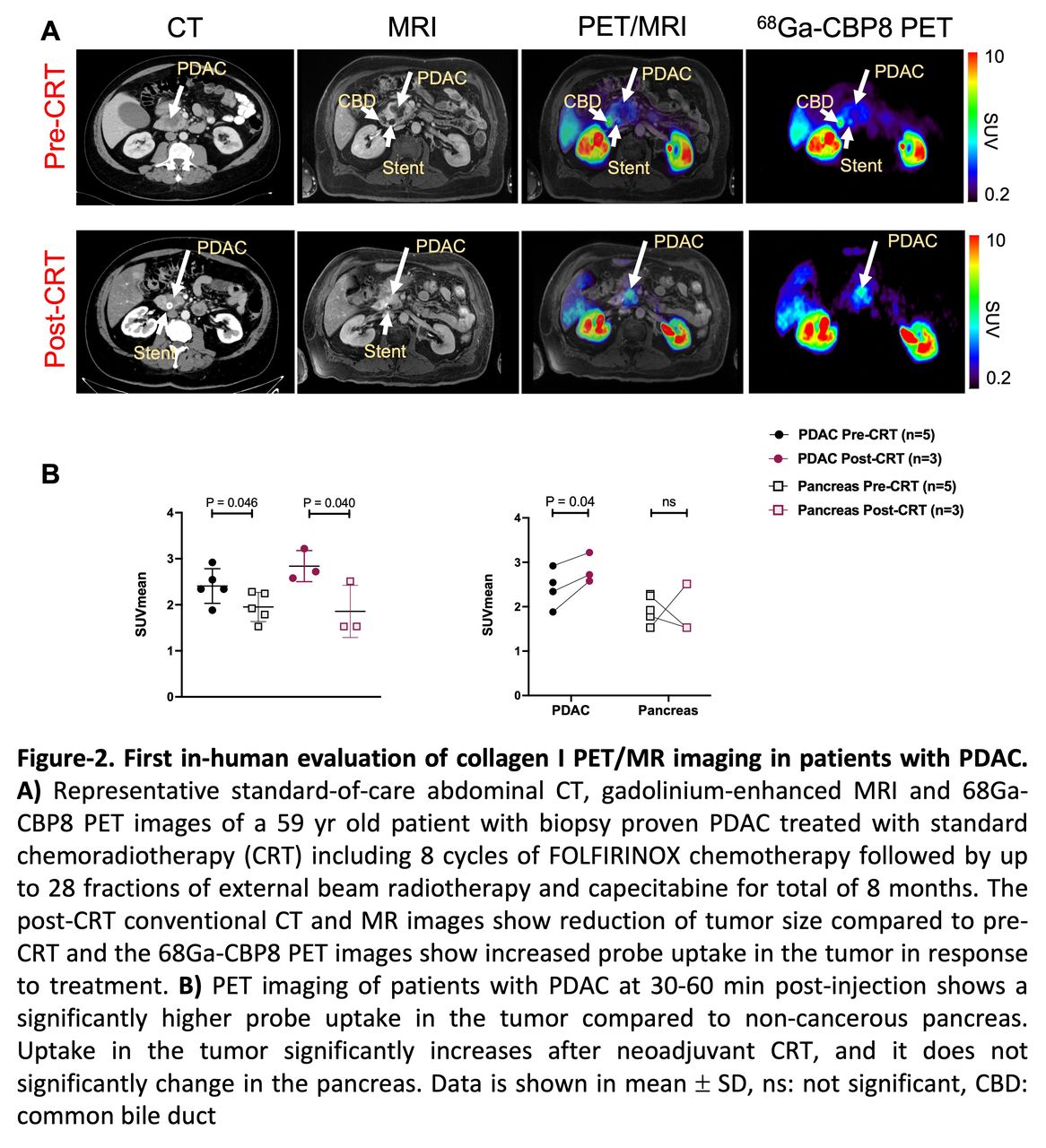
Results: PET imaging of PANC1 tumors showed significantly higher 68Ga-CBP8 uptake compared to 68Ga-CNBP at each time point (p < 0.05), demonstrating the specificity of the probe in targeting collagen (Fig 1A-B). PET values significantly increased over time in FOLFIRINOX-sensitive tumors (PANC1 %ID/cc: 0.8 ± 0.2 vs. 1.1 ± 0.3 vs. 1.9 ± 0.2, p < 0.05 in day-0 vs. day-7 vs. day-15, respectively, p = 0.0005), whereas no significant change was seen in the probe uptake in the vehicle or resistant tumors over time (p > 0.05) (Fig 1C-E). There was a significant tumor growth reduction in PANC1 and PDAC6 tumors in response to FOLFIRINOX compared to vehicle and resistant SU8686 tumors (Fig 1D). Picrosirius red staining showed continuous increase of collagen in responding PANC1 and PDAC6 tumors (% of collagen proportion area (CPA) in PANC1: 5.2 ± 3.8 vs. 16.2 ± 8.7 vs. 46.9 ± 6.0, p < 0.05, at Day-0 vs. Day-7 vs. Day-15, respectively, p < 0.0001), no change to slightly decreased collagen content of the resistant SU8686 (%CPA: 33.0 ± 8.9 vs. 30.0 ± 9.7 vs. 28.1 ± 7.2, at Day-0 vs. Day-7 vs. Day-15, respectively, p = 0.66) and unchanged minimal collagen in pancreas (Fig 1E-F). PET/MRI of PDAC patients showed a significantly higher SUVmean in the tumor compared to pancreas in all subjects (2.40 ± 0.37 vs. 1.95 ± 0.31, p = 0.043). The PET values significantly increased in the treated compared to untreated tumors (SUVmean: 2.83 ± 0.33 vs. 1.85 ± 0.56, p = 0.04) (Fig 2). Response to treatment was confirmed on post-surgical histological evaluations.
Conclusions: 68Ga-CBP8 PET probe specifically binds to collagen and quantifies PDAC-associated fibrosis. PET imaging of collagen could be used as a reliable non-invasive tool for monitoring response to neoadjuvant therapy in PDAC. This approach could guide clinicians with optimized treatment planning and reduced cost of care and save patients from side effects of non-effective therapies and repeated invasive procedures.


In this issue


Jump to section
Related Articles
Cited By...
- No citing articles found.