


Abstract
P623
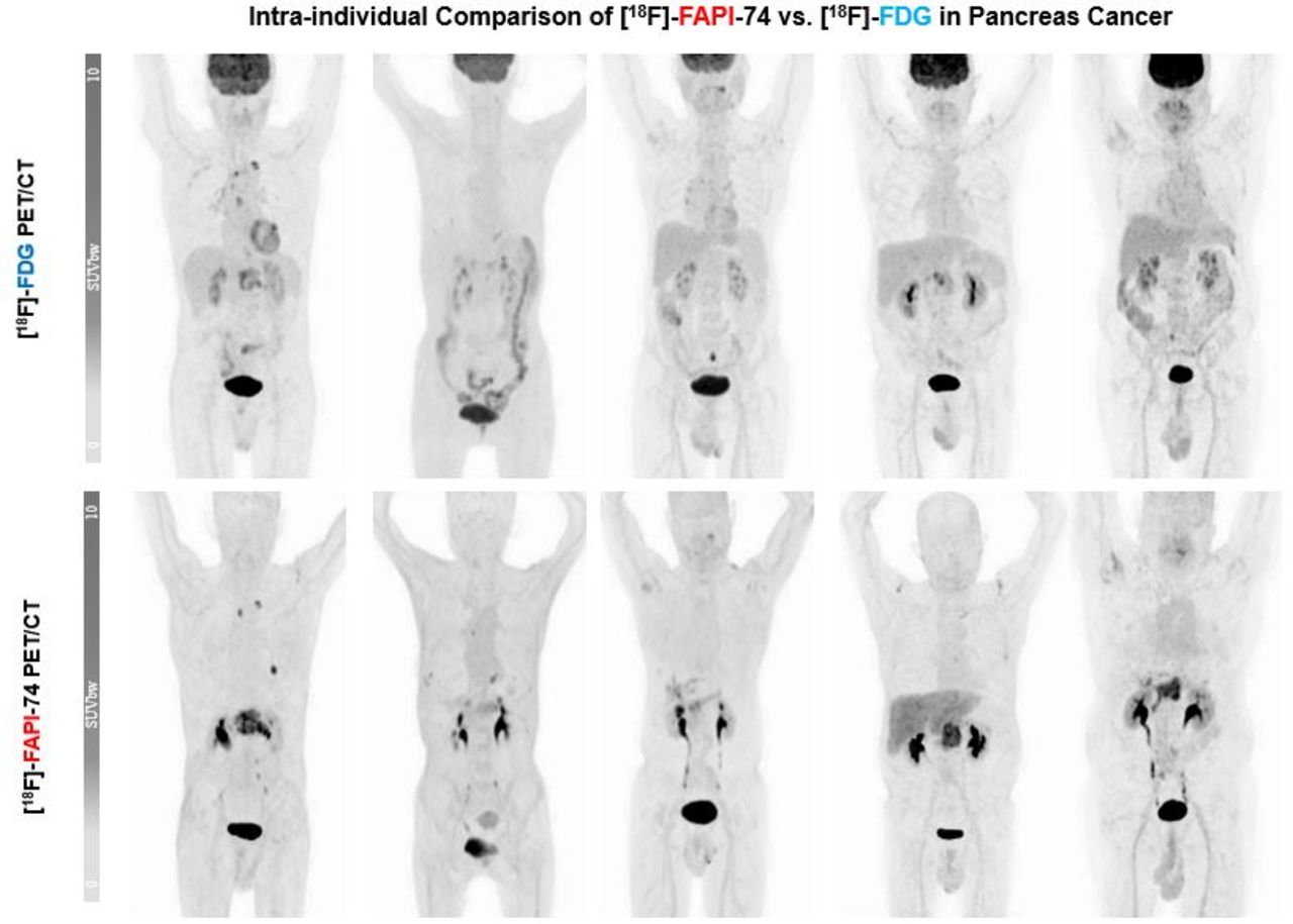
Introduction: Fibroblast activation protein-(FAP)-ligands, a novel class of tracers for PET/CT imaging, demonstrated very promising results in various oncological, especially epithelial malignancies, and also in some benign diseases with long-term potential, to surpass the current pan-cancer agent [18F]FDG PET/CT. Pancreatic ductal carcinoma (PDAC) belongs to the group of epithelial malignancies with a strong so-called "desmoplastic reaction", leading to a prominent tumor stroma with a marked overexpression of fibroblast activation protein (FAP). The first clinical experiences with 68Ga-labeled FAP ligands opened a new avenue for the utility of PET/CT modality in PDAC management. However, due to well-known limitations regarding the use of Ga-labeling of radiopharmaceuticals, great efforts have been made to employ 18F labeled FAP derivatives, i.e. [18F]FAPI-74. With this analysis of interim data from our ongoing prospective, proof-of-concept study, we sought to evaluate the biodistribution, tumor uptake, and lesion detectability in patients with PDAC using [18F]FAPI-74 compared to contrast-enhanced [18F]FDG PET/CT for PDAC staging on an intra-individual basis.
Methods: This interim analysis includes 5 patients (median age 66) who underwent both [18F]FDG with contrast-enhanced CT and [18F]FAPI-74 PET/CT whole-body imaging for primary staging (n=4) or re-staging (n=1) with a mean scan time interval of 11.6 ± 4.2 days (range 1 - 15 days) and without any change of treatment during the interval. [18F]FDG and [18F]FAPI-74 PET/CT scans were acquired at 64 ± 3.6 min (range 61 - 69 min) and 66 ± 4.8 min (range 60 - 76 min) after administration of 200 ± 78 MBq (range 79 - 307 MBq) and 235 ± 59 MBq (range 90 - 321 MBq), respectively. Quantification of tracer uptake was determined with SUVmax and SUVmean. Furthermore, the tumor-to-background ratio (TBR) was derived by dividing the SUVmax of tumor lesions by the SUVmax of adipose tissue, skeletal muscle, and blood pool.
Results: Overall, 20 lesions were detected in five patients including primary (n=5), lung (n= 7), bone (n=2), and lymph node (n=6) metastases. [18F]FAPI-74 detected 33 % more lesions compared to [18F]FDG with a better TBR and sharp image contrast. Concerning patient-based comparison, in one patient the primary lesion could be detected unequivocally with [18F]FAPI-74 that was missed by [18F]FDG imaging. Altogether, most of the lesions demonstrated markedly elevated uptake of [18F]FAPI-74 with a simultaneous lower uptake in the background, providing a very high visual contrast.
Conclusions: To our best knowledge, this is the first intra-individual investigation comparing [18F]FAPI-74 with [18F]FDG in PDAC with very promising results. These promising results support further investigation with larger, prospective-designed studies.

In this issue


Jump to section
Related Articles
Cited By...
- No citing articles found.