


Abstract
P608
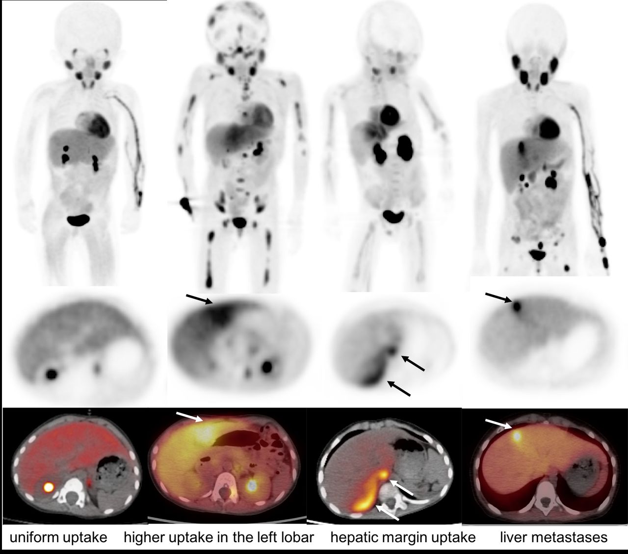
Introduction: 18F-meta-fluorobenzylguanidine (18F-MFBG) is a PET tracer which was reported to have a similar biodistribution with 123I-MIBG. 18F-MFBG has shown promising diagnostic efficiency in neural crest tumors, especially in neuroblastoma with short time consuming and improved images. However, the heterogeneous uptake of MFBG in the liver may interfere with image interpretation. We aimed to describe the liver uptake patterns and the incidence in 18F-MFBG PET/CT to avoid misdiagnosis.
Methods: Consecutive 18F-MFBG PET/CT for patients with neural crest tumors was retrospectively analyzed. Patterns of 18F-MFBG uptake and incidence were recorded. SUVmax of left and right lobar, left lobar to right lobar ratio (LRR) and increased sites to right lobar ratio (IRR) were recorded separately in adults and children.
Results: 187 patients (19.3 ± 20.1 years) were enrolled including 121 children (5.2 ± 3.5 years) with a history of neuroblastoma and 66 adults (43.3 ± 14.2 years) with paragangliomas/pheochromocytoma (PPGL). 13 patients with liver metastases show focal increased uptake with high IRR (5.4 ± 2.3) on 18F-MFBG PET/CT.
174 patients (116 children, 58 adults) had no history of liver disease or liver function abnormalities. The SUVmax of the left lobe was different from the right lobe (p<0.001). Three patterns of 18F-MFBG uptake were observed in normal liver tissue: (1) uniform uptake with LRR of 1.2 ± 0.1 (range, 1.0-1.4 ); (2) higher uptake in the left lobar but uniform in left and right lobes (LRR, 1.8 ± 0.3, range, 1.5-2.8 ); and (3) increased uptake in the hepatic margin with IRR of 2.0 ± 0.5 (range, 1.5-3.4) (Figure).
Uniform uptake in the whole liver was seen with similar proportions of 38.5% and 36.4% in adults and children respectively. 50.9% of adults showed higher uptake in the left lobar, and increased uptake in the hepatic margin was less common (12.7%). However, Hepatic margin uptake was more often shown in children (35.8%).
Conclusions: Physiological heterogeneous uptake of 18F-MFBG in the liver needs noted when interpreting the images. In particular, increased MFBG uptake in hepatic margins often seen in children should not be mistaken as lesions. In view of the high resolution of PET/CT with improved image quality, no false negatives or false positives lesions in the liver are shown.

In this issue


Jump to section
Related Articles
Cited By...
- No citing articles found.