


Abstract
P398
Introduction: To characterize the diagnostic performance of [18F]FDG dose de-escalation with shortened acquisition times using total-body PET/CT in pediatric tumor imaging in terms of the subjective image quality and quantification of tracer uptake.
Methods: In this single-center prospective study, 31 pediatric oncology patients under 14 years old were enrolled from November 2020 to August 2021 and underwent total-body PET/CT using the uEXPLORER PET/CT scanner. All patients were confirmed by postoperative pathologic examination or biopsy.
All patients were randomly assigned to one of four [18F]FDG dose groups: full-dose (3.7 MBq/kg), 1/2-dose (1.9 MBq/kg), 1/3-dose (1.2 MBq/kg), and 1/4-dose (0.9 MBq/kg). The full acquisition time of each patient was 20 min, and images with a shortened acquisition time frame (12 min, 10 min, 8 min, 6 min, 5 min, 4 min, 3 min, 2 min, 1.5 min, 1 min, and 0.5 min) were reconstructed.
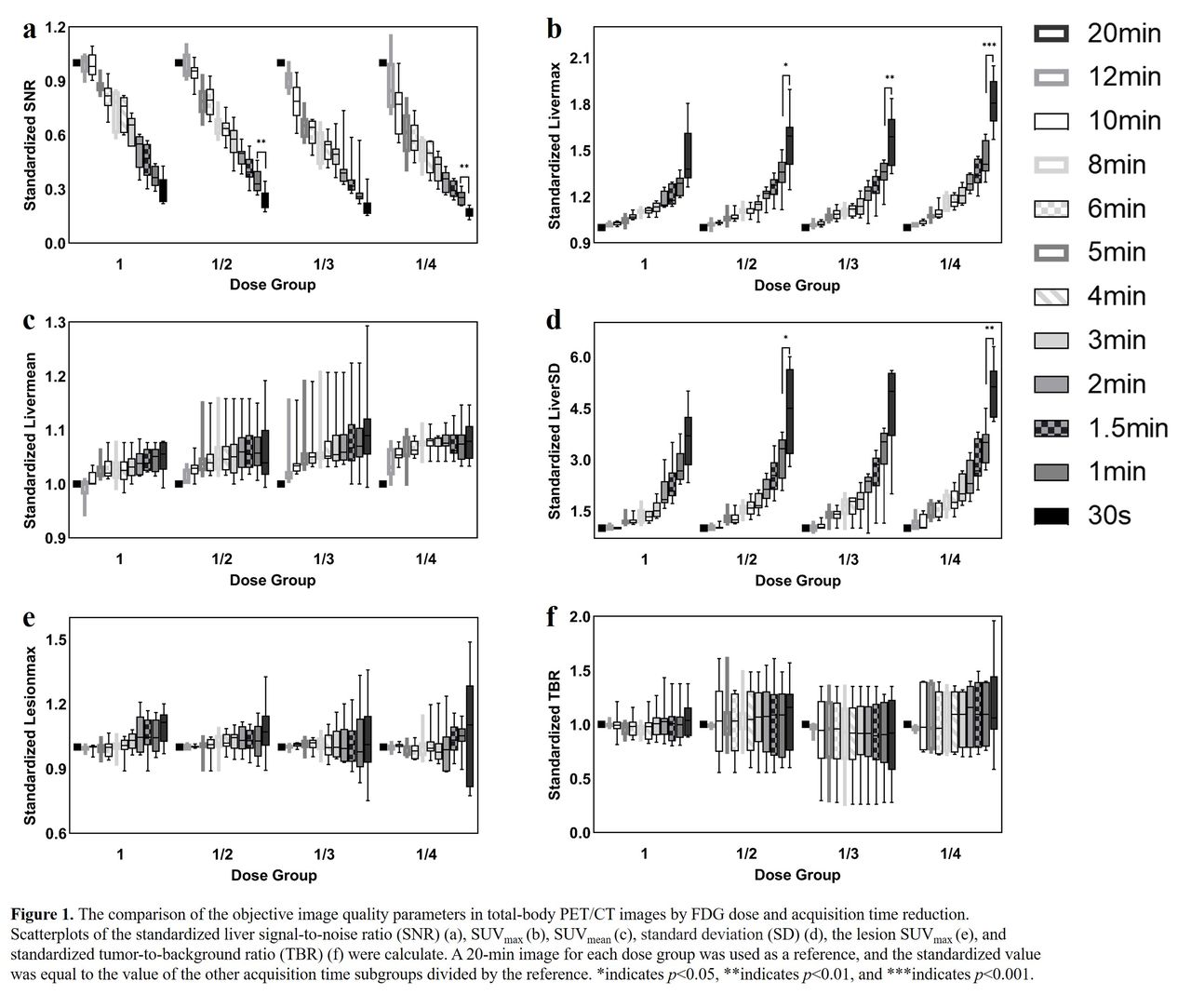
Image quality was evaluated by objective and subjective indicators. Objective uptake metrics were assessed using region-of-interest (ROI) analysis of healthy liver and suspected lesions, including liver maximum standard uptake value (SUVmax), SUVmean, standard deviation (SD), and signal-to-noise ratio (SNR). The variability in lesion SUVmax and tumor-to-background ratio (TBR) were also calculated. The subjective analysis was performed using a 5-point Likert scale. The full-time 20-min PET images served as the reference for other duration groups to evaluate lesion detectability and diagnostic confidence. Suspected major lesions and microlesions were recorded, while 3-point Likert scales were used for diagnostic confidence.
Results: With shortened acquisition times, the liver maximum SUVmax and SD increased in each dose group, while SNR was reduced with shortened acquisition time. The liver SUVmean, lesion SUVmax, and TBR showed no significant deviation (all p>0.05).
A sufficient subjective image quality score could be achieved in the full-, 1/2-, 1/3-, and 1/4-dose groups with at least 2-min, 4-min, 6-min, and 8-min acquisitions, respectively, where great overall image quality and brain delineation (scored 5.0) and superior organ boundaries and image noise (scored over 4.0) could be achieved, and all suspicious lesions found in 20-min images were detectable with high diagnostic confidence. The image noise was more deranged than the overall quality and lesion conspicuity.
Conclusions: The regimen of full-dose [18F] FDG with a 2-min scan, 1/2-dose with a 4-min scan, 1/3-dose with a 6-min scan, and 1/4-dose with an 8-min scan using total-body PET/CT can provide great image qualities, can maintain a desired diagnostic performance and is feasible for pediatric oncological clinical applications.


In this issue


Jump to section
Related Articles
Cited By...
- No citing articles found.