


Abstract
P1513
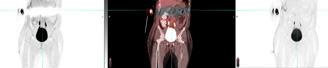
Introduction: Intense activity on PET images can cause an artifactual band of severe photopenia that obscures the same axial slices.
This artifact is most common around the urinary bladder, radiotracer injection or contamination sites.
This affects the attenuation-corrected images (AC), but not NAC images, and is caused by scatter correction processing. Technologists and Nuclear Radiologists may be not aware how to correct this artifact or why it occurs.
Methods: In the simulation method proposed by Watson, the tracer distribution is considered as the emission image and the scattering media as the attenuation image or mu map. The recorded image is based on both scattered and non-scattered photons. The estimate of the distribution of scattered events is computed by the simulation method that utilizes known physics of photon interaction with matter as photons traverse the body. This estimate of scattered photons is subtracted from the original image data. This is repeated iteratively to refine the result.
A limitation of this scatter-correction method is that it relies on the acquired images to estimate the origins of the scattered photons. Scatter events also occur outside of the scanner field of view, and some of these are recorded by the scanner, but the density, location and scattering activity of these structures is unknown.
The artifactual activity in the air outside of the patient is used to estimate scatter by external structures. Some isotopes emit a third prompt gamma photon, such as Rb-82, I-124 and Ga-68, which is in time coincidence with the positron and such photons will be accepted by the scanner’s coincidence window if these scatter to a similar energy of 511keV photons. These form an almost uniform background and will add more apparent counts to the air region outside the attenuation. In order to avoid an over-scaling of the scatter, the prompt gamma correction (PGC) will scale this flat background along with the simulated scatter using the counts in the air region.
Relative scaling –Usually the default setting utilized. Relative scaling includes the simulation, scaling and the PRC as described above. Since most scans have activity external to the scanner field of view, this scatter correction method will produce the most accurate images. However, scatter correction artifacts can result if patient movement degrades the attenuation map (mu map). To correct for the scatter correction artifacts, the scatter computation has to be either simplified or the scatter correction has to be completely turned off.
Absolute scaling – utilizes a simplified scatter algorithm. Absolute scaling does not rely on knowing the air region, as used for relative scaling. If true sources of activity are mistakenly included in the air region, this will be considered scatter, resulting in an over-scaling of scatter and a photopenic artifact. Isotopes that emit a prompt gamma ray with a low probability (13% for 82Rb), the flat background will be mostly ignored by the image reconstruction. The simulated scatter reflects scatter from the true coincidences. When the activity is mostly in the field of view, a reasonably accurate scatter correction will result with absolute scatter correction method. Isotopes with extremely low count level like 90Y, a more robust quantitation will result with absolute scaling as this scatter correction method does not rely on rescaling the simulation.
Results: Scatter correction processing can produce substantial artifacts that can result in non-diagnostic images in the presence of intense source of activity. Recognizing this artifact and understanding the available methods to correct it can salvage the affected PET scans and can avoid unnecessary re-scanning.
Conclusions: Artifacts caused by scatter correction can be effectively mitigated by applying relative scaling instead of absolute scaling method. Non-attenuation-corrected images are free of these artifacts and can be visually inspected.

In this issue


Jump to section
Related Articles
Cited By...
- No citing articles found.