


Abstract
P1473
Introduction: Alzheimer's disease (AD) is a global health problem characterized by progressive cognitive dysfunction and behavioral impairment, posing a heavy social and economic burden on the global public health system. It is known that microglia, astrocytes, etc can cause a neuroinflammatory cascade in brain and increase the progressive course of AD [1]. Studies revealed that Electro-puncture(EA)treatment may improve syndrome of AD through decreasing expression of microglia and astrocyte to improve AD,but the molecular mechanism remains to be explored [2-3]. Sphingosine 1 Phosphate (S1P) is an important active lipid molecule, also for neuronal survival and for the maintenance of neuronal functions in adult brains. The S1P receptor 1 (S1PR1) signaling pathways have important and diverse functions. Stage- and cell-specific sphingolipid metabolism and expression are crucial for brain development and maintenance toward adult age. Herein, we aimed to study the PET imaging based S1PR1 ([18F]-TZ4877) toevaluate EA therapy for transgenic mice model AD.
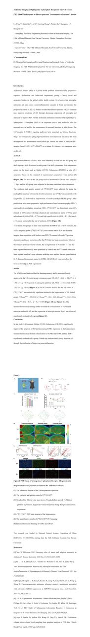
Methods: Eight-month-old-male APP/PS1 mice were randomly divided into the AD group and the EA group , with five mice in each group. For the EA treatment, five acupuncture points on the head, such as Baihui (GV20), Sishencong (EX-HN1, a total of 4 acupoints) based on the standard of experimental acupuncture were applied [4] (Figure 1A). The mice in the EA group received treatment for 20 min once a day for 15 days; and the AD group were subjected to the same conditions but not treatment.
The synthesis and quality control of [18F]TZ4877 were achieved by using the nucleophilic reaction between the tosylate precursor and [18F]KF in acetonitrile with Kryptofifix 222 followed by deprotection of methoxymethyl (MOM) group. After purification using semi-preparative high-performance liquid chromatography (HPLC) combined with solid-phase extraction (SPE), [18F]TZ4877 was formulated using 10% ethanol in 0.9% saline with high chemical and radiochemical purity (>98%), good radiochemical yields (28.8 ± 3.7%), and high specific activity (43.1 ± 8.3 GBq/mmol, n = 8, decay corrected to the end of synthesis, EOS) (Figure 1B).
To evaluate, two groups of mice were underwent the MWM test . For PET studies, the S1PR1 targeting probe ([18F]-TZ4877)[5] were carried out for 0-30 min dynamic imaging data acquisition with Mediso nanoPET/CT scanner followed CT position attenuation and decay correction; after the PET data has been reconstructed followed the standard protocol from the vendor, the co-registration of PET and CT,and the brain regional radioactivity were quantified by PMOD and the VT valued from each brain regions based on Logan reference modeling were applied for data quantification [6-7]. Immunofluorescence stains for S1PR1, GFAP, IBA-1 were carried out for tissue collected post-PET experiments.
Results: The MWM tested indicated that the y was significantly improved in the EA treated group (swimming speed EA, 19.39 ± 1.66 vs AD, 17.41 ± 1.78, n = 5, p < 0.05; period of seeking the platform EA, 30.42 ±13.11 vs AD, 48.33 ± 7.31, n = 5, p < 0.05) (Figure 1C). The PET studies showed that the VT values of [18F]-TZ4877 was statistically significant reduced in the hippocampus of EA treated groups (VTS1PR1EA-r, 1.25±0.26 vs VT S1PR1AD-r, 1.81 ± 0.42; VTS1PR1EA-l,1.24 ± 0.28 vs VT S1PR1AD-l, 1.72 ± 0.39,n = 5, p < 0.05) (Figure 1D and Figure 1E). The immunofluorescence staining further revealed the colocalization of S1PR1 and astrocyte marker GFAP, and the expression of microglia marker IBA-1 was observed significantly reduced in EA group(Figure 1F).
Conclusions: In this study, EA treatment (Baihui (GV20) -Sishencong (EX-HN1)) significantly improved the symptom of AD and decreasing S1PR1 expression in the hippocampus. Immunofluorescence showed colocalization of S1PR1 and GFAP; and the IBA-1 significantly reduced in EA group, Which may indicate that EA may improve AD through the mechanism of improving neuroinflamation.


In this issue


Jump to section
Related Articles
Cited By...
- No citing articles found.