


Abstract
P1418
Introduction: Back and neck pain are one of the major health problems in US and a leading cause of health care expenditure. Standard traditional imaging such as plain radiographs, CT and MRI can detect degenerative disease, but can be non-specific as degenerative changes are commonly seen in many asymptomatic individuals. Bone scintigraphy with SPECT as an imaging modality allows assessment of both physiological and morphological changes of active painful degenerative diseases of the spine. Additionally, fused CT or MRI imaging gives added advantage of precise anatomical localization and targeted therapeutic approach.
Methods: We present spectrum of characteristic imaging findings and patterns of bone SPECT in painful degenerative diseases of the spine. Bone SPECT findings with CT and MRI as hybrid imaging are also discussed.
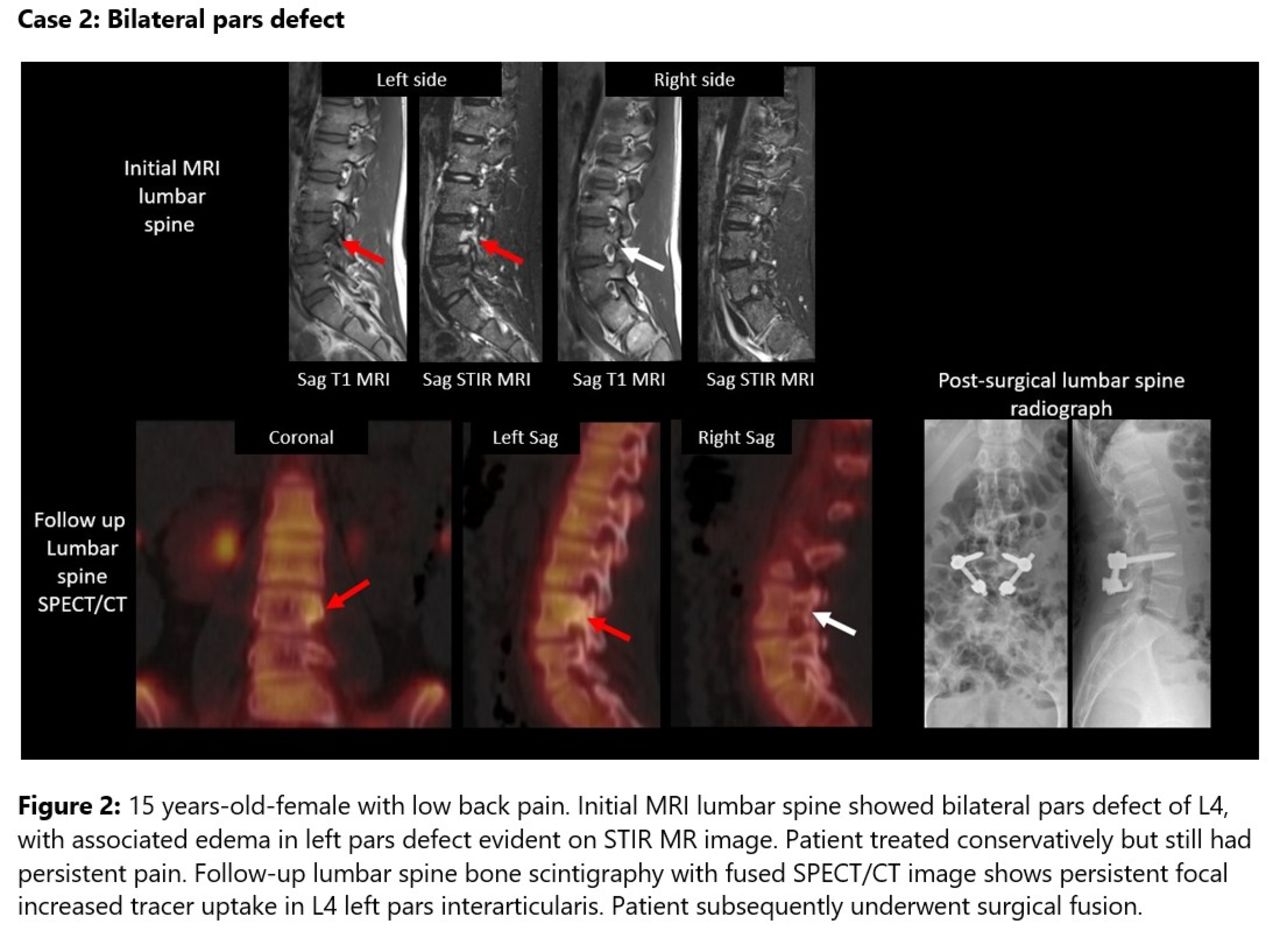
Results: We present a case based pictorial review highlighting the spectrum of cases of painful degenerative disease on bone scintigraphy with fused CT and/or MRI imaging wherever available. It’s role in young patients with pars defect is also discussed.
Conclusions: Traditional CT and/or MR imaging is non-specific as degenerative changes are also commonly seen in an asymptomatic individual. The presented bone scintigraphy with SPECT imaging and its fusion with CT/MR imaging provides precise anatomical localization of the source of neck or back pain and provides the treating physician a targeted therapeutic approach for resolving patients pain.


In this issue


Jump to section
Related Articles
Cited By...
- No citing articles found.