
Main menu

  • Home
  • Content
    • Current
    • Ahead of print
    • Past Issues
    • JNM Supplement
    • SNMMI Annual Meeting Abstracts
    • Continuing Education
    • JNM Podcasts
  • Subscriptions
    • Subscribers
    • Institutional and Non-member
    • Rates
    • Journal Claims
    • Corporate & Special Sales
  • Authors
    • Submit to JNM
    • Information for Authors
    • Assignment of Copyright
    • AQARA requirements
  • Info
    • Reviewers
    • Permissions
    • Advertisers
  • About
    • About Us
    • Editorial Board
    • Contact Information
  • More
    • Alerts
    • Feedback
    • Help
    • SNMMI Journals
  • SNMMI
    • JNM
    • JNMT
    • SNMMI Journals
    • SNMMI

User menu

  • Subscribe
  • My alerts
  • Log in
  • Log out
  • My Cart

Search

  • Advanced search
Journal of Nuclear Medicine
  • SNMMI
    • JNM
    • JNMT
    • SNMMI Journals
    • SNMMI
  • Subscribe
  • My alerts
  • Log in
  • Log out
  • My Cart
Journal of Nuclear Medicine

Advanced Search

  • Home
  • Content
    • Current
    • Ahead of print
    • Past Issues
    • JNM Supplement
    • SNMMI Annual Meeting Abstracts
    • Continuing Education
    • JNM Podcasts
  • Subscriptions
    • Subscribers
    • Institutional and Non-member
    • Rates
    • Journal Claims
    • Corporate & Special Sales
  • Authors
    • Submit to JNM
    • Information for Authors
    • Assignment of Copyright
    • AQARA requirements
  • Info
    • Reviewers
    • Permissions
    • Advertisers
  • About
    • About Us
    • Editorial Board
    • Contact Information
  • More
    • Alerts
    • Feedback
    • Help
    • SNMMI Journals
  • View or Listen to JNM Podcast
  • Visit JNM on Facebook
  • Join JNM on LinkedIn
  • Follow JNM on Twitter
  • Subscribe to our RSS feeds
Research ArticleClinical Investigation

Facial Anonymization and Privacy Concerns in Total-Body PET/CT

Aaron R. Selfridge, Benjamin A. Spencer, Yasser G. Abdelhafez, Keisuke Nakagawa, John D. Tupin and Ramsey D. Badawi
Journal of Nuclear Medicine August 2023, 64 (8) 1304-1309; DOI: https://doi.org/10.2967/jnumed.122.265280
Aaron R. Selfridge
1Department of Biomedical Engineering, University of California–Davis, Davis, California;
  • Find this author on Google Scholar
  • Find this author on PubMed
  • Search for this author on this site
Benjamin A. Spencer
1Department of Biomedical Engineering, University of California–Davis, Davis, California;
2Department of Radiology, University of California–Davis, Davis, California;
  • Find this author on Google Scholar
  • Find this author on PubMed
  • Search for this author on this site
Yasser G. Abdelhafez
2Department of Radiology, University of California–Davis, Davis, California;
3Radiotherapy and Nuclear Medicine Department, South Egypt Cancer Institute, Assiut University, Assiut, Egypt;
  • Find this author on Google Scholar
  • Find this author on PubMed
  • Search for this author on this site
Keisuke Nakagawa
4Cloud Innovation Center, University of California–Davis, Davis, California; and
  • Find this author on Google Scholar
  • Find this author on PubMed
  • Search for this author on this site
John D. Tupin
5IRB Administration, University of California–Davis, Davis, California
  • Find this author on Google Scholar
  • Find this author on PubMed
  • Search for this author on this site
Ramsey D. Badawi
1Department of Biomedical Engineering, University of California–Davis, Davis, California;
2Department of Radiology, University of California–Davis, Davis, California;
  • Find this author on Google Scholar
  • Find this author on PubMed
  • Search for this author on this site
  • Article
  • Figures & Data
  • Supplemental
  • Info & Metrics
  • PDF
Loading

Article Figures & Data

Figures

  • Tables
  • Additional Files
  • Figure
    • Download figure
    • Open in new tab
    • Download powerpoint
  • FIGURE 1.
    • Download figure
    • Open in new tab
    • Download powerpoint
    FIGURE 1.

    (Top) Faces rendered from CT images at 6 time points. Ninety-minute time point used clinical low-dose CT protocol. All others used research ultra-low-dose protocol. (Bottom) Rendering of defaced images at each time point.

  • FIGURE 2.
    • Download figure
    • Open in new tab
    • Download powerpoint
    FIGURE 2.

    (Top) Faces rendered from PET images at 6 time points. (Bottom) Rendering of defaced images at each time point.

  • FIGURE 3.
    • Download figure
    • Open in new tab
    • Download powerpoint
    FIGURE 3.

    (A) Facial embeddings for 15 full-dose cohort subjects at 6 time points, plotted in 2 dimensions using t-SNE. Before defacing, facial embeddings are highly clustered. (B) After defacing, data are no longer clustered.

  • FIGURE 4.
    • Download figure
    • Open in new tab
    • Download powerpoint
    FIGURE 4.

    (A) Facial embeddings for all 30 full- and low-dose cohort subjects at first 3 time points. Before defacing, clusters are present for each participant. (B) After defacing, participants are not uniquely associated with clusters.

  • FIGURE 5.
    • Download figure
    • Open in new tab
    • Download powerpoint
    FIGURE 5.

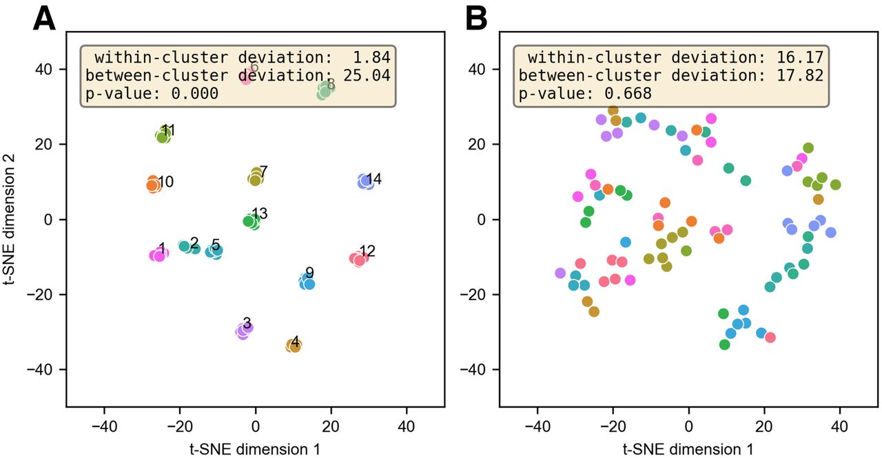
    (A) Facial embeddings for 15 full-dose cohort subjects at 6 time points. Clustered facial embeddings for PET images indicate modest but significant (P < 0.05) degree of identifiability. (B) No clusters are present after defacing. Within-cluster deviation is significantly greater than between-cluster deviation.

  • FIGURE 6.
    • Download figure
    • Open in new tab
    • Download powerpoint
    FIGURE 6.

    (A) Grid showing CT slices before and after defacing. PET images are reconstructed from same raw data, with different CT scans for attenuation correction. (B) Normalized difference image showing percentage change in PET activity. High-intensity region in brain largely overlaps ventricles, which have low [18F]FDG uptake. (C) Absolute difference in PET SUV.

  • FIGURE 7.
    • Download figure
    • Open in new tab
    • Download powerpoint
    FIGURE 7.

    (A) Percentage change when using defaced CT for PET attenuation correction. Values were measured at 5 spheric ROIs along cerebral cortex. Error bars correspond to SD within ROI. (B) Difference image and corresponding CT slice, overlaid with 5 ROIs.

Tables

  • Figures
  • Additional Files
    • View popup
    TABLE 1.

    Scan Details for 30 Subjects Imaged at 3 or 6 Time Points

    CohortParticipants (n)ModalityInitial*90 min3 h6 h9 h12 h
    Low-dose cohort15CT parameter140 kVp/ 5 mAs140 kVp/ 50 mAs140 kVp/ 5 mAsNANANA
    PET activity†15 MBq11 MBq6.3 MBqNANANA
    Full-dose cohort15CT parameter140 kVp/ 5 mAs140 kVp/ 50 mAs140 kVp/ 5 mAs140 kVp/ 5 mAs140 kVp/ 5 mAs140 kVp/ 5 mAs
    PET activity†288 MBq209 MBq117 MBq36.8 MBq11.6 MBq3.65 MBq
    • ↵* Initial CT and PET images were at 0 and 40 min, respectively.

    • ↵† Reported PET activity is actual injected dose averaged over all subjects. Later time points indicate remaining activity after decay of [18F] (not accounting for excreted activity).

    • NA = not applicable.

    • View popup
    TABLE 2.

    Identifiability of PET Images Using Classifier Trained with PET (PET-to-PET) or CT (PET-to-CT) Images

    Identifiability (%)Initial90 min3 h6 h9 h12 h
    PET-to-PET64.350.057.157.128.67.1
    PET-to-CT50.042.928.621.428.614.3

Additional Files

  • Figures
  • Tables
  • Supplemental Data

    Files in this Data Supplement:

    • Supplemental Data
PreviousNext
Back to top

In this issue

Journal of Nuclear Medicine: 64 (8)
Journal of Nuclear Medicine
Vol. 64, Issue 8
August 1, 2023
  • Table of Contents
  • Table of Contents (PDF)
  • About the Cover
  • Index by author
  • Complete Issue (PDF)
Print
Download PDF
Article Alerts
Sign In to Email Alerts with your Email Address
Email Article

Thank you for your interest in spreading the word on Journal of Nuclear Medicine.

NOTE: We only request your email address so that the person you are recommending the page to knows that you wanted them to see it, and that it is not junk mail. We do not capture any email address.

Enter multiple addresses on separate lines or separate them with commas.
Facial Anonymization and Privacy Concerns in Total-Body PET/CT
(Your Name) has sent you a message from Journal of Nuclear Medicine
(Your Name) thought you would like to see the Journal of Nuclear Medicine web site.
Citation Tools
Facial Anonymization and Privacy Concerns in Total-Body PET/CT
Aaron R. Selfridge, Benjamin A. Spencer, Yasser G. Abdelhafez, Keisuke Nakagawa, John D. Tupin, Ramsey D. Badawi
Journal of Nuclear Medicine Aug 2023, 64 (8) 1304-1309; DOI: 10.2967/jnumed.122.265280

Citation Manager Formats

  • BibTeX
  • Bookends
  • EasyBib
  • EndNote (tagged)
  • EndNote 8 (xml)
  • Medlars
  • Mendeley
  • Papers
  • RefWorks Tagged
  • Ref Manager
  • RIS
  • Zotero
Share
Facial Anonymization and Privacy Concerns in Total-Body PET/CT
Aaron R. Selfridge, Benjamin A. Spencer, Yasser G. Abdelhafez, Keisuke Nakagawa, John D. Tupin, Ramsey D. Badawi
Journal of Nuclear Medicine Aug 2023, 64 (8) 1304-1309; DOI: 10.2967/jnumed.122.265280
Twitter logo Facebook logo LinkedIn logo Mendeley logo
  • Tweet Widget
  • Facebook Like
  • Google Plus One
Bookmark this article

Jump to section

  • Article
    • Visual Abstract
    • Abstract
    • MATERIALS AND METHODS
    • RESULTS
    • DISCUSSION
    • CONCLUSION
    • DISCLOSURE
    • Footnotes
    • REFERENCES
  • Figures & Data
  • Supplemental
  • Info & Metrics
  • PDF

Related Articles

  • PubMed
  • Google Scholar

Cited By...

  • No citing articles found.
  • Google Scholar

More in this TOC Section

  • First-in-Human Study of 18F-Labeled PET Tracer for Glutamate AMPA Receptor [18F]K-40: A Derivative of [11C]K-2
  • Detection of HER2-Low Lesions Using HER2-Targeted PET Imaging in Patients with Metastatic Breast Cancer: A Paired HER2 PET and Tumor Biopsy Analysis
  • [11C]Carfentanil PET Whole-Body Imaging of μ-Opioid Receptors: A First in-Human Study
Show more Clinical Investigation

Similar Articles

Keywords

  • total-body PET/CT
  • facial anonymization
  • uEXPLORER
  • facial recognition
SNMMI

© 2025 SNMMI

Powered by HighWire