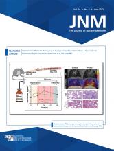
Visual Abstract
Abstract
PET/CT with the new 68Ga-labeled minigastrin analog DOTA-dGlu-Ala-Tyr-Gly-Trp-(N-Me)Nle-Asp-1-Nal-NH2 (68Ga-DOTA-MGS5) was performed on patients with advanced medullary thyroid cancer (MTC) to evaluate cholecystokinin-2 receptor expression status. Methods: Six patients with advanced MTC underwent PET/CT with 68Ga-DOTA-MGS5. From the images acquired 1 and 2 h after injection, preliminary data on the biodistribution and tumor-targeting properties were evaluated in a retrospective analysis. Results: In total, 87 lesions with increased radiotracer uptake considered malignant were detected (2 local recurrences, 8 lymph node lesions, 27 liver lesions, and 50 bone lesions). In general, radiotracer accumulation in lesions was higher at 2 h than at 1 h after injection (mean SUVmax, 7.2 vs. 6.0, respectively; mean SUVmean, 4.4 vs. 3.6, respectively). Conclusion: The preliminary results clearly demonstrate the potential of 68Ga-DOTA-MGS5 PET/CT in detecting local recurrence and metastases in patients with advanced MTC.
Medullary thyroid cancer (MTC) is a rare disease arising from the parafollicular C cells of the thyroid and accounts for 1%–2% of thyroid cancers. Calcitonin is routinely used as a tumor marker for MTC. After primary treatment, additional imaging procedures are recommended when calcitonin levels rise above 150 pg/mL (1). Besides conventional radiologic imaging procedures (ultrasound, CT, MRI), PET with different radiotracers is performed to detect and localize persistent or recurrent disease. In patients with suspected MTC recurrence, 18F-FDG PET/CT has a reported detection rate of 59%–69%, whereas 6-[18F]fluoro-l-3,4-dihydroxyphenylalanine (18F-DOPA) PET/CT shows a detection rate of 66%–72%, which increases to 86% in patients with a calcitonin doubling time of less than 24 mo. PET/CT using 68Ga-labeled somatostatin analogs has demonstrated variable sensitivity, with an overall detection rate of 63.5%, and allows selection of patients for peptide receptor radionuclide therapy (2).
Cholecystokinin-2 receptors (CCK2Rs) are overexpressed in more than 90% of MTC cases (3). We recently reported development of the new minigastrin analog DOTA-dGlu-Ala-Tyr-Gly-Trp-(N-Me)Nle-Asp-1-Nal-NH2 (DOTA-MGS5) with improved stability in vivo and enhanced tumor targeting (4). CCK2R targeting with DOTA-MGS5, therefore, offers a promising new diagnostic and therapeutic approach for patients with advanced MTC.
We here report our initial clinical experience with 68Ga-labeled DOTA-MGS5 for PET/CT, with a primary goal of evaluating the potential of the new radiotracer to detect tumor lesions in patients with proven recurrent or residual metastatic disease.
MATERIALS AND METHODS
Six patients with histologically proven MTC and confirmed metastatic disease from previously performed diagnostic contrast-enhanced CT (ceCT) and PET with 18F-DOPA or 68Ga-labeled DOTATOC underwent PET/CT with 68Ga-labeled DOTA-MGS5. The patient characteristics are in Supplemental Table 1 (supplemental materials are available at http://jnm.snmjournals.org). The patients had not undergone tumor-specific treatment at the time of imaging and were selected individually to evaluate whether radionuclide therapy targeting CCK2R is a potential option. The examination was performed on a named-patient basis, and written informed consent was obtained from all patients as part of the standard practice before any nuclear medicine examination. All procedures were in accordance with the principles of the 1964 Declaration of Helsinki and its subsequent amendments. The retrospective analysis of the data was approved by the Ethics Committee of the Medical University of Innsbruck (approval 1162/2022).
Radiopharmaceutical
68Ga-labeled DOTA-MGS5 was prepared according to the Austrian Medicines Act (Arzneimittelgesetz §8 and §62) as described elsewhere (5) and was administered as a slow intravenous injection (∼2 min) with a mean administered mass of 16 ± 6 μg (range, 12–28 μg) and a mean administered activity of 177 ± 16 MBq (range, 163–208 MBq).
Imaging Protocol
Scans were obtained on a dedicated PET/CT system in time-of-flight mode (Discovery; GE Healthcare). A whole-body PET scan (skull vertex to upper thighs) in 3-dimensional mode was acquired 1 and 2 h after tracer injection (emission time, 2 min per bed position, with an axial field of view of 20 cm). Five patients underwent diagnostic ceCT 1 h after injection. A CT scan of the thorax, abdomen, and pelvis (shallow breathing) was acquired 40–70 s after injection of contrast agent (60–120 mL of iomeprol, 400 mg J/mL [Iomeron; Bracco], depending on patient body weight), followed by a CT scan of the thorax in deep inhalation. In 1 patient, with ceCT available from another PET/CT examination, only low-dose CT was performed 1 h after injection. All patients underwent low-dose CT 2 h after injection. Low-dose CT was used for attenuation correction of the PET emission data. Images were corrected for random events, scatter, and decay. Reconstruction was performed on a GE Healthcare Advantage Workstation with the iterative reconstruction method VUE Point FX (GE Healthcare), no z-axis filter, and the software package Q.Clear (β = 1,000; GE Healthcare), a fully convergent iterative reconstruction method with noise control.
Image Analysis
All PET/CT images were analyzed with dedicated commercially available software (Advantage Workstation Server, version 3.2-2.0; GE Healthcare), which allowed the review of PET, CT, and fused imaging data in axial, coronal, and sagittal slices. The intensity of tracer accumulation in organs and tissues with physiologic tracer uptake was measured using SUVmean and SUVmax. For SUV calculations, volumes of interest were generated automatically with the default isocontour threshold of 42% centered on organs and tissues of interest. SUV calculations 1 and 2 h after injection were performed in the blood pool (aortic arch), gluteal muscle, brain, bone (thoracic vertebra), lung, liver, gallbladder, pancreas, stomach, adrenal gland, spleen, small bowel, large bowel, kidney, renal pelvis, and urinary bladder. For bowel activity, the area with the highest uptake was selected. In addition, PET images were analyzed visually, and lesions with increased radiotracer uptake judged as pathologic were counted with respect to their localization. The SUVs of these lesions were measured on images 1 and 2 h after injection. Furthermore, the tumor-to-background (T/B) ratio was determined, dividing the SUVmax of tumor lesions by the SUVmean of the surrounding tissue (SUVmean of blood pool for local recurrence and lymph nodes; SUVmean of normal tissue for liver and bone lesions).
RESULTS
The administration of 68Ga-DOTA-MGS5 was well tolerated, with no adverse effects. In all 6 patients with metastatic MTC confirmed by diagnostic ceCT and PET imaging with 18F-DOPA or 68Ga-labeled DOTA-TOC (Supplemental Table 1), metastatic spread was also shown with 68Ga-DOTA-MGS5. CCK2R-positive local recurrence was detected in 2 patients. Eight CCK2R-positive lymph nodes with pathologic uptake were found in 5 patients, 27 liver lesions with increased uptake suggestive of metastases were found in 3 patients, and 50 tracer-avid bone lesions were found in 2 patients. Semiquantitative assessment of tumor lesions showed a slight increase in radiotracer accumulation between 1 and 2 h after injection in lymph nodes and in liver and bone metastases, remaining stable in local recurrences. An overview of the SUVs and T/B ratios of lesions judged as malignant is given in Table 1 and Supplemental Tables 2 and 3.
68Ga-DOTA-MGS5 SUVmax in Lesions Considered Malignant
With regard to the physiologic biodistribution of 68Ga-DOTA-MGS5, physiologic tracer uptake and detailed information on SUVs in normal tissue and organs is presented in Table 2, Supplemental Table 4, and Supplemental Figure 1. An increase in median SUVmax 2 h after injection compared with 1 h after injection was observed in the brain, gallbladder, urinary bladder, renal pelvis, small bowel, large bowel, and stomach. In contrast, a decrease in median SUVmax between 1 and 2 h after injection was detected in the blood pool, bone, adrenal gland, lung, spleen, liver, kidney and pancreas, whereas the median SUVmax of background activity (gluteal muscle) remained stable. Apart from 1 bone lesion, T/B ratio was higher 2 h after injection than 1 h after injection in all lesions, irrespective of tumor site. When the T/B ratios of the different tumor sites were compared, mean T/B ratios of 3.3 at 1 h after injection and 4.1 at 2 h after injection were found for local recurrence, whereas for lymph nodes values of 2.4 and 3.3 were found, respectively. In comparison, higher mean T/B ratios were observed in liver lesions, with values of 5.1 and 7.1 at 1 and 2 h after injection, respectively, as well as in bone lesions, with values of 5.4 and 7.6 at 1 and 2 h after injection, respectively (Supplemental Table 3). Example images of a patient with different sites of metastasis are shown in Figure 1.
68Ga-DOTA-MGS5 SUVmax in Organs and Tissues with Physiologic Tracer Uptake
Maximum-intensity projection and axial PET/CT images 1 h (A–E) and 2 h (F–J) after injection of 68Ga-DOTA-MGS5 into metastatic MTC patient (calcitonin, >2,000 ng/L). Images show local recurrence in right paratracheal region, with SUVmax of 7.8 and 8.5 at 1 and 2 h, respectively (B and G; arrows). A cervical lymph node metastasis is seen in left cervical region, with SUVmax of 7.1 and 8.1, respectively (C and H; arrows). Three liver metastases are seen, with SUVmax of 5.5, 6.3, and 5.3 vs. 6.7, 7.1, and 6.7, respectively (D and I; arrows). Bone metastases are also seen, in left iliac bone (SUVmax of 7.6 and 9.7, respectively) and left femur (SUVmax of 3.6 and 5.1, respectively) (E and J; arrows).
DISCUSSION
The potential of CCK2R-targeting peptide analogs for imaging and therapy had already been highlighted by the late 1990s. However, the first 111In-labeled minigastrin analogs suffered from low diagnostic performance, and PET/CT with 68Ga-labeled minigastrin analogs has been reported in only 2 patients so far (6). New clinical trials have been initiated evaluating the diagnostic performance and dosimetry of alternative peptide derivatives by scintigraphic imaging (7,8). Because DOTA-MGS5 showed increased in vivo stability and enhanced tumor targeting in preclinical investigations, we have performed PET/CT with 68Ga-DOTA-MGS5. All 6 patients revealed at least 1 CCK2R-positive lesion consistent with malignancy. Lesions rated as positive for local recurrence, as well as local and distant metastases (lymph nodes, liver, and bone), could be visualized, as demonstrated in Figure 1. In most lesions (87.4%), a trend was found toward a higher SUVmax at 2 h than 1 h after injection. Higher T/B ratios were present 2 h after injection in 98.9% of the lesions. The low radiotracer uptake in normal tissue resulted in high contrast, especially in hepatic and skeletal lesions.
However, the preliminary data have to be interpreted with caution because of the small number of patients and the selection bias, as all patients presented with tumor lesions known from previously performed imaging. In addition, the data do not provide information on the diagnostic accuracy of the new radiotracer, because we did not perform a systematic direct comparison with standard imaging procedures such as 18F-DOPA PET, 68Ga-SSTR PET, and ceCT. Therefore, further studies are needed to confirm the preliminary results in more patients.
CONCLUSION
The preliminary results of this small series of patients clearly demonstrate that 68Ga-DOTA-MGS5 PET/CT has the potential to detect local recurrence and metastases in advanced MTC. 68Ga-DOTA-MGS5 PET/CT further allows evaluation of the feasibility of peptide receptor radionuclide therapy targeting CCK2R. To provide data on the diagnostic performance of 68Ga-DOTA-MGS5 PET/CT in patients with locally advanced or metastatic MTC, we have recently initiated a prospective study (EudraCT number 2020-003932-26; approval 1336/2020) at our center, comparing 68Ga-DOTA-MGS5 PET/CT with 18F-DOPA PET, 68Ga-SSTR PET, and ceCT as reference standard.
DISCLOSURE
Elisabeth von Guggenberg and Maximilian Klingler were named in a patent application (EP3412303) for peptide analogs with improved pharmacokinetics and CCK2R targeting for diagnosis and therapy. No other potential conflict of interest relevant to this article was reported.
KEY POINTS
QUESTION: In the diagnostic follow-up of patients with advanced MTC, is there a potential role for 68Ga-DOTA-MGS5 PET/CT targeting the expression of the CCK2R?
PERTINENT FINDINGS: In 6 patients with advanced MTC, 68Ga-DOTA-MGS5 PET/CT visualized local recurrence, as well as metastasis of the lymph nodes, liver, and bone. The low physiologic liver uptake of the radiotracer allowed for high contrast of hepatic lesions.
IMPLICATIONS FOR PATIENT CARE: 68Ga-DOTA-MGS5 PET/CT is an interesting new tool in the diagnostic follow-up of patients with advanced MTC. In addition to localizing tumor lesions, 68Ga-DOTA-MGS5 PET/CT can evaluate the feasibility of peptide receptor radionuclide therapy targeting CCK2R.
ACKNOWLEDGMENTS
We thank all staff members for their help in performing the PET imaging studies.
Footnotes
Published online Jan. 19, 2023.
- © 2023 by the Society of Nuclear Medicine and Molecular Imaging.
REFERENCES
- Received for publication September 29, 2022.
- Revision received January 9, 2023.


