
Main menu

  • Home
  • Content
    • Current
    • Ahead of print
    • Past Issues
    • JNM Supplement
    • SNMMI Annual Meeting Abstracts
    • Continuing Education
    • JNM Podcasts
  • Subscriptions
    • Subscribers
    • Institutional and Non-member
    • Rates
    • Journal Claims
    • Corporate & Special Sales
  • Authors
    • Submit to JNM
    • Information for Authors
    • Assignment of Copyright
    • AQARA requirements
  • Info
    • Reviewers
    • Permissions
    • Advertisers
  • About
    • About Us
    • Editorial Board
    • Contact Information
  • More
    • Alerts
    • Feedback
    • Help
    • SNMMI Journals
  • SNMMI
    • JNM
    • JNMT
    • SNMMI Journals
    • SNMMI

User menu

  • Subscribe
  • My alerts
  • Log in
  • My Cart

Search

  • Advanced search
Journal of Nuclear Medicine
  • SNMMI
    • JNM
    • JNMT
    • SNMMI Journals
    • SNMMI
  • Subscribe
  • My alerts
  • Log in
  • My Cart
Journal of Nuclear Medicine

Advanced Search

  • Home
  • Content
    • Current
    • Ahead of print
    • Past Issues
    • JNM Supplement
    • SNMMI Annual Meeting Abstracts
    • Continuing Education
    • JNM Podcasts
  • Subscriptions
    • Subscribers
    • Institutional and Non-member
    • Rates
    • Journal Claims
    • Corporate & Special Sales
  • Authors
    • Submit to JNM
    • Information for Authors
    • Assignment of Copyright
    • AQARA requirements
  • Info
    • Reviewers
    • Permissions
    • Advertisers
  • About
    • About Us
    • Editorial Board
    • Contact Information
  • More
    • Alerts
    • Feedback
    • Help
    • SNMMI Journals
  • View or Listen to JNM Podcast
  • Visit JNM on Facebook
  • Join JNM on LinkedIn
  • Follow JNM on Twitter
  • Subscribe to our RSS feeds
Research ArticleClinical Investigation

A Pilot Study of 68Ga-PSMA11 and 68Ga-RM2 PET/MRI for Evaluation of Prostate Cancer Response to High-Intensity Focused Ultrasound Therapy

Heying Duan, Pejman Ghanouni, Bruce Daniel, Jarrett Rosenberg, Guido A. Davidzon, Carina Mari Aparici, Christian Kunder, Geoffrey A. Sonn and Andrei Iagaru
Journal of Nuclear Medicine April 2023, 64 (4) 592-597; DOI: https://doi.org/10.2967/jnumed.122.264783
Heying Duan
1Division of Nuclear Medicine and Molecular Imaging, Department of Radiology, Stanford University, Stanford, California;
  • Find this author on Google Scholar
  • Find this author on PubMed
  • Search for this author on this site
Pejman Ghanouni
2Division of Body MRI, Department of Radiology, Stanford University, Stanford, California;
  • Find this author on Google Scholar
  • Find this author on PubMed
  • Search for this author on this site
Bruce Daniel
2Division of Body MRI, Department of Radiology, Stanford University, Stanford, California;
  • Find this author on Google Scholar
  • Find this author on PubMed
  • Search for this author on this site
Jarrett Rosenberg
1Division of Nuclear Medicine and Molecular Imaging, Department of Radiology, Stanford University, Stanford, California;
  • Find this author on Google Scholar
  • Find this author on PubMed
  • Search for this author on this site
Guido A. Davidzon
1Division of Nuclear Medicine and Molecular Imaging, Department of Radiology, Stanford University, Stanford, California;
  • Find this author on Google Scholar
  • Find this author on PubMed
  • Search for this author on this site
Carina Mari Aparici
1Division of Nuclear Medicine and Molecular Imaging, Department of Radiology, Stanford University, Stanford, California;
  • Find this author on Google Scholar
  • Find this author on PubMed
  • Search for this author on this site
Christian Kunder
3Department of Pathology, Stanford University, Stanford, California; and
  • Find this author on Google Scholar
  • Find this author on PubMed
  • Search for this author on this site
Geoffrey A. Sonn
2Division of Body MRI, Department of Radiology, Stanford University, Stanford, California;
4Department of Urology, Stanford University, Stanford, California
  • Find this author on Google Scholar
  • Find this author on PubMed
  • Search for this author on this site
Andrei Iagaru
1Division of Nuclear Medicine and Molecular Imaging, Department of Radiology, Stanford University, Stanford, California;
  • Find this author on Google Scholar
  • Find this author on PubMed
  • Search for this author on this site
  • Article
  • Figures & Data
  • Info & Metrics
  • PDF
Loading

Article Figures & Data

Figures

  • Tables
  • Figure
    • Download figure
    • Open in new tab
    • Download powerpoint
  • FIGURE 1.
    • Download figure
    • Open in new tab
    • Download powerpoint
    FIGURE 1.

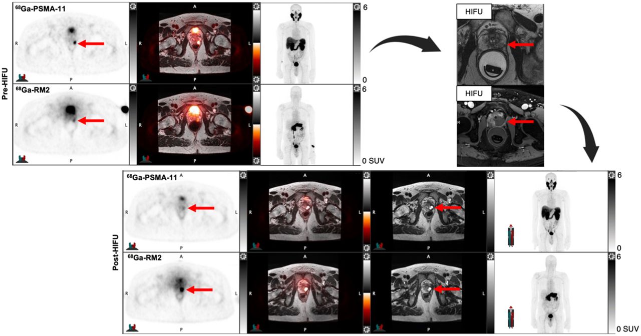
    A 62-y-old man with Gleason 3 + 4 PC in right lateral base and Gleason 3 + 3 PC in right posterior base (A shows color-coded needle tracks from biopsy; green: benign, yellow: Gleason 3 + 3, red: Gleason ≥ 3 + 4). He presented with PSA of 7.0 ng/mL and PSA density of 0.24 ng/mL2. Pretherapy 68Ga-RM2 (B) and 68Ga-PSMA-11 (C) axial PET/MRI and PET, respectively, show focal uptake in right prostate lesion (red arrows). This was treated with HIFU, and 6 mo later, uptake resolved on 68Ga-RM2 (D) and 68Ga-PSMA11 (E) axial PET/MRI and PET, respectively. Focal uptake in left prostate (blue arrows) was subsequently biopsied and showed nonaggressive PC. U = urethra with excreted 68Ga-RM2.

Tables

  • Figures
    • View popup
    TABLE 1.

    Patients’ Characteristics Before HIFU Ablation

    CharacteristicData
    n14
    Age (y)64.50 ± 8.00 (48.00–78.00)
    PSA (ng/mL)8.41 ± 3.47 (1.22–15.90)
    PSA density (ng/mL2)0.23 ± 0.09 (0.07–0.31)
    mpMRI18 lesions
     PI-RADS 53 (17%)
     PI-RADS 411 (61%)
     PI-RADS 34 (22%)
    Biopsy, Gleason grade18 lesions
     13 (17%)
     25 (28%)
     37 (39%)
     42 (11%)
     51 (5%)
    Risk
     Intermediate13
     High1
    Clinical stage
     T1c5
     T2a2
     T2b4
     T2c3
    68Ga-PSMA11
     Injected activity (MBq)151.33 ± 44.80 (70.30–222.00)
     Uptake time (min)46.50 ± 3.50 (44.00–57.00)
     Length of PET/MRI (min)49.00 ± 16.96 (30.00–83.00)
     Delay to pelvic PET/MRI (min)23.00 ± 9.19 (22.00–49.00)
    68Ga-RM2
     Injected activity (MBq)138.80 ± 4.61 (132.98–150.20)
     Uptake time (min)45.50 ± 2.12 (43.00–52.00)
     Length of PET/MRI (min)47.00 ± 6.58 (36.00–60.00)
     Delay to pelvic PET/MRI (min)25.00 ± 5.54 (11.00–37.00)
    Time between scans (d)5.50 ± 2.50 (2.00–9.00)
    • Qualitative data are number and percentage; continuous data are median ± SD and range.

    • View popup
    TABLE 2.

    Patients’ Characteristics After HIFU Ablation

    CharacteristicData
    n14
    PSA (ng/mL)2.83 ± 1.65 (0.02–5.79)
    PSA density (ng/mL2)0.07 ± 0.04 (0.00–0.17)
    PSA nadir (ng/mL)2.80 ± 1.48 (0.01–5.79)
    Time to PSA nadir (mo)6.55 ± 5.92 (2.90–24.83)
    Biopsy (n = 13)
    Residual lesions
     Clinically significant1
     Clinically insignificant3
    Recurrent lesions
     Clinically significant3
     Clinically insignificant6
    68Ga-PSMA11
     Injected activity (MBq)145.60 ± 37.75 (82.51–221.26)
     Uptake time (min)47.50 ± 2.40 (41.00–49.00)
     Length of PET/MRI (min)45.50 ± 5.90 (33.00–62.00)
     Delay to pelvic PET/MRI (min)26.00 ± 6.53 (22.00–48.00)
    68Ga-RM2
     Injected activity (MBq)139.77 ± 5.04 (133.32–149.67)
     Uptake time (min)46.00 ± 3.14 (39.00–52.00)
     Length of PET/MRI (min)51.50 ± 9.53 (41.00–73.00)
     Delay to pelvic PET/MRI (min)26.00 ± 6.22 (21.00–47.00)
    Time between scans (d)5.00 ± 40.66 (2.00–172.00)
    Time between pre- and post-HIFU scans (mo)7.43 ± 2.37 (5.93–12.60)
    • Qualitative data are number and percentage; continuous data are median ± SD and range.

    • View popup
    TABLE 3.

    Direct Comparison of mpMRI, 68Ga-PSMA11, and 68Ga-RM2 PET/MRI Findings Before and After HIFU Ablation

    mpMRI68Ga-PSMA1168Ga-RM2
    ParameterBefore HIFUAfter HIFUBefore HIFUAfter HIFUBefore HIFUAfter HIFU
    All lesions (n)18 (PI-RADS 3: 4; PI-RADS 4: 11; PI-RADS 5: 3)5 (PI-RADS 3: 4; PI-RADS 4: 1)239 (2 residual; 7 recurrent)239 (1 residual; 8 recurrent)
    Target lesions149/13 patients: negative; 3/13 patients: PI-RADS 3 (1 csPC, 2 ncsPC)142 residual (1 csPC; 2 ncsPC)121 residual (1 csPC)
    Sensitivity43%81%70%
    Specificity98%89%88%
    • View popup
    TABLE 4.

    SUVmax and SUVpeak of Target Lesions in Whole Body and Delayed Pelvic 68Ga-PSMA11 and 68Ga-RM2 PET/MRI Before and After HIFU Ablation

    68Ga-PSMA1168Ga-RM2
    ParameterWhole bodyDelayed pelvicWhole bodyDelayed pelvic
    Before HIFU, SUVmax9.51 (6.63–18.50)8.91 (6.66–18.94)7.70 (5.67–11.05)7.48 (4.97–11.51)
    After HIFU
     SUVmax2.27 (1.80–2.78)2.03 (1.80–2.51)2.55 (2.07–3.48)2.61 (1.68–2.74)
     P0.0010.0010.0050.006
    Before HIFU, SUVpeak5.04 (3.97–8.84)5.16 (4.27–9.50)5.22 (4.15–8.05)4.96 (4.02–8.57)
    After HIFU
     SUVpeak1.96 (1.89–2.31)2.11 (1.83–2.39)3.06 (2.85–3.49)2.81 (2.22–3.08)
     P0.0120.0680.0260.084
    • Data are median and interquartile range.

PreviousNext
Back to top

In this issue

Journal of Nuclear Medicine: 64 (4)
Journal of Nuclear Medicine
Vol. 64, Issue 4
April 1, 2023
  • Table of Contents
  • Table of Contents (PDF)
  • About the Cover
  • Index by author
  • Complete Issue (PDF)
Print
Download PDF
Article Alerts
Sign In to Email Alerts with your Email Address
Email Article

Thank you for your interest in spreading the word on Journal of Nuclear Medicine.

NOTE: We only request your email address so that the person you are recommending the page to knows that you wanted them to see it, and that it is not junk mail. We do not capture any email address.

Enter multiple addresses on separate lines or separate them with commas.
A Pilot Study of 68Ga-PSMA11 and 68Ga-RM2 PET/MRI for Evaluation of Prostate Cancer Response to High-Intensity Focused Ultrasound Therapy
(Your Name) has sent you a message from Journal of Nuclear Medicine
(Your Name) thought you would like to see the Journal of Nuclear Medicine web site.
Citation Tools
A Pilot Study of 68Ga-PSMA11 and 68Ga-RM2 PET/MRI for Evaluation of Prostate Cancer Response to High-Intensity Focused Ultrasound Therapy
Heying Duan, Pejman Ghanouni, Bruce Daniel, Jarrett Rosenberg, Guido A. Davidzon, Carina Mari Aparici, Christian Kunder, Geoffrey A. Sonn, Andrei Iagaru
Journal of Nuclear Medicine Apr 2023, 64 (4) 592-597; DOI: 10.2967/jnumed.122.264783

Citation Manager Formats

  • BibTeX
  • Bookends
  • EasyBib
  • EndNote (tagged)
  • EndNote 8 (xml)
  • Medlars
  • Mendeley
  • Papers
  • RefWorks Tagged
  • Ref Manager
  • RIS
  • Zotero
Share
A Pilot Study of 68Ga-PSMA11 and 68Ga-RM2 PET/MRI for Evaluation of Prostate Cancer Response to High-Intensity Focused Ultrasound Therapy
Heying Duan, Pejman Ghanouni, Bruce Daniel, Jarrett Rosenberg, Guido A. Davidzon, Carina Mari Aparici, Christian Kunder, Geoffrey A. Sonn, Andrei Iagaru
Journal of Nuclear Medicine Apr 2023, 64 (4) 592-597; DOI: 10.2967/jnumed.122.264783
Twitter logo Facebook logo LinkedIn logo Mendeley logo
  • Tweet Widget
  • Facebook Like
  • Google Plus One
Bookmark this article

Jump to section

  • Article
    • Visual Abstract
    • Abstract
    • MATERIALS AND METHODS
    • RESULTS
    • DISCUSSION
    • CONCLUSION
    • DISCLOSURE
    • Footnotes
    • REFERENCES
  • Figures & Data
  • Info & Metrics
  • PDF

Related Articles

  • No related articles found.
  • PubMed
  • Google Scholar

Cited By...

  • A Vision for Gastrin-Releasing Peptide Receptor Targeting for Imaging and Therapy: Perspective from Academia and Industry
  • Google Scholar

More in this TOC Section

  • SNMMI Procedure Standard/EANM Practice Guideline for Brain [18F]FDG PET Imaging, Version 2.0
  • Meeting Upcoming Clinical and Diagnostic Needs in Oncologic Imaging: A Structured Reporting System for Fibroblast-Activation-Protein–Targeted Imaging—FAP-RADS Version 1.0
  • Imaging Efficacy of [18F]CTT1057 PET for the Detection of PSMA-Positive Tumors Using Histopathology as Standard of Truth: Results from the GuideView Phase 2/3 Prospective Multicenter Study
Show more Clinical Investigation

Similar Articles

Keywords

  • 68Ga-RM2
  • 68Ga-PSMA11
  • PET
  • prostate cancer
  • HIFU
SNMMI

© 2025 SNMMI

Powered by HighWire