
Main menu

  • Home
  • Content
    • Current
    • Ahead of print
    • Past Issues
    • JNM Supplement
    • SNMMI Annual Meeting Abstracts
  • Subscriptions
    • Subscribers
    • Institutional and Non-member
    • Rates
    • Corporate & Special Sales
    • Journal Claims
  • Authors
    • Submit to JNM
    • Information for Authors
    • Assignment of Copyright
    • AQARA requirements
  • Info
    • Permissions
    • Advertisers
    • Continuing Education
  • About
    • About Us
    • Editorial Board
    • Contact Information
  • More
    • Alerts
    • Feedback
    • Help
    • SNMMI Journals
  • SNMMI
    • JNM
    • JNMT
    • SNMMI Journals
    • SNMMI

User menu

  • Subscribe
  • My alerts
  • Log in
  • My Cart

Search

  • Advanced search
Journal of Nuclear Medicine
  • SNMMI
    • JNM
    • JNMT
    • SNMMI Journals
    • SNMMI
  • Subscribe
  • My alerts
  • Log in
  • My Cart
Journal of Nuclear Medicine

Advanced Search

  • Home
  • Content
    • Current
    • Ahead of print
    • Past Issues
    • JNM Supplement
    • SNMMI Annual Meeting Abstracts
  • Subscriptions
    • Subscribers
    • Institutional and Non-member
    • Rates
    • Corporate & Special Sales
    • Journal Claims
  • Authors
    • Submit to JNM
    • Information for Authors
    • Assignment of Copyright
    • AQARA requirements
  • Info
    • Permissions
    • Advertisers
    • Continuing Education
  • About
    • About Us
    • Editorial Board
    • Contact Information
  • More
    • Alerts
    • Feedback
    • Help
    • SNMMI Journals
  • Follow JNM on Twitter
  • Visit JNM on Facebook
  • Join JNM on LinkedIn
  • Subscribe to our RSS feeds
Meeting ReportInstrumentation & Data Analysis

The Utility of an Injection Monitoring System in Diagnostic Imaging and Therapy: A Technologist perspective

Shelley Acuff, Kristen Smith and Dustin Osborne
Journal of Nuclear Medicine June 2022, 63 (supplement 2) 4109;
Shelley Acuff
1UT Medical Center
  • Find this author on Google Scholar
  • Find this author on PubMed
  • Search for this author on this site
Kristen Smith
2University of Tennessee
  • Find this author on Google Scholar
  • Find this author on PubMed
  • Search for this author on this site
Dustin Osborne
3University of Tennessee: Graduate School of Medicine
  • Find this author on Google Scholar
  • Find this author on PubMed
  • Search for this author on this site
  • Article
  • Figures & Data
  • Info & Metrics
Loading

Abstract

4109

Introduction: When an IV needs to be set and an injection is given there is always concern for an infiltration. In molecular imaging, the responsibility for the injection quality falls on the technologist. Although some infiltrations are obvious when they occur, some are more subtle with minor leakage out of the vessel. Until recently, the only way to check for a suspected infiltration after a radiotracer injection was to place the injection site in the imaging system field of view. This type of quality check can indicate some activity was left behind but can only be used when the technologist is aware of the infiltration. This post-injection technique: 1) gives no information on initially retained activity at the injection site, 2) no data on how long the extravasation persisted, and 3) gives no ability to intervene during the injection. For PET imaging, infiltration assessment is especially challenging as patients are typically imaged with their arms outside the field of view and any rescan of the injection site for infiltration confirmation requires a full scan of at least one bed position. It is known that a compromised injection in molecular imaging or a therapy administration can have a big impact on the outcome and can also have lasting effects. The purpose of this abstract is to discuss how an injection monitoring device can be helpful in monitoring infiltrations.

Methods: Over a 6-month period, an injection monitoring device was used to monitor multiple injections in a PET-CT department and multiple Y90 therapy injections in Interventional Radiology.

For the PET-CT injections each patient had 2 sensors monitoring the injection. Sensor #1 distal to IV location and sensor #2 on opposite arm in the same location for control. Upon completion of injection, the monitoring system generates an injection curve and score. For the Y90 therapy injections 4 sensors were used. One place on the injection line, one on the dose vial, one on the liver, and one on the patient’s foot. Upon completion of the injection the monitoring system generates an injection curve. No score is given for this type of injection.

A retrospective analysis was completed on both types of injection. In the PET-CT department, the injections were analyzed to see if the administering technologist suppected a comprised injection. In interventional Radiology, the injections were analyzed to see if the monitoring system could detect the therapy leaving the vial and entering the liver for use in real-time confirmation of successful dose delivery.

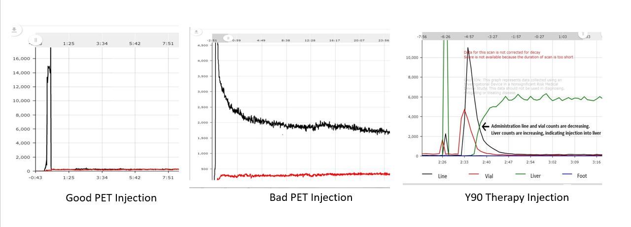
Results: In the PET-CT department, 590 injections were monitored. 61 injections were flagged by the monitoring system as less than idea injections all falling within minor to major range. Out of those 61 injections only 7 (11%) were noted by the technologist administering the injection to possibly be compromised at the time of injection before seeing the generated curve or score. This indicates that the remaining 89% of injections that were compromised to some degree would potentially have gone undetected without monitoring. In figure1 there is an example of a compromised injection curve and a non-compromised injection curve for reference.

In the Interventional Radiology department, the monitoring system was used on 15 Y90 therapies. The curves generated by the monitoring device indicated that on all 15 therapies the dose exited the vial and entered the liver with the added ability to measure residual activities prior to the patient leaving the procedure room. Figure 1 shows a good injection, bad injection, and a Y90 therapy injection.

Conclusions: Even with years of IV experience, a compromised injection can occur and go undetected. This may be more problematic for injections that take place outside of the traditional Nuclear Medicine setting. Actively monitoring injections can provide timely information on infiltrations and provide physicians with key information regarding possible injection issues to guide clinical decision making.

Figure
  • Download figure
  • Open in new tab
  • Download powerpoint
Previous
Back to top

In this issue

Journal of Nuclear Medicine
Vol. 63, Issue supplement 2
June 1, 2022
  • Table of Contents
  • Index by author
Article Alerts
Sign In to Email Alerts with your Email Address
Email Article

Thank you for your interest in spreading the word on Journal of Nuclear Medicine.

NOTE: We only request your email address so that the person you are recommending the page to knows that you wanted them to see it, and that it is not junk mail. We do not capture any email address.

Enter multiple addresses on separate lines or separate them with commas.
The Utility of an Injection Monitoring System in Diagnostic Imaging and Therapy: A Technologist perspective
(Your Name) has sent you a message from Journal of Nuclear Medicine
(Your Name) thought you would like to see the Journal of Nuclear Medicine web site.
Citation Tools
The Utility of an Injection Monitoring System in Diagnostic Imaging and Therapy: A Technologist perspective
Shelley Acuff, Kristen Smith, Dustin Osborne
Journal of Nuclear Medicine Jun 2022, 63 (supplement 2) 4109;

Citation Manager Formats

  • BibTeX
  • Bookends
  • EasyBib
  • EndNote (tagged)
  • EndNote 8 (xml)
  • Medlars
  • Mendeley
  • Papers
  • RefWorks Tagged
  • Ref Manager
  • RIS
  • Zotero
Share
The Utility of an Injection Monitoring System in Diagnostic Imaging and Therapy: A Technologist perspective
Shelley Acuff, Kristen Smith, Dustin Osborne
Journal of Nuclear Medicine Jun 2022, 63 (supplement 2) 4109;
Twitter logo Facebook logo LinkedIn logo Mendeley logo
  • Tweet Widget
  • Facebook Like
  • Google Plus One
Bookmark this article

Jump to section

  • Article
  • Figures & Data
  • Info & Metrics

Related Articles

  • No related articles found.
  • Google Scholar

Cited By...

  • No citing articles found.
  • Google Scholar

More in this TOC Section

  • Partial injection technique for real time infiltration assessment
  • Explore the Functions of Super Iterative Reconstruction Technology on 68Ga-FAPI PET/MR Imaging
Show more Instrumentation & Data Analysis

Similar Articles

SNMMI

© 2023 Journal of Nuclear Medicine

Powered by HighWire