


Abstract
3333
Introduction: Heart failure (HF) is a chronic and progressive clinical syndrome in which the heart structure or function is abnormal due to multiple factors. Fibroblast activation protein (FAP) is robustly expressed by activated cardiac fibroblasts and minimal expression in normal hearts, which has been a potential candidate for targeting of pathological cardiac fibroblasts and the progression of HF. Currently, the 68Ga-labeled fibroblast activating protein inhibitor (68Ga-FAPI), as an outstanding target to FAP, is proved to display the activated fibroblast response noninvasively with the advantage of high target to background ratios. The purpose of this study was to use 68Ga-FAPI image to continuously visualize the dynamic change process of HF, monitor the progress of HF, and try to help noninvasive visual diagnosis of HF.
Methods: The rat model of heart failure was established by subcutaneous injection of isoproterenol in 5 mg/kg/d for 14 days continuously. The rats were performed echocardiographic tests the day before FAPI imaging. At the level of the left ventricle papillary muscles, M-mode images guided by two-dimensional images of parasternal short-axis views were acquired. left ventricular ejection fraction (LVEF); left ventricular fraction shortening (LVFS); systole interventricular septal thickness (IVSs); diastole interventricular septal thickness (IVSd); systole posterior wall thickness (LVPWs); diastole posterior wall thickness (LVPWd) were measured. 68Ga-FAPI imaging was performed at one-week intervals from the start of the model preparation until four weeks. Then, isolated hearts were taken weekly for biodistribution, autoradiography, Hematoxylin-eosin (H&E) staining and Masson's trichrome staining. Anova or T-test were used to compare the differences between groups. P < 0.05 indicates statistical significance.
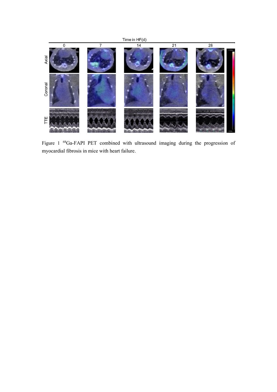
Results: Extensive myocardial uptake of 68Ga-FAPI-04 peaked 7 days after the onset of cardiac failure modeling, and was primarily seen in the left ventricular wall. Additionally, the contractile ability of myocardium was also enhanced. With modeling time extended, HF developed further, and the intensity of myocardial uptake and ventricular wall movement decreased by Echocardiographic examination. Compared with the control group, LVEF, LVFS, LVPWd, LVPWs, IVSd and IVSs significantly increased at 7 days after injection of isoproterenol, and then these parameters gradually decreased and had a significantly statistical difference at 21 days and 28 days (p<0.05). Additionally, Biological distribution of isolated heart tissue showed that the concentration of myocardial imaging agent was strongest at the apex on the seventh day of modeling (p < 0.01). At the same time, the H&E, Masson, and IHC staining showed that slightly fibrotic changes were found on day 7 but the expression of FAP protein was most pronounced. As the disease progressed, fibrosis was most severe at 28 days when FAP protein failed to be detected.
Conclusions: From continuous imaging of animal models of heart failure, In the early stage of heart failure, the uptake of imaging agent was obvious, suggesting that the expression of activated fibrin was significantly increased. Nevertheless, the uptake of 68Ga-FAPI in myocardium gradually decreased to almost invisible with the time extension, which may be related to the gradual decrease to near cessation of the activated fiber expression. These images were confirmed by immunohistochemistry and Masson staining. The preliminary study suggested that 68Ga-FAPI PET may be used to show active fibrosis, which may have clinical significance for guiding anti-fibrosis drug treatment decisions.


In this issue


Jump to section
Related Articles
Cited By...
- No citing articles found.