


Abstract
3209
Introduction: Multiple myeloma (MM) is typically characterized by an infinite proliferation of malignant plasma cells and an activated protein synthesis process.11C-Methionine (MET), as a radiolabeled amio acid tracer, has been widely used for MM imaging. Previously we have conducted a 60 min dynamic 11C-Methionine PET/CT study. In this work, we aimed to further simplify the dynamic scanning process using shorter scaning time.
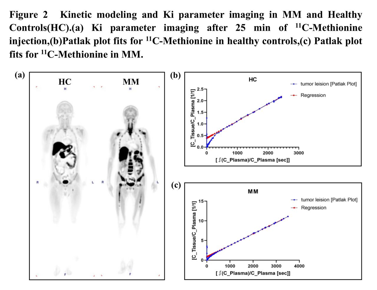
Methods: 7 patients with pathologically proven MM were included as well as 2 healthy as controls. The dynamic 11C-Methionine PET/CT was performed on the total-body PET/CT (uEXPLORER, United-Imaging Healthcare). Data were scanned for 60 min after injection and reconstructed into 92 frames according to the acquisition protocol below: 30 × 2 s, 12 × 5 s, 6 × 10 s, 4 × 30 s, 25 × 60 s, and 15 × 120 s (as shown in Figure1). The volume of interest (VOIs) within muscle and pathologically tumor leision (the right hipbone) were drawn manually using a commercial software (PMOD, Version 4.2, Zürich, Switzerland). The descending aorta was used as the input function for the total body kinetic modeling in the Image derived input function (IDIF) method. The transport kinetics of the 25min 11C-Methionine post-injection was well described by patlak plot model, and then the net influx constant (Ki) was obtained. The correlations between standard uptake value (SUVmean/max) and Ki were analyzed by Spearman correlation analysis. All the statistical analyses were performed using SPSS Statistics for Windows, Version 13.0 (SPSS Inc., Chicago, IL, USA). Figures were generated by GraphPad Prism 8 (GraphPad Software Inc., San Diego, CA, USA).
Results: At the 25 min after injection, a patlak plot could well fit the 11C-Methionine dynamics in pathological tumor lesions as shown in Figure 2. The net influx rate Ki derived from the patlak plot model was confirmed to have a strong positive correlation with the SUVmean/max of tumor lesions (as shown Figure 3).The correlation coefficients between SUVmean/max and Ki were 0.950 (P<0.01), 0.783 (P<0.05), respectively. The SUVmean values of pathological tumor lesions at 20–25 min and 50-60 min post-injection were similar (Figure 4). Comparing Ki parametric imaging with SUVbw (standardized uptake value based on body weight), we found that Ki parametric imaging improved the lesion visibility (Figure 5).
Conclusions: Our study has confirmed that the clinical scanning time of 11C-Methionine PET/CT can be reasonably shorten to 25 min. Appropriate pharmacokinetic models can be selected to simplify clinical protocols.
Acknowledgements: The study was sponsored by the National Natural Science Foundation of China (Grant Nos.82102089).


In this issue


Jump to section
Related Articles
Cited By...
- No citing articles found.