


Abstract
3125
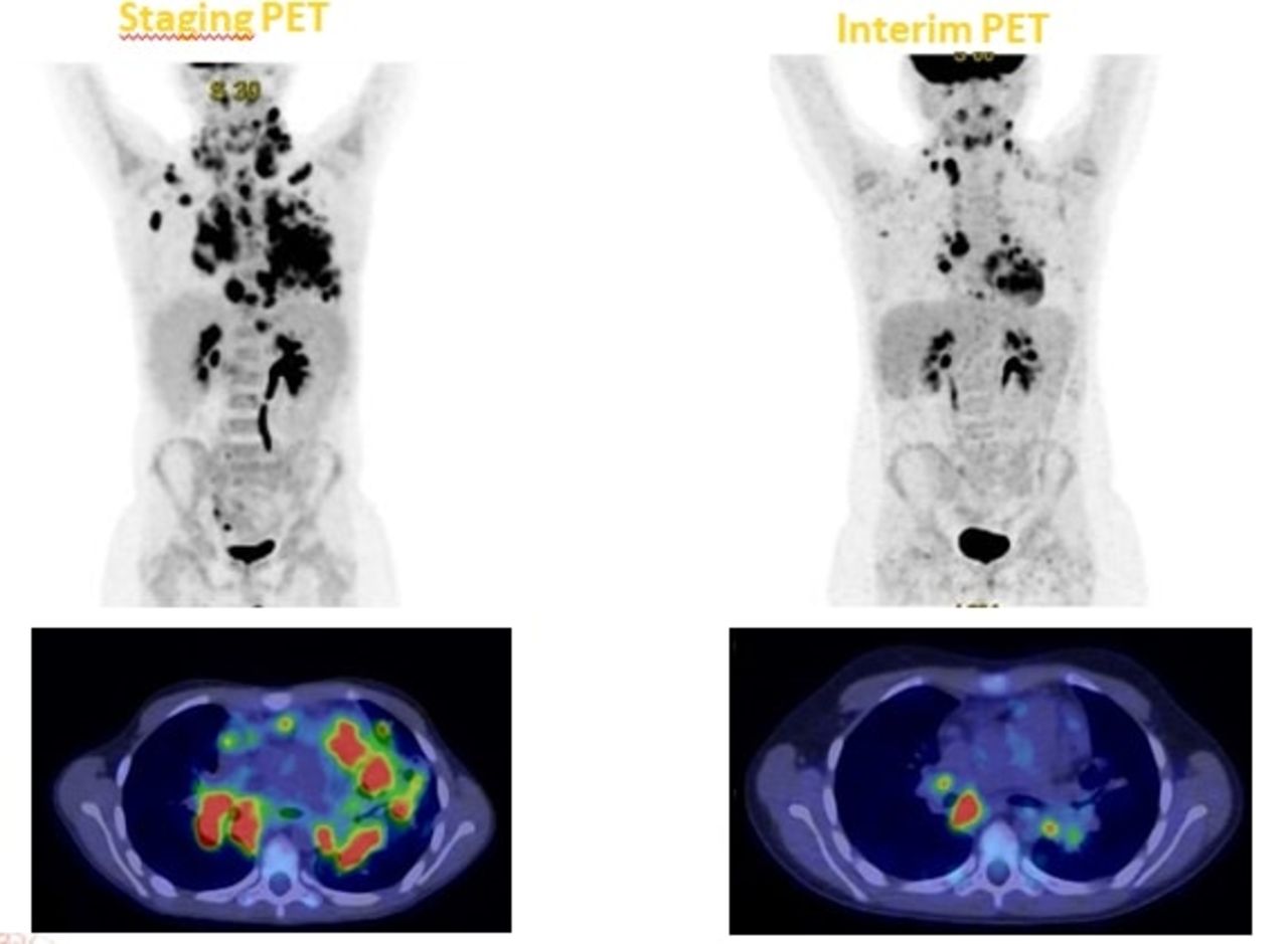
Introduction: 18-Fluorodeoxyglucose (FDG) positron emission tomography/computed tomography (PET/CT) is an useful imaging modality for staging and for the evaluation of response to therapy in Hodgkin lymphoma (HL). It has been demonstrated that interim-PET has also a prognostic role. This study aimed to explore the prognostic role of i-PET in paediatric patients with HL with the calculation of qPET , that is the ratio of SUVpeak assessed in a target lesion to the liver SUVmean.
Methods: We retrospectively evaluated 63 children (32 males, mean age 13, range 5-17) with newly diagnosed HL, stage I-IV disease (14 stage I- IIA, 49 stage IIB-IVB). All patients were studied with 18F-FDG PET/CT prior to treatment(baseline PET) and after two, three or four cycles of chemotherapy (i-PET). Lesions with the highest FDG uptake on i-PET were selected as ‘‘reference lesion’’. qPET was calculated for each patient; we considered the study positive with a cut-off of qPET ≥ 1.3 (Hasenclever et al. 2014). Correlation between the values of qPET and clinical outcome of patients in terms of DFS and OS was evaluated using the Fisher exact text, with a p value < 0.05 considered as significant.
Results: The median follow-up was 44 months for DFS and 55 months for OS; 2 patients had progression of disease and died, while 8 patients relapsed (at last follow-up all of them are still alive and 1/8 has still affected by disease). i-PET scan resulted negative in 56 patients (89%) and positive in 7 patients (11%).The values of qPET of patients with persistence, relapse or progression of disease were 1.32 (average value; median value 0.86, range 0.46-3.93); the corresponding values of qPET in not relapsed patients were 0.91 (average value; median value 0.84, range 0.34-5.27). No significant correlation was found between qPET and DFS (p-value 0.07). qPET values of patients that are still alive and of those who died for progression of disease were respectively 0.91 (average value; median value 0.83; range 0.34-5.27) and 3.11 (average value; median value 3.11; range 2.29-3.93. In this case we found a correlation between qPET values and OS (p-value 0.01); the positive (PPV) and negative predictive values (NPV) of i-PET were 100 % and 92%, respectively. Patients with a i-PET with a qPET value ≥1.3 had a worst prognosis in terms of OS.
Conclusions: Our study suggested that a cut-off of qPET of 1.3 better discriminated patients with poor prognosis from the others in terms of OS and it could be the proof of the prognostic value of interim PET in paediatric Hodgkin lymphoma. Further studies are needed to confirm our results.

In this issue


Jump to section
Related Articles
Cited By...
- No citing articles found.