


Abstract
2970
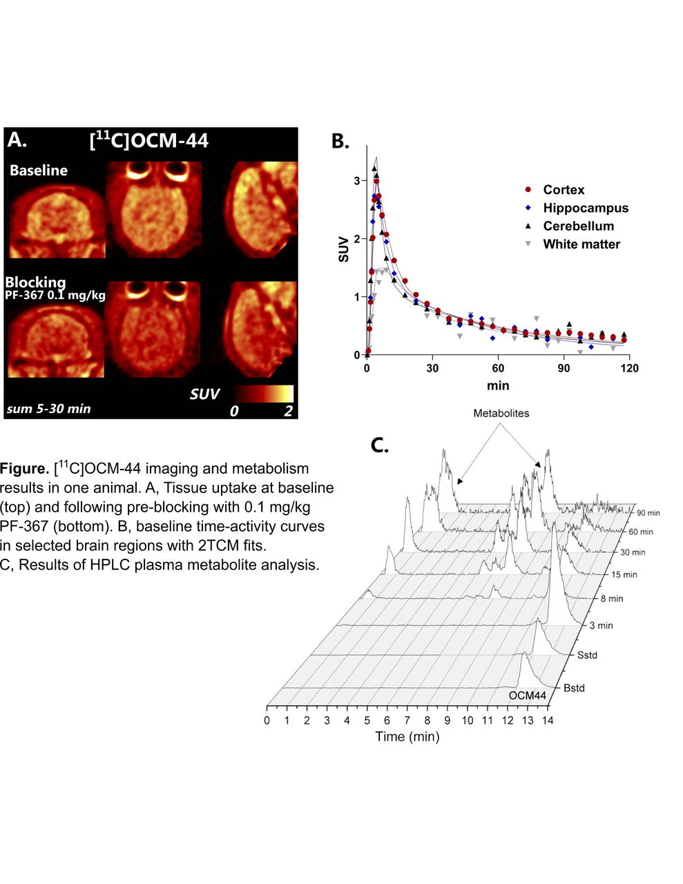
Introduction: Glycogen synthase kinase 3 (GSK-3) is a key enzyme in intracellular signaling pathways mediating physiological functions throughout the central nervous system and periphery. GSK-3 dysfunction is implicated in neuropsychiatric disorders including Alzheimer’s disease and mood disorders. Several candidate compounds for GSK-3 labeling were recently developed by the Vasdev lab based on oxazole-4-carboxamide inhibitors, with excellent potency and selectivity profiles. Preliminary PET imaging in non-human primates revealed [11C]OCM-44 as having the highest brain uptake [1]. The goal of this work is to characterize the in vivo imaging properties of [11C]OCM-44 using arterial sampling and quantitative kinetic modeling.
Methods: [11C]OCM-44 (412 ± 119 MBq/nmol) was prepared as described previously [1]. A total of five PET scans were conducted in two rhesus macaques using a Focus 220 scanner. [11C]OCM-44 scans (178 ± 2.1 MBq) were performed at baseline for each animal, along with three blocking scans acquired after injection of the GSK-3 inhibitor PF-367 (0.03 mg/kg, 0.1 mg/kg, or 0.25 mg/kg). Dynamic PET data were acquired for 120 minutes. Arterial blood samples were drawn to measure radioactivity concentration in plasma, perform metabolite analysis by HPLC, and determine free fraction in plasma (fp). Regional brain time-activity curves (TACs) were extracted and fitted with the one- and two-tissue compartment models (1TCM and 2TCM) using metabolite-corrected arterial input functions. Reduction in specific binding was assessed using occupancy plots.
Results: [11C]OCM-44 metabolism was relatively rapid, with ~20% parent compound remaining after 30 minutes. HPLC analysis in plasma showed radiometabolites emerging over the duration of the scan, including some with similar lipophilicity to the parent molecule present after approximately 15 minutes. Baseline fp was 3% in both animals, increasing to 7-10% in blocking scans. Radiotracer uptake was widespread across grey matter regions and lower in white matter. Peak SUV across regions was 1.9 ± 0.2 and 3.0 ± 0.2 in the two animals.
In baseline scans, the 2TCM produced good model fits and stable parameter estimates. Fits were generally poor with the 1TCM based on visual inspection and inferior based on the Akaike Information Criterion. Volume of distribution (VT) estimates from the 2TCM were in the range of 1.7-3.1 mL/cm3, with highest values in frontal and cingulate cortices and globus pallidus. K1 values were 0.1-0.5 mL∙cm-3∙min-1. However, stable parameter estimates could not be determined in all blocking scans (VT relative standard error >20% in most regions). To evaluate binding specificity, data from each scan were fitted again using only the first 60 minutes, which produced acceptable 2TCM model fits and parameter estimates. VT values from the truncated baseline scans were 14 ± 18% lower than those generated from full scan data. Absolute VT values were reduced in blocking scans following 0.03 mg/kg and 0.1 mg/kg PF-367 but not the 0.25 mg/kg dose. Using VT/fP to correct for the observed increase in fP during blocking, binding reductions were estimated to be 64%, 90%, and 81%, respectively, in blocking scans with 0.03, 0.1, and 0.25 mg/kg dose of PF-367.
Conclusions: Consistent with prior work, [11C]OCM-44 shows good brain uptake and specific binding to GSK-3 in non-human primates. However, metabolite profiles and tissue kinetics reveal the possibility of brain-penetrant metabolites, which limit the accuracy of quantitative parameters and, if confirmed, translational potential. Parallel experiments in mice have corroborated the metabolic instability of [11C]OCM-44 in vivo [3] and identified rapid formation of radiometabolites in tumor-bearing mice. Kinetic analysis of [11C]OCM-44 in non-human primates is an informative step forward in the effort to develop a GSK-3-selective radiotracer for neuroimaging. Kinetic evaluation of two promising 18F-labeled analogs of OCM-44 [1,2] is underway.


In this issue


Jump to section
Related Articles
Cited By...
- No citing articles found.