


Abstract
2952
Introduction: Alzheimer’s Disease (AD) is a neurodegenerative disorder characterized by the accumulation of amyloid-β plaques and aggregated tau tangles in the brain. The progression of tau neuropathology typically starts in the entorhinal cortex (ERC) before spreading throughout the temporal lobe. PET imaging is an important method for imaging early, pre-symptomatic AD development. [F-18]MK6240 is a next-generation radiotracer that binds to neurofibrillary tangles with high affinity and is useful for analyzing and staging AD progression. However, off-target binding in the close-proximity regions of the meninges and sinus can confound the ability to accurately quantify specific MK6240 binding due to “spill-in" signal to the reference and target regions of the brain. In this study, we simulate different MK6240 off-target binding distributions from PET scans at our site to evaluate how they influence detection limits of early-stage AD pathology within the ERC.
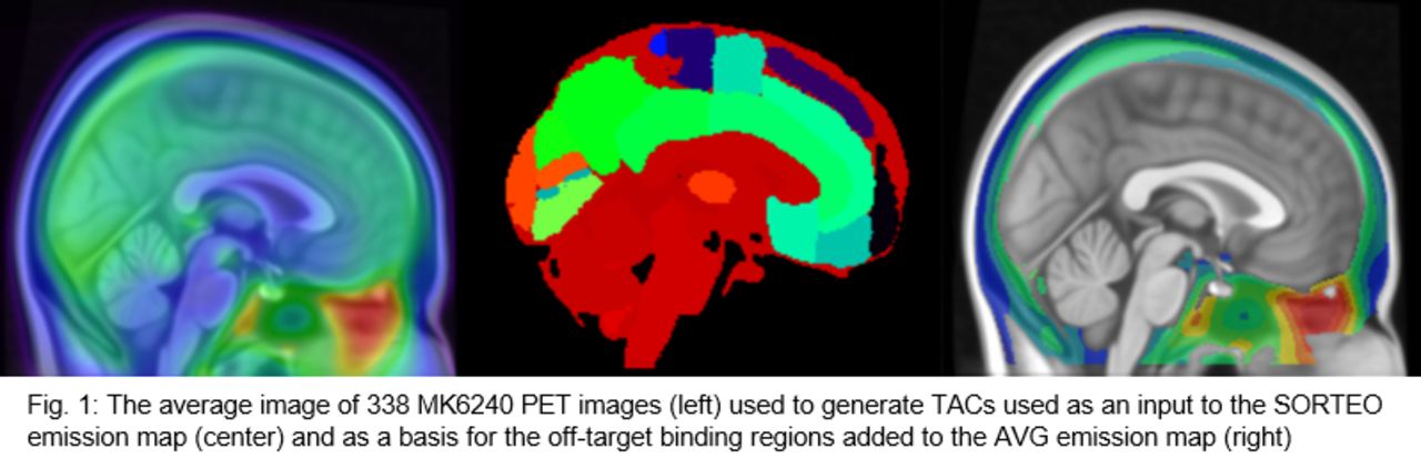
Methods: PET brain images in this study were simulated using PET-SORTEO, a Monte-Carlo based simulation software that generates realistic PET data from an input emission brain map with associated time activity curves (TAC). Our emission map was based on the spatial distribution of the Harvard-Oxford atlas with additional regions including white matter, ventricles and areas of increased tauopathy in the ERC consistent with Braak stage I. Further regions of off-target binding in the meninges and sinus around Meckel’s cave were also included based on MK6240 scans from our center. Four emission maps were used in our simulations with a range of off-target binding profiles: one with no off-target binding or signal (NONE), one based on the average of 338 MK6240 scans from the same scanner (AVG) and two based on representative PET scans with mean meninges and sinus binding that were 10% below average (LOW) and 60% above average (HIGH). TACs for cortical brain regions were based on averages of all MK6240 scans from our site. Ten simulations were generated for each group consisting of 4x5minute frames starting 70 minutes post-injection (i.d.= 370MBq) and then reconstructed using filtered back projection with a ramp filter. The frames of the reconstructed images were then summed, registered into standard space using a rigid body transform and converted into an SUVR image using inferior cerebellar grey matter as the reference region. The Harvard-Oxford atlas anterior parahippocampal gyrus region was used to evaluate the ERC with an SUVR ≥1.27 in this region considered tau positive. To evaluate detection sensitivity of these different cohorts we varied the input activity concentration in the ERC between 6 and 9.5 times the activity concentration in the reference region and then determined the detection limit of a group of simulations as the input activity (representing the concentration of tau pathology) necessary to have an average ERC SUVR at least two standard deviations above the tau(+) threshold.
Results: The detection limits for the NONE, LOW, AVG and HIGH cohorts were at activity concentrations of 6.8, 7.8, 8.1 and 8.3 times the reference region respectively. At the NONE detection limit of 6.8 times reference the AVG cohort has an ERC SUV 8.2% higher, compared to an ERC SUV 10.5% and 15.0% for LOW and HIGH. However, the cerebellar SUV for AVG, LOW and HIGH were 12.8%, 11.5% and 17.5% higher, decreasing the global SUVR and leading to a decrease in the ERC SUVR of 4.1%, 3.8% and 5.6% respectively.
Conclusions: While there is a large gap between the simulations with and without off-target binding in the meninges and sinus, the difference between the LOW, AVG and HIGH groups is marginal. The effects of activity from the sinus increasing the ERC SUV and the meninges increasing the cerebellar SUV act in different directions on the SUVR outcome, indicating that for accurate quantification of early tau accumulation and tau(+/-) stratification measured with MK6240 the pattern of off-target binding needs to be taken into account.


In this issue


Jump to section
Related Articles
Cited By...
- No citing articles found.