
Main menu

  • Home
  • Content
    • Current
    • Ahead of print
    • Past Issues
    • JNM Supplement
    • SNMMI Annual Meeting Abstracts
    • Continuing Education
    • JNM Podcasts
  • Subscriptions
    • Subscribers
    • Institutional and Non-member
    • Rates
    • Journal Claims
    • Corporate & Special Sales
  • Authors
    • Submit to JNM
    • Information for Authors
    • Assignment of Copyright
    • AQARA requirements
  • Info
    • Reviewers
    • Permissions
    • Advertisers
  • About
    • About Us
    • Editorial Board
    • Contact Information
  • More
    • Alerts
    • Feedback
    • Help
    • SNMMI Journals
  • SNMMI
    • JNM
    • JNMT
    • SNMMI Journals
    • SNMMI

User menu

  • Subscribe
  • My alerts
  • Log in
  • My Cart

Search

  • Advanced search
Journal of Nuclear Medicine
  • SNMMI
    • JNM
    • JNMT
    • SNMMI Journals
    • SNMMI
  • Subscribe
  • My alerts
  • Log in
  • My Cart
Journal of Nuclear Medicine

Advanced Search

  • Home
  • Content
    • Current
    • Ahead of print
    • Past Issues
    • JNM Supplement
    • SNMMI Annual Meeting Abstracts
    • Continuing Education
    • JNM Podcasts
  • Subscriptions
    • Subscribers
    • Institutional and Non-member
    • Rates
    • Journal Claims
    • Corporate & Special Sales
  • Authors
    • Submit to JNM
    • Information for Authors
    • Assignment of Copyright
    • AQARA requirements
  • Info
    • Reviewers
    • Permissions
    • Advertisers
  • About
    • About Us
    • Editorial Board
    • Contact Information
  • More
    • Alerts
    • Feedback
    • Help
    • SNMMI Journals
  • View or Listen to JNM Podcast
  • Visit JNM on Facebook
  • Join JNM on LinkedIn
  • Follow JNM on Twitter
  • Subscribe to our RSS feeds
Meeting ReportOutcomes/Comparative Effectiveness Research & Radiation Safety

18F-FDG Fetal Dosimetry Calculated with PET/MRI

Paolo Zanotti Fregonara, Tatsuya Ishiguro, Kosuke Yoshihara, Shiro Ishii and Takayuki Enomoto
Journal of Nuclear Medicine August 2022, 63 (supplement 2) 2775;
Paolo Zanotti Fregonara
1Molecular Imaging Branch, NIMH-NIH, Bethesda, MD, USA
  • Find this author on Google Scholar
  • Find this author on PubMed
  • Search for this author on this site
Tatsuya Ishiguro
1Molecular Imaging Branch, NIMH-NIH, Bethesda, MD, USA
  • Find this author on Google Scholar
  • Find this author on PubMed
  • Search for this author on this site
Kosuke Yoshihara
1Molecular Imaging Branch, NIMH-NIH, Bethesda, MD, USA
  • Find this author on Google Scholar
  • Find this author on PubMed
  • Search for this author on this site
Shiro Ishii
1Molecular Imaging Branch, NIMH-NIH, Bethesda, MD, USA
  • Find this author on Google Scholar
  • Find this author on PubMed
  • Search for this author on this site
Takayuki Enomoto
1Molecular Imaging Branch, NIMH-NIH, Bethesda, MD, USA
  • Find this author on Google Scholar
  • Find this author on PubMed
  • Search for this author on this site
  • Article
  • Figures & Data
  • Info & Metrics
Loading

Abstract

2775

Introduction: The fetal absorbed dose from 18F-FDG administration to the mother is the most important piece of information when considering the use of PET to stage cancers during pregnancy. However, the few existing human case reports were obtained using either PET-only or PET/CT machines, which may not accurately identify the soft tissues of the fetus for dosimetric calculations. This study presents data from 11 women injected with 18F-FDG for cancer staging during the first two trimesters of pregnancy. This series significantly expands the pool of available human dosimetric data and is the first to be entirely acquired with PET/MRI.

Methods: Eleven pregnant women (12 scans) with cervical cancer were imaged with 18F-FDG PET/MRI, and their images were retrospectively analyzed for this study. The fraction of injected activity concentrated by the fetus was derived from manually drawing regions of interest on the MRI slices. From the activity fraction, the fetal time-integrated coefficients were derived and combined with the standard coefficients of the mothers' organs. The absorbed doses were calculated with OLINDA/EXM 1.1 and a dynamic bladder model.

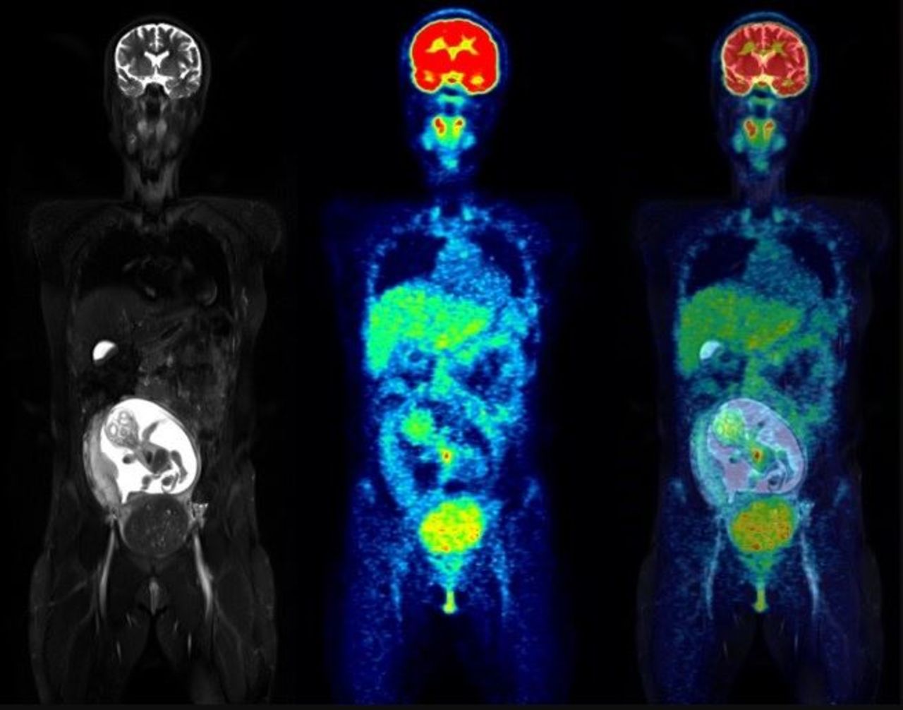
Results: All fetuses after early pregnancy could be accurately delineated due to the coregistered MRI scans. 18F-FDG activity was unevenly distributed in the fetal body. The hearts and the bladders were generally visible, while the brain showed lower uptake. The estimated doses were 2.21E-02 mGy/MBq for one woman imaged in early pregnancy, 7.38 ± 0.25 E-03 mGy/MBq for three women imaged at the end of the first trimester, and 4.92 ± 1.53 E-03 mGy/MBq for eight women imaged during the second trimester.

Conclusions: PET/MRI allows precise delineation of fetal bodies in women injected with 18F-FDG during pregnancy. The resulting dosimetric values, which are likely more accurate than those obtained with PET only or PET/CT, confirm that the fetal 18F-FDG dose is very low. Therefore, clinically appropriate 18F-FDG scans in women with cancer should not be withheld because of pregnancy.

Figure
  • Download figure
  • Open in new tab
  • Download powerpoint
Previous
Back to top

In this issue

Journal of Nuclear Medicine
Vol. 63, Issue supplement 2
August 1, 2022
  • Table of Contents
  • Index by author
Article Alerts
Sign In to Email Alerts with your Email Address
Email Article

Thank you for your interest in spreading the word on Journal of Nuclear Medicine.

NOTE: We only request your email address so that the person you are recommending the page to knows that you wanted them to see it, and that it is not junk mail. We do not capture any email address.

Enter multiple addresses on separate lines or separate them with commas.
18F-FDG Fetal Dosimetry Calculated with PET/MRI
(Your Name) has sent you a message from Journal of Nuclear Medicine
(Your Name) thought you would like to see the Journal of Nuclear Medicine web site.
Citation Tools
18F-FDG Fetal Dosimetry Calculated with PET/MRI
Paolo Zanotti Fregonara, Tatsuya Ishiguro, Kosuke Yoshihara, Shiro Ishii, Takayuki Enomoto
Journal of Nuclear Medicine Aug 2022, 63 (supplement 2) 2775;

Citation Manager Formats

  • BibTeX
  • Bookends
  • EasyBib
  • EndNote (tagged)
  • EndNote 8 (xml)
  • Medlars
  • Mendeley
  • Papers
  • RefWorks Tagged
  • Ref Manager
  • RIS
  • Zotero
Share
18F-FDG Fetal Dosimetry Calculated with PET/MRI
Paolo Zanotti Fregonara, Tatsuya Ishiguro, Kosuke Yoshihara, Shiro Ishii, Takayuki Enomoto
Journal of Nuclear Medicine Aug 2022, 63 (supplement 2) 2775;
Twitter logo Facebook logo LinkedIn logo Mendeley logo
  • Tweet Widget
  • Facebook Like
  • Google Plus One
Bookmark this article

Jump to section

  • Article
  • Figures & Data
  • Info & Metrics

Related Articles

  • No related articles found.
  • Google Scholar

Cited By...

  • No citing articles found.
  • Google Scholar

More in this TOC Section

  • Cost-effectiveness of F-18 FDG PET/CT in lung and colorectal cancer: a systematic review and narrative synthesis
  • Diagnostic efficacy of 99mTc-sulfur colloid lymphoscintigraphy in chyle leak and incremental value of SPECT-CT in localizing the site of chyle leak
Show more Outcomes/Comparative Effectiveness Research & Radiation Safety

Similar Articles

SNMMI

© 2025 SNMMI

Powered by HighWire