
Main menu

  • Home
  • Content
    • Current
    • Ahead of print
    • Past Issues
    • JNM Supplement
    • SNMMI Annual Meeting Abstracts
  • Subscriptions
    • Subscribers
    • Institutional and Non-member
    • Rates
    • Corporate & Special Sales
    • Journal Claims
  • Authors
    • Submit to JNM
    • Information for Authors
    • Assignment of Copyright
    • AQARA requirements
  • Info
    • Permissions
    • Advertisers
    • Continuing Education
  • About
    • About Us
    • Editorial Board
    • Contact Information
  • More
    • Alerts
    • Feedback
    • Help
    • SNMMI Journals
  • SNMMI
    • JNM
    • JNMT
    • SNMMI Journals
    • SNMMI

User menu

  • Subscribe
  • My alerts
  • Log in
  • My Cart

Search

  • Advanced search
Journal of Nuclear Medicine
  • SNMMI
    • JNM
    • JNMT
    • SNMMI Journals
    • SNMMI
  • Subscribe
  • My alerts
  • Log in
  • My Cart
Journal of Nuclear Medicine

Advanced Search

  • Home
  • Content
    • Current
    • Ahead of print
    • Past Issues
    • JNM Supplement
    • SNMMI Annual Meeting Abstracts
  • Subscriptions
    • Subscribers
    • Institutional and Non-member
    • Rates
    • Corporate & Special Sales
    • Journal Claims
  • Authors
    • Submit to JNM
    • Information for Authors
    • Assignment of Copyright
    • AQARA requirements
  • Info
    • Permissions
    • Advertisers
    • Continuing Education
  • About
    • About Us
    • Editorial Board
    • Contact Information
  • More
    • Alerts
    • Feedback
    • Help
    • SNMMI Journals
  • Follow JNM on Twitter
  • Visit JNM on Facebook
  • Join JNM on LinkedIn
  • Subscribe to our RSS feeds
Meeting ReportGeneral Clinical Specialties

Multimodality imaging in calcific uremic arteriolopathy

Pokhraj Suthar, Khushboo Gupta, Rahul Jadhav, Sindhuja Sivanandham and Rameshwar Prasad
Journal of Nuclear Medicine June 2022, 63 (supplement 2) 2673;
Pokhraj Suthar
1Rush University Medical Center
  • Find this author on Google Scholar
  • Find this author on PubMed
  • Search for this author on this site
Khushboo Gupta
2Mount Sinai Radiology
  • Find this author on Google Scholar
  • Find this author on PubMed
  • Search for this author on this site
Rahul Jadhav
  • Find this author on Google Scholar
  • Find this author on PubMed
  • Search for this author on this site
Sindhuja Sivanandham
1Rush University Medical Center
  • Find this author on Google Scholar
  • Find this author on PubMed
  • Search for this author on this site
Rameshwar Prasad
1Rush University Medical Center
  • Find this author on Google Scholar
  • Find this author on PubMed
  • Search for this author on this site
  • Article
  • Figures & Data
  • Info & Metrics
Loading

Abstract

2673

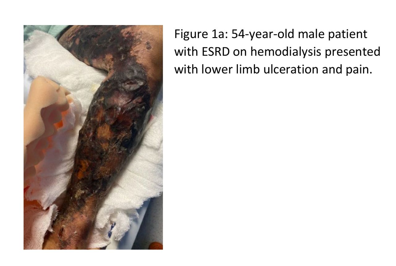
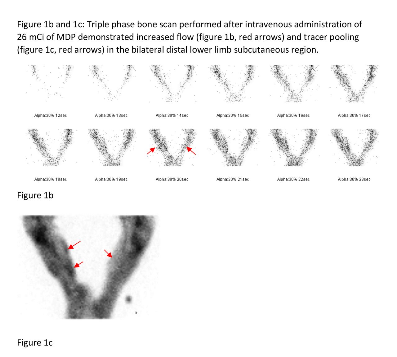
Introduction: Calcific uremic arteriolopathy, also known as calcific vasculopathy or calciphylaxis, is characterized by necrosis of the skin and fatty tissues. The cause is largely related to calcium deposition in soft tissues causing small blood vessel occlusion and skin necrosis. The disease is associated with renal failure and has high 1-year mortality rate of more than 50% (1). Diagnosis is based on clinical suspicion, imaging with radiographs, CT and bone scans, and tissue biopsy. This educational exhibit highlights the role of imaging modalities in early diagnosis and localization of this mortal disease.

Methods: We retrospectively evaluated referred cases to our department for bone scan with high clinical suspicion for calciphylaxis. Multi-imaging data was collected and their role in evaluation of the disease was assessed. Finally the histopathology results were used to confirm the calcific vasculopathy.

Results: Presented in the exhibit are picturesque illustration of calcific vasculopathy lesions (clinical presentation), evaluated on Bone scan, radiographs and CT scan. The cases include peripheral (truncal and appendicular) as well as visceral calciphylaxis and their pathology confirmation.

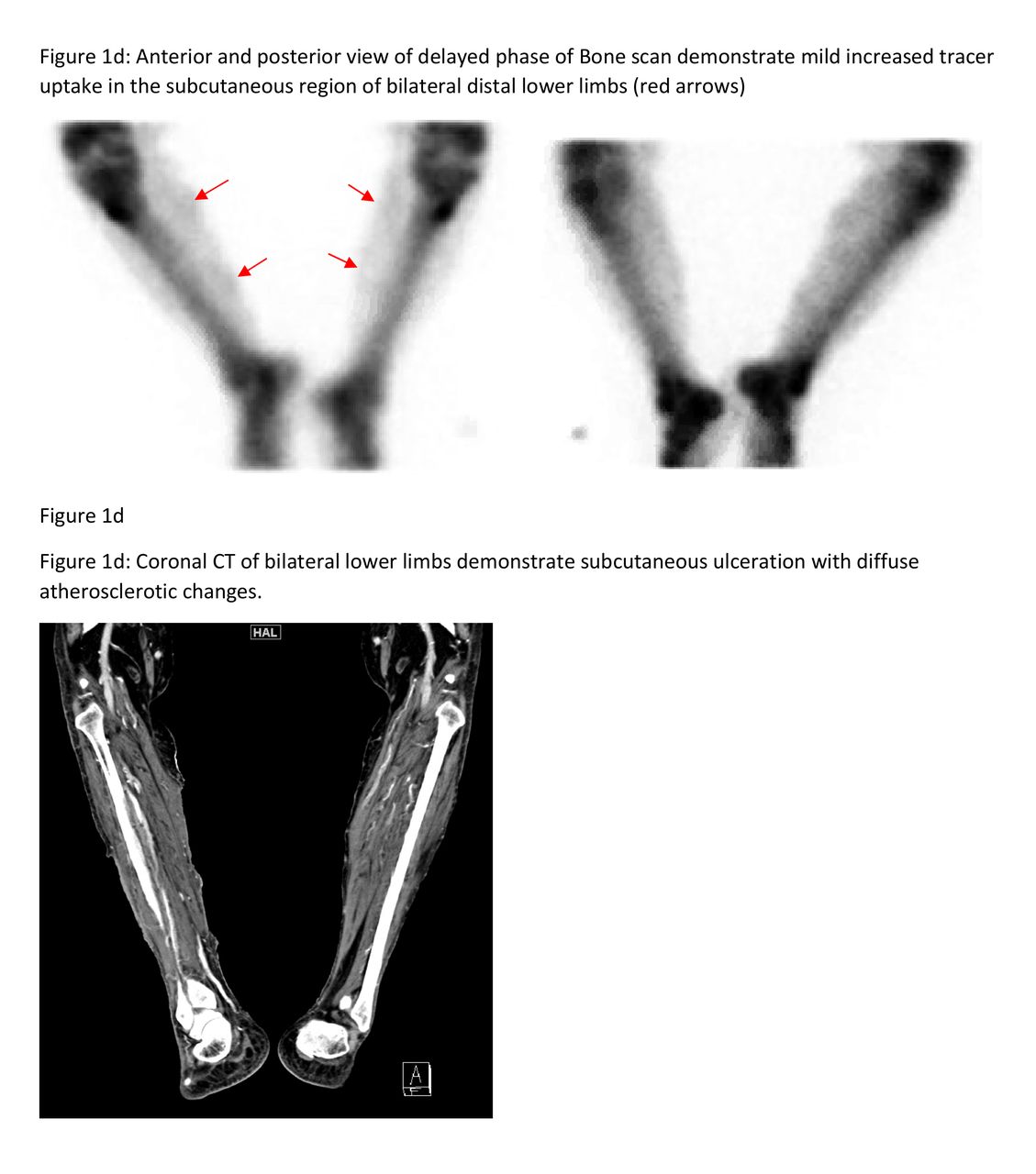
Conclusions: Calcific uremic arteriolopathy aka calcific vasculopathy is an uncommon and serious disease. Recent data shows about 1% of patients on dialysis being affected. Patients usually present with imbalance in calcium metabolism, with calcium deposition in the wall of the arterioles causing occlusion and necrosis of the overlying skin, leading to painful ulcers (2). Skin biopsy is the standard for diagnosis, however, can cause non-healing and worsening of the ulcers. Imaging modalities play an important role in localization and diagnosis formation. Radiographs demonstrate vascular calcification within the skin, however cannot differentiate from the renal patients without calciphylaxis. Bone scintigraphy demonstrate increased tracer uptake in calciphylaxis affected regions with enhanced uptake in indurated plaques. CT scan works similar to radiographs with higher resolution and may help in evaluation of visceral lesions. Treatment is directed towards normalizing the calcium and phosphate blood levels and meticulous wound management. Calcific vasculopathy is a challenging disease and early diagnosis is imperative for regulating calcium and improving overall quality of life for this subset of patients.

References:

Mazhar AR, Johnson RJ, Gillen D, et al. Risk factors and mortality associated with calciphylaxis in end-stage renal disease. Kidney Int. 2001;60(1):324–32. Mochel MC, Arakaki RY, Wang G, Kroshinsky D, Hoang MP. Cutaneous calciphylaxis: a retrospective histopathologic evaluation. Am J Dermatopathol. 2013;35(5):582–6.

Figure
  • Download figure
  • Open in new tab
  • Download powerpoint
Figure
  • Download figure
  • Open in new tab
  • Download powerpoint
Figure
  • Download figure
  • Open in new tab
  • Download powerpoint
Previous
Back to top

In this issue

Journal of Nuclear Medicine
Vol. 63, Issue supplement 2
June 1, 2022
  • Table of Contents
  • Index by author
Article Alerts
Sign In to Email Alerts with your Email Address
Email Article

Thank you for your interest in spreading the word on Journal of Nuclear Medicine.

NOTE: We only request your email address so that the person you are recommending the page to knows that you wanted them to see it, and that it is not junk mail. We do not capture any email address.

Enter multiple addresses on separate lines or separate them with commas.
Multimodality imaging in calcific uremic arteriolopathy
(Your Name) has sent you a message from Journal of Nuclear Medicine
(Your Name) thought you would like to see the Journal of Nuclear Medicine web site.
Citation Tools
Multimodality imaging in calcific uremic arteriolopathy
Pokhraj Suthar, Khushboo Gupta, Rahul Jadhav, Sindhuja Sivanandham, Rameshwar Prasad
Journal of Nuclear Medicine Jun 2022, 63 (supplement 2) 2673;

Citation Manager Formats

  • BibTeX
  • Bookends
  • EasyBib
  • EndNote (tagged)
  • EndNote 8 (xml)
  • Medlars
  • Mendeley
  • Papers
  • RefWorks Tagged
  • Ref Manager
  • RIS
  • Zotero
Share
Multimodality imaging in calcific uremic arteriolopathy
Pokhraj Suthar, Khushboo Gupta, Rahul Jadhav, Sindhuja Sivanandham, Rameshwar Prasad
Journal of Nuclear Medicine Jun 2022, 63 (supplement 2) 2673;
Twitter logo Facebook logo LinkedIn logo Mendeley logo
  • Tweet Widget
  • Facebook Like
  • Google Plus One
Bookmark this article

Jump to section

  • Article
  • Figures & Data
  • Info & Metrics

Related Articles

  • No related articles found.
  • Google Scholar

Cited By...

  • No citing articles found.
  • Google Scholar

More in this TOC Section

  • COMMON PEDIATRIC NUCLEAR MEDICINE IMAGING STUDIES: A COMPREHENSIVE REVIEW
  • COVID-19 and Nuclear Medicine: what we know so far…
  • Utility of Depth Correction Calculator for Split Renal Function Studies utilizing prior CT or MRI
Show more General Clinical Specialties

Similar Articles

SNMMI

© 2023 Journal of Nuclear Medicine

Powered by HighWire