
Main menu

  • Home
  • Content
    • Current
    • Ahead of print
    • Past Issues
    • JNM Supplement
    • SNMMI Annual Meeting Abstracts
    • Continuing Education
    • JNM Podcasts
  • Subscriptions
    • Subscribers
    • Institutional and Non-member
    • Rates
    • Journal Claims
    • Corporate & Special Sales
  • Authors
    • Submit to JNM
    • Information for Authors
    • Assignment of Copyright
    • AQARA requirements
  • Info
    • Reviewers
    • Permissions
    • Advertisers
  • About
    • About Us
    • Editorial Board
    • Contact Information
  • More
    • Alerts
    • Feedback
    • Help
    • SNMMI Journals
  • SNMMI
    • JNM
    • JNMT
    • SNMMI Journals
    • SNMMI

User menu

  • Subscribe
  • My alerts
  • Log in
  • My Cart

Search

  • Advanced search
Journal of Nuclear Medicine
  • SNMMI
    • JNM
    • JNMT
    • SNMMI Journals
    • SNMMI
  • Subscribe
  • My alerts
  • Log in
  • My Cart
Journal of Nuclear Medicine

Advanced Search

  • Home
  • Content
    • Current
    • Ahead of print
    • Past Issues
    • JNM Supplement
    • SNMMI Annual Meeting Abstracts
    • Continuing Education
    • JNM Podcasts
  • Subscriptions
    • Subscribers
    • Institutional and Non-member
    • Rates
    • Journal Claims
    • Corporate & Special Sales
  • Authors
    • Submit to JNM
    • Information for Authors
    • Assignment of Copyright
    • AQARA requirements
  • Info
    • Reviewers
    • Permissions
    • Advertisers
  • About
    • About Us
    • Editorial Board
    • Contact Information
  • More
    • Alerts
    • Feedback
    • Help
    • SNMMI Journals
  • View or Listen to JNM Podcast
  • Visit JNM on Facebook
  • Join JNM on LinkedIn
  • Follow JNM on Twitter
  • Subscribe to our RSS feeds
Meeting ReportGU

Diagnostic accuracy of 99mTc-Sestamibi SPECT/CT with visual and quantitative interpretation for the characterization of solid renal lesions

Ashwin Singh Parihar, Joyce Mhlanga, Carrie Ronstrom, Lisa Schmidt, Robert Figenshau, Farrokh Dehdashti and Richard Wahl
Journal of Nuclear Medicine August 2022, 63 (supplement 2) 2617;
Ashwin Singh Parihar
1Mallinckrodt Institute of Radiology
  • Find this author on Google Scholar
  • Find this author on PubMed
  • Search for this author on this site
Joyce Mhlanga
1Mallinckrodt Institute of Radiology
  • Find this author on Google Scholar
  • Find this author on PubMed
  • Search for this author on this site
Carrie Ronstrom
2Washington University in St. Louis
  • Find this author on Google Scholar
  • Find this author on PubMed
  • Search for this author on this site
Lisa Schmidt
  • Find this author on Google Scholar
  • Find this author on PubMed
  • Search for this author on this site
Robert Figenshau
2Washington University in St. Louis
  • Find this author on Google Scholar
  • Find this author on PubMed
  • Search for this author on this site
Farrokh Dehdashti
3Mallinckrodt Inst.Of Rad, Nuclear Medicine Dept
  • Find this author on Google Scholar
  • Find this author on PubMed
  • Search for this author on this site
Richard Wahl
1Mallinckrodt Institute of Radiology
  • Find this author on Google Scholar
  • Find this author on PubMed
  • Search for this author on this site
  • Article
  • Figures & Data
  • Info & Metrics
Loading

Abstract

2617

Introduction: Incidental detection of solid renal lesions has been increasing with the widespread use of cross-sectional abdominal imaging for other indications. The features of aggressive tumors cannot be reliably differentiated from those of benign and indolent histologies on conventional imaging. 99mTc-Sestamibi preferentially localizes in the mitochondria-rich cells of the oncocytic neoplasms and can differentiate oncocytic tumors from the aggressive renal cell carcinomas (RCC), as the latter have negligible radiotracer uptake. We conducted this study to estimate the diagnostic accuracy of 99mTc-Sestamibi SPECT/CT for characterizing solid renal lesions and comparing it with that of contrast-enhanced CT (CECT).

Methods: Imaging and clinical records of patients who underwent 99mTc-Sestamibi SPECT/CT for work-up of their solid renal lesions, from September 2018 to October 2021 were retrospectively reviewed. Histopathology results obtained either from biopsy or after surgical excision of the lesions formed the reference standard. The histopathologic diagnoses were segregated as malignant/ clinically concerning (RCCs other than chromophobe histology) and benign/ clinically non-concerning (oncocytic renal neoplasms, other benign diagnoses) to calculate the sensitivity and specificity of SPECT/CT and CECT. The clinical reads of the SPECT/CT images were noted for the visual classification of the lesions. Additionally, the images were manually segmented to obtain maximum and mean counts of the lesion and adjacent renal cortex, and lesional maximum and mean HU.

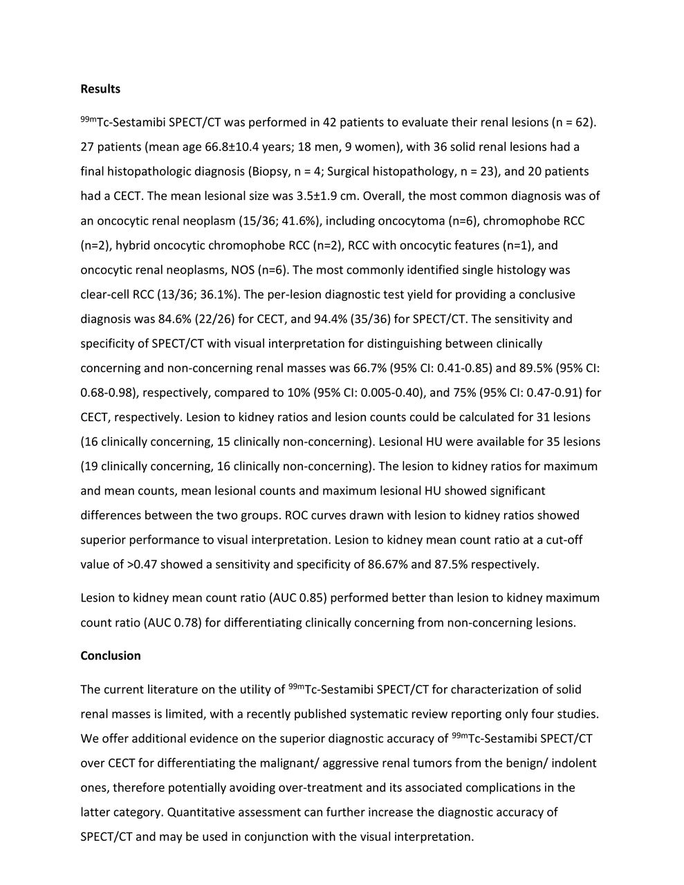
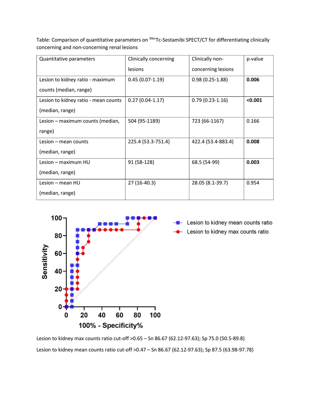
Results: 99mTc-Sestamibi SPECT/CT was performed in 42 patients to evaluate their renal lesions (n = 62). 27 patients (mean age 66.8±10.4 years; 18 men, 9 women), with 36 solid renal lesions had a final histopathologic diagnosis (Biopsy, n = 4; Surgical histopathology, n = 23), and 20 patients had a CECT. The mean lesional size was 3.5±1.9 cm. Overall, the most common diagnosis was of an oncocytic renal neoplasm (15/36; 41.6%), including oncocytoma (n=6), chromophobe RCC (n=2), hybrid oncocytic chromophobe RCC (n=2), RCC with oncocytic features (n=1), and oncocytic renal neoplasms, NOS (n=6). The most commonly identified single histology was clear-cell RCC (13/36; 36.1%). The per-lesion diagnostic test yield for providing a conclusive diagnosis was 84.6% (22/26) for CECT, and 94.4% (35/36) for SPECT/CT. The sensitivity and specificity of SPECT/CT with visual interpretation for distinguishing between clinically concerning and non-concerning renal masses was 66.7% (95% CI: 0.41-0.85) and 89.5% (95% CI: 0.68-0.98), respectively, compared to 10% (95% CI: 0.005-0.40), and 75% (95% CI: 0.47-0.91) for CECT, respectively. Lesion and kidney counts could be calculated for 31 lesions (16 concerning, 15 non-concerning). Lesional HU was available for 35 lesions (19 concerning, 16 non-concerning). The lesion to kidney ratios for maximum and mean counts, mean lesional counts and maximum lesional HU showed significant differences between the two groups. ROC curves drawn with lesion to kidney ratios showed superior performance to visual interpretation (sensitivity and specificity of 86.67% and 87.5% at a cut-off of >0.47). Lesion to kidney mean count ratio (AUC 0.85) performed better than lesion to kidney maximum count ratio (AUC 0.78) for differentiating clinically concerning from non-concerning lesions.

Conclusions: The current literature on the utility of 99mTc-Sestamibi SPECT/CT for characterization of solid renal masses is limited, with a recently published systematic review reporting only four studies. We offer additional evidence on the superior diagnostic accuracy of 99mTc-Sestamibi SPECT/CT over CECT for differentiating the malignant/ aggressive renal tumors from the benign/ indolent ones, therefore potentially avoiding over-treatment and its associated complications in the latter category. Quantitative assessment can further increase the diagnostic accuracy of SPECT/CT and may be used in conjunction with visual interpretation.

Figure
  • Download figure
  • Open in new tab
  • Download powerpoint
Figure
  • Download figure
  • Open in new tab
  • Download powerpoint
Figure
  • Download figure
  • Open in new tab
  • Download powerpoint
Previous
Back to top

In this issue

Journal of Nuclear Medicine
Vol. 63, Issue supplement 2
August 1, 2022
  • Table of Contents
  • Index by author
Article Alerts
Sign In to Email Alerts with your Email Address
Email Article

Thank you for your interest in spreading the word on Journal of Nuclear Medicine.

NOTE: We only request your email address so that the person you are recommending the page to knows that you wanted them to see it, and that it is not junk mail. We do not capture any email address.

Enter multiple addresses on separate lines or separate them with commas.
Diagnostic accuracy of 99mTc-Sestamibi SPECT/CT with visual and quantitative interpretation for the characterization of solid renal lesions
(Your Name) has sent you a message from Journal of Nuclear Medicine
(Your Name) thought you would like to see the Journal of Nuclear Medicine web site.
Citation Tools
Diagnostic accuracy of 99mTc-Sestamibi SPECT/CT with visual and quantitative interpretation for the characterization of solid renal lesions
Ashwin Singh Parihar, Joyce Mhlanga, Carrie Ronstrom, Lisa Schmidt, Robert Figenshau, Farrokh Dehdashti, Richard Wahl
Journal of Nuclear Medicine Aug 2022, 63 (supplement 2) 2617;

Citation Manager Formats

  • BibTeX
  • Bookends
  • EasyBib
  • EndNote (tagged)
  • EndNote 8 (xml)
  • Medlars
  • Mendeley
  • Papers
  • RefWorks Tagged
  • Ref Manager
  • RIS
  • Zotero
Share
Diagnostic accuracy of 99mTc-Sestamibi SPECT/CT with visual and quantitative interpretation for the characterization of solid renal lesions
Ashwin Singh Parihar, Joyce Mhlanga, Carrie Ronstrom, Lisa Schmidt, Robert Figenshau, Farrokh Dehdashti, Richard Wahl
Journal of Nuclear Medicine Aug 2022, 63 (supplement 2) 2617;
Twitter logo Facebook logo LinkedIn logo Mendeley logo
  • Tweet Widget
  • Facebook Like
  • Google Plus One
Bookmark this article

Jump to section

  • Article
  • Figures & Data
  • Info & Metrics

Related Articles

  • No related articles found.
  • Google Scholar

Cited By...

  • No citing articles found.
  • Google Scholar

More in this TOC Section

  • Can primary PET/CT staging using [68Ga]Ga-RM2 and [18F]Choline predict recurrence in prostate cancer?
  • Initial outcome of phase I study of Lu-177 Ludotadipep treatment in metastatic castration-resistant prostate cancer patients
  • [68Ga]-PSMA-11 can clarify equivocal lesions on conventional imaging and change management decisions among men with previously treated prostate cancer
Show more GU

Similar Articles

SNMMI

© 2025 SNMMI

Powered by HighWire