


Abstract
2604
Introduction: Down syndrome (DS) is characterized by the triplication of chromosome 21, which contains the gene that encodes for amyloid precursor protein (APP). The additional copy of this gene leads to earlier beta-amyloid (Aβ) plaque accumulation and an increased prevalence of Alzheimer’s disease (AD) in the DS population. While the age of AD onset is earlier in DS, AD pathology progression in non-DS sporadic AD follows similar trends to the DS cohort. The goal of this study is to compare the accumulation rates of Aβ measured with [C-11]PiB PET at a single site using the same scanner as participants become Aβ positive in order to evaluate the influence of the APP gene in Aβ deposition in the aging DS and non-DS populations.
Methods: For this study we selected individuals with four or more [C-11]PiB scans at our site at the University of Wisconsin spanning 11 years that had at least one Aβ positive [C-11]PiB scan, identified as having a grey matter SUVR greater than 1.40, and at least one Aβ negative scan. Using these criteria, we identified 11 out of 58 DS participants in the Neurodegeneration in Aging DS (NiAD) study and 10 out of 246 non-DS subjects from the PREDICT sub-study of the WRAP (Wisconsin Registry for Alzheimer’s Prevention) cohort that observes people with familial history of AD. An identical imaging procedure was conducted for DS and non-DS subjects imaged on an ECAT HR+ scanner from 50-70 minutes after injection with a nominal injected dose of 15mCi [C-11]PiB. Reconstructed PET images were aligned and summed before being normalized into MNI-152 space and converted into an SUVR image using cerebellar grey matter as a reference region. Global SUVR composed of grey matter regions in the anterior cingulate, frontal cortex, parietal cortex, precuneus, temporal cortex, and striatum from the AAL atlas was used as a metric for Aβ deposition. Rate of Aβ accumulation was quantified as the change in global SUVR per year between each scan with a focus on the difference seen between the last Aβ negative scan and the first Aβ positive scan for members of our cohorts.
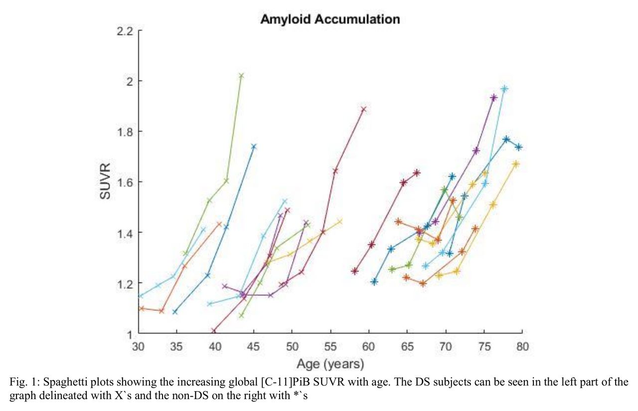
Results: The mean observed age of Aβ positivity for the DS cohort was 46.3±5.9 years and 69.0±3.6 years for the non-DS group. In a spaghetti plot of global [C-11]PiB SUVR and age (Fig 1) there is no age overlap between the two groups with the oldest DS subject becoming Aβ positive at age 54.1 and the youngest non-DS subject becoming positive at 61.4. The average rate of amyloid accumulation per year when becoming Aβ positive in the DS group was 0.060±0.026 compared to 0.056±0.026 for the non-DS sample. While there is a large variability in the accumulation rates of both image groups, the average and standard deviation of both datasets are quite similar. These two groups also have similar ranges of 0.020-0.103 and 0.019-0.114 respectively, however, the non-DS population group has less variance about the mean. This can be seen in box plots of the data (Fig 2), which have an interquartile range of 0.040 for the DS subjects and 0.019 for non-DS.
Conclusions: While we see a difference in the age of onset of Aβ between DS and non-DS, the rates of accumulation in the DS and general populations follow similar patterns as they transition to Aβ positivity. Although the overall prevalence of Ab pathology is near universal in adults with DS, these findings do not support the hypothesis that increased expression of the APP gene in DS results in more rapid deposition of Aβ in the brain. Related research showing that APOE4, an allele associated with earlier onset and a higher burden of Aβ, does not change Aβ accumulation rate (Lopresti 2020) also aligns with our results. However, as in the non-DS population, consideravle variability in the rate of accumulation is observed requiring further investigation towards understanding this process.


In this issue


Jump to section
Related Articles
Cited By...
- No citing articles found.